ABSTRACT:The purpose of the present study was to assess the correlation between transverse expansion and the increase in upper arch perimeter, after maxillary expansion. Dental casts of eighteen patients were obtained before treatment and again ive months after maxillary expansion. Measurements of intermolar width, intercanine width, arch length and arch perimeter were made with a digital caliper on photocopies taken from the dental casts. After assessment of the method error, a multiple regression model was developed following the identiication of the best subset of variables. The resulting equation led to the conclusion that the increase in arch perimeter is approxi-mately given by the addition of 0.54 times the intercanine expansion, and 0.87 times the arch length alteration. DESCRIPTORS:Orthodontics; Palatal expansion technique; Dental arch.
RESUMO: O presente estudo avaliou a correlação entre expansão transversal e aumento no perímetro do arco dentário superior, após disjunção maxilar, em dezoito pares de modelos de gesso, obtidos antes e depois de apro-ximadamente cinco meses da disjunção maxilar. Os modelos foram fotocopiados e as variáveis largura intermo-lares, largura intercaninos, comprimento e perímetro do arco dentário superior foram mensuradas por meio de paquímetro digital. Depois de veriicado o erro do método, um modelo de regressão múltipla foi desenvolvido em seqüência à identiicação do melhor conjunto de variáveis. A equação resultante permitiu concluir que o aumen -to no perímetro do arco é dado pela adição de 0,54 vezes a alteração intercaninos e de 0,87 vezes a alteração no comprimento do arco.
DESCRITORES: Ortodontia; Técnica de expansão palatina; Arcada dentária.
INTRODUCTION
The reduction of the transverse dimension of the mandible and the maxilla is one of the teeth-crowding etiological factors. Therefore, expansion will not only restore the proper dental arch range, but it will also provide additional space for sub-sequent alignment. However, forecasting the arch perimeter increase amount, resulting from expan-sion procedures, is questionable.
Inference of the dental arch perimeter in-crease resulting from transverse expansion is a common practice, usually found in planning and treatment protocols9,23. Treatment of transverse discrepancies is supposed to promote arch perim-eter increase, often allowing overall teeth-crowding correction. Despite the lack of unanimity found
in the related literature on the ratios between an increase in transverse dimensions and changes to arch perimeter and arch length, these ratios are used in orthodontic treatment planning and are often associated with the decision to extract or not. However, given the fact that different values have been found, the increase in arch perimeter can be overestimated or underestimated.
Some researchers have studied perimeter chang-es due to lower dental arch expansion5,7,17-20, and some due to expansion of the maxilla1,2,4,11,19; while other authors have utilized a linear regression equa-tion to estimate the perimeter incremental amount due to expansion1,2,4, and some have inferred this amount based on mean results of the variables5.
* Doctorate Students; **Associate Professor; ***PhD, Professor – Department of Orthodontics, School of Dentistry, University of São Paulo.
Correlation between transverse expansion and increase in the
upper arch perimeter after rapid maxillary expansion
Correlação entre expansão transversal e aumento no perímetro
do arco dentário após disjunção maxilar
Cristiane Aparecida de Assis Claro* Jorge Abrão**
Ricketts et al.21 (1982)suggested an increase of 0.25 mm in the perimeter for every 1 mm added to the intermolar distance, a ratio of 1:1 regarding the intercanine distance; and a ratio of 2:1 regarding the incisor proclination. The equation established by Adkins et al.1 (1990) estimates an arch perim-eter increase of 0.7 mm to every millimperim-eter added to the inter-first premolar distance. However, the equation proposed by Gandini et al.4(1997) states a ratio of 0.88 between perimeter increase and intermolar change.
The mathematical model developed by Ger-mane et al.5 (1991) led to the conclusion that an in-crement of 1 mm in the lower intermolar distance promotes a gain of 0.27 mm in arch perimeter. The combination of the increments of 1 mm in the intermolar distance and 1 mm in the interca-nine distance results in an increase of 0.93 mm in perimeter; while the simultaneous increase of 1 mm in the intercanine distance and 1 mm in arch length leads to a gain of 1.71 mm in arch perimeter.
Rapid palatal expansion increases arch perim-eter, providing space for correction of moderate (3 to 4 mm) crowding15, and allowing spontaneous lower arch expansion13. However, the outcome is more favorable when lower expansion is obtained with an appliance prior to rapid palatal expansion, increasing lower arch perimeter and additionally increasing upper expansion capacity, and con-sequently the upper arch perimeter15. The valid-ity of carrying out rapid palatal expansion in the absence of crossbites has lately been questioned, based on the assumption that in such situations it becomes necessary to work on the mandibular arch to coordinate treatment effects. However, ex-pansion of the mandibular arch is unstable6.
Rapid palatal expansion followed by fixed ap-pliances promoted an increase of 6.0 mm in upper arch perimeter and 4.5 mm in lower arch perim-eter14.
The aim of this study was to establish a cor-relation between transverse expansion and an increase in upper arch perimeter after maxillary expansion.
MATERIAL AND METHODS
The present work used upper arch study casts from eighteen patients with ages ranging from sev-en to tsev-en years, treated in a private practice. The sample size was defined after statistical analysis of a pilot group, and resulted in eighteen pairs
of upper casts obtained before and roughly five months after rapid palatal expansion. The study included only casts provided with first permanent molars, permanent central incisors and decidu-ous canines, in pretreatment and post-treatment stages, which were considered reference points for the measurement process.
Patients were treated with a bonded acrylic splint expander, featuring the utilization of a hy-rax-type screw, with a high opening capacity, and posterior occlusal acrylic coverage.
Measurement method
Before and after treatment, casts were pho-tocopied based on the paper by Simplício et al.22 (1995), which confirmed the reliability of this procedure in the study of occlusogram tracings. Aiming to identify the magnitude of distortion in-troduced by the copying process, the casts were photocopied side-by-side with a coin, used as a dimensional standard for image size adjustment of the copy machine, so that the coin diameter meas-ured with a digital caliper (accuracy: 0.01 mm) on the paper matched the actual coin diameter.
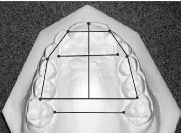
The selected reference points were the same adopted by Adkins et al.1 (1990), except for the first bicuspids, which were not included in the present study due to the sample age group. Ref-erence points were marked with a pen (Paper-mate, ultrafine model, Diadema, Brazil) to assist in locating and reducing potential measurement errors (Figure 1). The reference points were the following:
a) Inner lingual points on the gingival margin of the first upper molars were taken for the
intermolar width measurement.
b) Inner lingual points on the gingival margin of the deciduous canines were taken for the intercanine width measurement.
c) Points on the mesial aspect of the permanent first molars, on the distal aspect of the ca-nines and of the central incisors were taken for measurement of the arch perimeter. d) Points on the mesial aspect of the first molars
and on the mesial aspect of the central inci-sors were taken for the measurement of arch length.
Measurements were carried out with a digital caliper (Mitutoyo-Digimatic, Kawasaki, Japan) ac-curacy of 0.01 mm.
Measurement error
The same examiner repeated the measure-ments on 27.77% of the sample. Random errors of the measurement process were assessed using the Dahlberg test (1940)8 followed by the pairwise means t test for identification of systematic errors, if any.
Statistical method
A significance level of 5% was adopted. The number of samples was determined after a pi-lot study and it was similar to the sample size employed by other researchers. Data normality was proved by the Kolmogorov-Smirnov test. The regression model was constructed by employing the best subset practice. Correlation among ex-planatory variables was identified and properly treated to avoid the undesirable effect of multi-collinearity.
RESULTS
Measurement error assessment
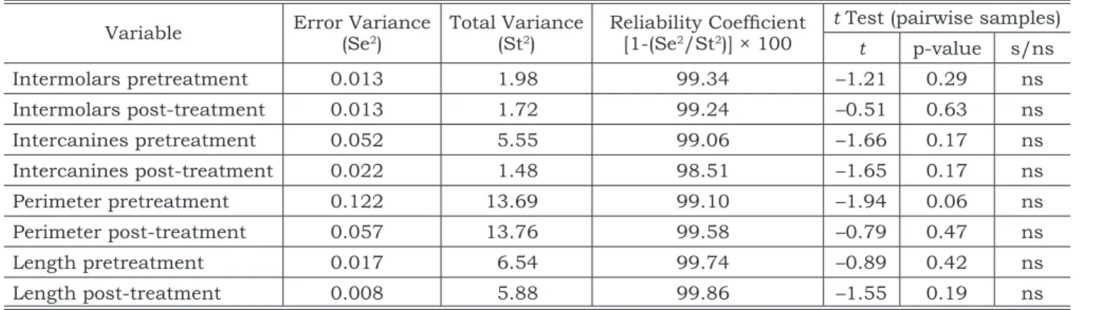
To evaluate reliability of the measurements, five pairs of casts (27.77% of the total sample) were measured twice. All variables were submitted to the Dahlberg test and to the t test. Results, shown in Table 1, revealed that the random error ranged from 0.14% to 1.49% (reliability coefficient from 98.51% to 99.86%) and that the systematic error was non-significant, confirming the data repeatability.
Sample size calculation
Before estimating sample size, a pilot lot of 5 samples was taken, resulting in a standard devia-tion (s) of 0.83 mm in perimeter change (response variable). By applying the equation n = (16s2/ d2) + 1, with a test power of 0.75 and a difference worth detecting (d) of 0.8 mm, a total of 18 samples was obtained.
Descriptive statistics
Table 2 shows the variables descriptive sta-tistics.
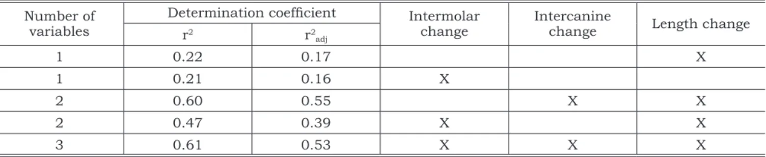
As dental arch perimeter can be influenced by several factors, the estimate of the contribution of each one to the regression model was investigated by the method of the BestSubsets, and the results are shown in Table 3.
The highest correlations values (r2 = 0.61 and r2adj = 0.53) and (r2 = 0.60 and r2adj = 0.55) have respectively led to the following multiple regres-sion equations:
Perimeter change = 0.37 + 0.12 of the intermolar change + 0.46 of the intercanine change + 0.84 of
arch length. TABLE 1 - Results of the Dahlberg test and of the t test.
Variable Error Variance (Se2)
Total Variance
(St2) Reliability Coeficient [1-(Se2/St2)] × 100
t Test (pairwise samples)
t p-value s/ns
Intermolars pretreatment 0.013 1.98 99.34 –1.21 0.29 ns
Intermolars post-treatment 0.013 1.72 99.24 –0.51 0.63 ns
Intercanines pretreatment 0.052 5.55 99.06 –1.66 0.17 ns
Intercanines post-treatment 0.022 1.48 98.51 –1.65 0.17 ns
Perimeter pretreatment 0.122 13.69 99.10 –1.94 0.06 ns
Perimeter post-treatment 0.057 13.76 99.58 –0.79 0.47 ns
Length pretreatment 0.017 6.54 99.74 –0.89 0.42 ns
Length post-treatment 0.008 5.88 99.86 –1.55 0.19 ns
Perimeter change = 0.77 + 0.54 of the intercanine change + 0.87 of arch length.
Both equations above are significant at a p level of p = 0.003 and p = 0.001, respectively. How-ever, the second equation was considered more suitable because no multicollinearity was found in the model; that is, the explanatory variables (intermolar change, intercanine change and arch length) were tested one against the other to identify the eventual correlation between them. By test-ing intermolar and intercanine distance change, a significant correlation was found (r = 0.66 and p = 0.002), which was not observed in the inter-molar distance change and arch length change (r = –0.06 and p = 0.797), nor in the intercanine distance change and arch length change (r = –0.36 and p = 0.134).
DISCUSSION
The present work assessed patients with mixed dentitions, as it is known that disjunction orthopedic results are more effective within this time span. The studies by Melsen12 (1975) and by Ennes, Consolaro3 (2004) have revealed a relation-ship between the complexity increase of the
mid-palatal suture with subject age hindering maxilla separation. Studies to estimate arch perimeter in-crease from transverse changes after disjunction have dealt with patients in the late transitional or early permanent dentition stage1,2.
In this study, an assessment after 5 months was based on the prescription by McNamara and Brudon15 which claims that, at this time interval, the midpalatal suture reossification and reorgani-zation have already taken place. The purpose of the present study was to establish the magnitude of the increment in arch perimeter due to transverse changes, and to use this information in treatment planning. For the estimate of the space available for future alignment, only the values related to the end of the appliance stabilization period (5 months) were considered. No other device was simultane-ously used during this time. However, each patient received orthodontic treatment according to their individual needs after the retention period.
This study did not include a control group, as the authors considered that changes resulting from growing in the time gap of nearly 5 months would be negligible. However, given the statement by Moorrees, Reed16 (1964) that the greater inter-canine distance change takes place during the re-placement of the incisors, it is important to clarify that only in 2 out of the 18 observed patients did the eruption of the lateral incisors take place dur-ing this assessment period. The literature review has shown that a control group was also not em-ployed in studies with a purpose similar to that of the present study1,2,4.
Research in the literature has shown a notable lack of agreement among authors (see Chart 1), with the application of distinct methodologies.
Previous works by Adkins et al.1 (1990) and Ak-kaya et al.2 (1998) have shown that an increase of 1 mm in the region of the first premolars promotes, respectively, a gain of 0.7 mm and 0.60-0.65 mm
TABLE 3 - Correlation between explanatory variables and response variable by means of the Best Subsets meth-od.
Number of variables
Determination coeficient Intermolar change
Intercanine
change Length change
r2 r2
adj
1 0.22 0.17 X
1 0.21 0.16 X
2 0.60 0.55 X X
2 0.47 0.39 X X
3 0.61 0.53 X X X
TABLE 2 - Descriptive statistics for the casts of the pa-tients submitted to rapid palatal expansion (mm).
Variable Mean SD Min. Max.
Intermolar
distance change 5.48 1.2 3.4 7.2 Intercanine
distance change 3.59 1.39 1.05 6.8
Arch perimeter
change 2.41 1.14 0.63 4.59
Arch length
in arch perimeter. In the above-mentioned stud-ies, results of orthopedic expansion were evalu-ated. However, Akkaya et al.2 (1998) obtained two equations, one being for rapid maxillary expansion (perimeter change = 0.62 + 0.65 times the first pre -molars width change) and the other for slow max-illary expansion (perimeter change = 0.03 + 0.60 times the first premolar width change), with deter-mination coefficients of r2adj = 0.67 and r2adj = 0.43, respectively. Equations for the changes after the retention period were developed leading to a pe-rimeter increase ratio of 0.54 times the change in premolar interdistance with the rapid expansion procedure, and 0.52 times the change in premolar interdistance with slow maxillary expansion.
Gandini et al.4 (1997) developed a study to as-sess the results of the orthodontic expansion with a removable expander appliance that led to the following equation: perimeter change = 0.52 + 0.88 of the intermolars change with r = 0.57, therefore with r2 = 0.32, significant at a level of 5%.
It seems controversial that the arch perimeter increase resulting from an increase in intermo-lar distance (0.88 of the intermointermo-lar change in the study by Gandini et al.4, 1997) is greater than that resulting from a change in the first premolar dis-tance (0.7 in the study by Adkins et al.1, 1990, and 0.6 to 0.65 in the work by Akkaya et al.2, 1998). Be-sides, it is expected that rapid maxillary expansion promotes a greater increase in arch perimeter than expansion with a removable expansion plate, since rapid maxillary expansion promotes the opening of the midpalatal suture with the greatest separation occurring anteriorly.
Ricketts et al.21 (1982) forecasts that an in-crease of 1 mm in the intermolar distance will re-sult in an increase of 0.25 mm in arch perimeter, and that an increase of 1 mm in the intercanine
distance will increase arch perimeter by 1 mm, and that an increase of 1 mm in arch length will promote a gain of 2 mm in arch perimeter. Such values are practically two times greater than the values found in the present study, that identified a gain of 0.12 mm in perimeter for every increase of 1 mm in the intermolar distance. Furthermore, each increase of 1 mm in the intercanine distance promoted a gain of 0.46 mm in perimeter, and each increase of 1 mm in arch length led to a gain of 0.84 mm in arch perimeter, and eliminating the multicollinearity factor by removing the intermolar distance from the equation, the gain in perimeter was 0.54 times the intercanine distance and 0.87 times the arch length.
The comparison of ratios obtained by the dif-ferent studies is compromised due to distinct meth-odologies. Some studies were developed using cast models1,2,4, and others used computerized simula-tions and math analysis5,17-19; some evaluated the upper arch1,2,4, and others, the lower arch5,17-19.
It is known that the establishment of regres-sion equations is strongly dependent on a large sample size. Most of the works, including the pres-ent one, have been done with a relatively small number of subjects due to the inclusion criteria (age group, presence of specific teeth, appliances of the same type etc). Because of this, the employ-ment of meta-analysis in future studies will be very helpful to the scientific community by increasing sample size, testing statistical power, dealing with the uncertainties of controversial studies and fi-nally responding to questions poorly clarified in individual tests.
Initial arch shape is probably a factor to be considered in subsequent studies, considering that there are indications linking arch shape to distinct space gains in arch perimeter. By means CHART 1 - Perimeter arch relative gain values (mm) resulting from a 1 mm increase in the intermolar distance, interpremolar distance, intercanine distance and arch length.
Source Gain ratio from the increase of 1 mm in the distance:
Inter molars Inter premolars Inter canines Length
Ricketts et al.21 (1982) 0.25 1 2
Adkins et al.1 (1990) 0.7
Germane et al.5 (1991) 0.27 Gandini et al.4 (1997) 0.88
Akkaya et al.2 (1998) 0.6 to 0.65
Noroozi et al.19 (2002) 0.3 0.6 1
of a mathematic-geometric model, Mutinelli et al.18 (2000) identified the changes promoted in arch length by proclinating the lower incisor by 1 mm, keeping the intercanine distance, in dis-tinct arch forms. The length increment in the par-abolic arch was 1.51 mm, in the elliptical arch, 1.21 mm, in the hyperbolic arch, 1.61 mm, in the circular arch, 1.21 mm, and in the catenary arch, 2.07 mm.
Catenary forms were predominantly found describing the curvature of the upper and lower arches, regardless of facial types10.
Future studies will be able to compare the re-lationship between the increase in arch perimeter
and transverse expansion considering the use of different appliances.
CONCLUSIONS
Based on the evidence found, and considering the conditions under which the present study was conducted, it could be concluded that the increase in upper arch perimeter was 0.54 times the inter-canine expansion and 0.87 times the change in arch length.
The above conclusions may be used during treat-ment planning as a guideline to estimate upper arch perimeter gain as a consequence of expansion.
REFERENCES
1. Adkins MD, Nanda RS, Currier GF. Arch perimeter change on rapid palatal expansion. Am J Orthod Dentofac Orthop 1990;97(3):194-9.
2. Akkaya S, Lorenzon S, Ucem TT. Comparison of dental arch and arch perimeter changes between bonded rapid and slow maxillary expansion procedures. Eur J Orthod 1998;20(3):255-61.
3. Ennes J, Consolaro A. Sutura palatina mediana: avaliação do grau de ossificação em crânios humanos. R Dent Press Ortodon Ortop Facial 2004;9(5):64-73.
4. Gandini Jr LG, Oriqui OR, Riethmueller M, Santos Pinto A, Loffredol CM. Alterações dimensionais dos arcos den-tários no tratamento ortodôntico com aparelho expansor removível. Ortodontia 1997;30(1):39-44.
5. Germane N, Lindauer SJ, Rubenstein LK, Revere JH, Isaac-son RJ. Increase in arch perimeter due to orthodontic expan-sion. Am J Orthod Dentofacial Orthop 1991;100(5):421-7. 6. Gianelly AA. Rapid palatal expansion in the absence of
crossbites: Added value? Am J Orthod Dentofacial Orthop 2003;124(4):362-5.
7. Houlsley JA, Nanda RS, Currier F, McCune DE. Stability of transverse expansion in the mandibular arch. Am J Orthod Dentofacial Orthop 2003;124(3):288-93.
8. Houston WJB. The analysis of errors in orthodontic measure-ments. Am J Orthod Dentofacial Orthop 1983;83:382-90. 9. Interlandi S. Gráfico Vetorial Ortodôntico (Versão 2002).
R Dent Press Ortodon Ortop Facial 2002;7(4):23-42. 10. Kanashiro LK, Vigorito JW. Estudo das formas e
dimen-sões das arcadas dentárias superiores e inferiores em leu-codermas, brasileiros, com maloclusão de classe II, divisão 1 e diferentes tipos faciais. Ortodontia 2000;33(2):8-18. 11. Maruyama NE, Paiva JB, Rino Neto J, Novelli MD.
Análise do perímetro do arco dentário e da área palatina após expansão rápida da maxila [resumo]. RPG Rev Pos Grad 2002;9(3):275.
12. Melsen B. Palatal growth studied on human autopsy material. A histologic microradiographic study. Am J Or-thod 1975;68:42-54.
13. McLuckie WC. Effects of slow maxillary expansion on mandibular arch width. Am J Orthod Dentofacial Orthop 1994;105(3):317-8.
14. McNamara JA, Baccetti T, Franchi L, Herberger TA. Rapid maxillary expansion followed by fixed appliances: a long-term evaluation of changes in arch dimensions. Angle Orthod 2003;73(4):344-53.
15. McNamara JA, Brudon WL. Tratamiento ortodóncico y orto-pédico en la dentición mixta. Ann Arbor: Needham; 1995. 16. Moorrees CF, Reed RB. Changes in dental arch
di-mensions expressed on the basis of tooth eruption as a measure of biologic age. J Dent Res 1964;44:129-41. 17. Motoyoshi M, Hirabayashi M, Shimazaki T, Namura S.
An experimental study on mandibular expansion: increases in arch width and perimeter. Eur J Orthod 2002;24:125-30.
18. Mutinelli S, Manfredi M, Cozzani M. A mathematic-geometric model to calculate variation in mandibular arch form. Eur J Orthod 2000;22:113-25.
19. Noroozi H, Djavid GE, Moeinzad H, Teimouri AP. Predic-tion of arch perimeter changes due to orthodontic treatment. Am J Orthod Dentofacial Orthop 2002;122(6):601-7. 20. Osborn WS, Nanda RS, Currier GF. Mandibular arch
perimeter changes with lip bumper treatment. Am J Orthod Dentofacial Orthop 1991;99(6):527-32.
21. Ricketts RM, Roth RH, Chaconas SJ, Schulhof RJ, Engel GA. Orthodontic diagnosis and planning. USA: Rocky Mountain Data Systems; 1982.
22. Simplício AHM, Souza LA, Sakima MT, Martins JCR, Saki-ma T. Confiabilidade de xerox de modelos de estudo para o traçado de oclusogramas. Ortodontia 1995;28(3):62-7. 23. Vigorito JW. Ortodontia preventiva: pequenos
movi-mentos dentários. Ganhos e manutenção de espaços. In: Vigorito JW. Ortodontia clínica: diagnóstico e terapêuticas. São Paulo: Santa Madonna; 2004. cap. 8. p. 293-339.