INTRODUCTION
Congenital diaphragmatic hernia (CDH) is a severe developmental anom-aly, with a mean incidence of 1:2,500 live births, in which etiology remains poorly understood (1,2). This congenital anomaly is characterized by a diaphrag-matic defect that allows intrathoracic herniation of abdominal organs, and consequently, maldevelopment of the alveoli and pulmonary vessels. For
many years, this malformation was thought to be a surgical emergence, solely related to a diaphragmatic defect, and potentially curable by surgical clo-sure of this defect after birth, which al-lowed lung expansion. However, during 90 years, CDH pathophysiology pro-gressed for a physiological emergence (3,4). It is now clear that lung hypopla-sia and consecutive persistent pul-monary hypertension (PH) associated
with this disorder are the key determi-nants of mortality (1–4). Despite the im-provements in understanding CDH pathophysiology and advances in neo-natal care, such as the use of extra cor-poreal membrane oxygenation and in-haled nitric oxide, the mortality (50%) and morbidity rate in CDH newborns remains exceedingly high (1–4). In hu-mans, CDH can be accurately diagnosed at second trimester during routine ultra-sound examination. Therefore it is amenable to antenatal therapies. Hence, antenatal therapies that promote fetal lung growth remain an appealing ap-proach to improve survival of CDH fe-tuses. However, fetal surgical interven-tions, such as fetal tracheal occlusion, are invasive, technically demanding, limited by the maternal and fetal risks, and their efficacy is still not determined, with controversial results in survival
Treat Congenital Diaphragmatic Hernia
Cristina Nogueira-Silva,
1,2,3Emanuel Carvalho-Dias,
1,2,4Paulina Piairo,
1,2Susana Nunes,
5Maria J Baptista,
1,2,6Rute S Moura,
1,2and Jorge Correia-Pinto
1,2,71Life and Health Sciences Research Institute (ICVS), School of Health Sciences, University of Minho, Braga, Portugal; 2ICVS/3B’s - PT Government Associate Laboratory, Braga/Guimarães, Portugal; 3Department of Obstetrics and Gynecology, Hospital de Braga, Braga, Portugal; Departments of 4Urology, 5Pediatrics, and 6Pediatric Cardiology, Hospital de São João, Porto, Portugal; and 7Department of Pediatric Surgery, Hospital de Braga, Braga, Portugal
Antenatal stimulation of lung growth is a reasonable approach to treat congenital diaphragmatic hernia (CDH), a disease characterized by pulmonary hypoplasia and hypertension. Several evidences from the literature demonstrated a possible in-volvement of renin-angiotensin system (RAS) during fetal lung development. Thus, the expression pattern of renin, angiotensin-con-verting enzyme, angiotensinogen, type 1 (AT1) and type 2 (AT2) receptors of angiotensin II (ANGII) was assessed by immunohisto-chemistry throughout gestation, whereas the function of RAS in the fetal lung was evaluated using fetal rat lung explants. These were morphometrically analyzed and intracellular pathway alterations assessed by Western blot. In nitrofen-induced CDH model, pregnant rats were treated with saline or PD-123319. In pups, lung growth, protein/DNA ratio, radial saccular count, epithelial dif-ferentiation and lung maturation, vascular morphometry, right ventricular hypertrophy and overload molecular markers, gasom-etry and survival time were evaluated. Results demonstrated that all RAS components were constitutively expressed in the lung during gestation and that ANGII had a stimulatory effect on lung branching, mediated by AT1receptor, through p44/42 and Akt phosphorylation. This stimulatory effect on lung growth was mimicked by AT2-antagonist (PD-123319) treatment. In vivo antenatal PD-123319 treatment increased lung growth, ameliorated indirect parameters of pulmonary hypertension, improved lung function and survival time in nonventilated CDH pups, without maternal or fetal deleterious effects. Therefore, this study demonstrated a local and physiologically active RAS during lung morphogenesis. Moreover, selective inhibition of AT2receptor is presented as a putative antenatal therapy for CDH.
Online address: http://www.molmed.org doi: 10.2119/molmed.2011.00210
Address correspondence toJorge Correia-Pinto, Life and Health Sciences Research Insti-tute (ICVS), School of Health Sciences, University of Minho, Campus de Gualtar; 4710-057 Braga, Portugal. Phone: +351 253 604 910; Fax: +351 253 604 847; E-mail:
and morbidity rates (5,6). Therefore, less invasive approaches such as antenatal pharmacological treatment to stimulate lung growth before birth and to treat PH are also under investigation (3,7–10).
The renin–angiotensin system (RAS) is a classical endocrine system regulating blood pressure, electrolyte and fluid ho-meostasis, involving several key compo-nents, namely angiotensinogen (the hepatic-derived precursor), two critical enzymes, renin (secreted from the juxta-glomerular apparatus of the kidney) and angiotensin-converting enzyme (ACE, pulmonary-bound metalloproteinase), whose sequential actions produce an-giotensin I and the physiologically ac-tive, angiotensin II (ANGII), respectively (11). ANGII operates on two G protein-coupled receptors, the ANGII type 1 (AT1) and type 2 (AT2) receptors (11). During the last two decades, in addition to this classic endocrine system, evidence has demonstrated the presence of a local RAS with autocrine/paracrine actions in several developing organs, such as fetal kidney, heart, vasculature and adrenal development (12).
Regarding lung morphogenesis, there is some evidence that lung expresses ACE as well as AT1and AT2receptors during fetal development (13–15). How-ever, RAS components expression pat-tern, as well as effects during lung mor-phogenesis, is largely unknown.
In this study, we assessed the expres-sion pattern and function of RAS during fetal lung development. Moreover, we assessed the role of RAS as a putative target for treatment of fetal lung hy-poplasia in CDH.
MATERIALS AND METHODS
Animal experiments were performed according to the Portuguese law for ani-mal welfare (16; Diário da República, Por-taria 1005/92). Animals were housed in an accredited mouse house and treated as specified by the recommendations of the Helsinki convention (17) and the
Guide for the Care and Use of Laboratory Animals, published by the National Academy Press (18).
Experimental Design and Animal Model
In vitrostudies were carried out to as-sess expression and function of RAS in fetal lung, whereas in vivostudies were performed to explore RAS as a target to treat fetal lung hypoplasia using the nitrofen-induced CDH rat model (19).
Sprague Dawley female rats (225 g; Charles River; Barcelona, Spain) were maintained in appropriate cages under controlled conditions and fed with com-mercial solid food. The rats were mated and checked daily for vaginal plug. The day of plugging was defined as gesta-tional d 0.5 for time-dating purposes. According to the nitrofen-induced CDH rat model (19), at 9.5 d postconception (dpc), randomly selected pregnant rats were exposed to 100 mg nitrofen (2,4-dichlorophenyl-p-nitrophenylether). At different time points, fetuses were har-vested by cesarean section. After fetal de-capitation, a toraco-laparotomy was per-formed under a binocular surgical microscope (Leica, Wild M651.MSD, Heerbrugg, Switzerland) to inspect the diaphragm and harvest the organs. Fe-tuses were assigned to three experimen-tal groups: (i) Control group (C), fetuses not exposed to nitrofen; (ii) Nitrofen group (N), fetuses exposed to nitrofen without CDH; (iii) CDH group, fetuses exposed to nitrofen with CDH.
In Vitro Studies
Normal fetuses removed by cesarean section at 13.5, 15.5, 17.5, 19.5 and 21.5 dpc were killed by decapitation, and lungs dissected and processed for im-munohistochemistry (IHC). Lungs of fe-tuses with 13.5 dpc were also dissected to perform fetal lung explants cultures and posterior western blot analysis.
IHC
IHC was performed on formalin-fixed and paraffin-embedded lungs, as previ-ously described by Nogueira-Silva et al. (7). Renin antibody (sc-27320; Santa Cruz Biotechnology, Santa Cruz, CA, USA) was used in a 1:25 dilution, ACE antibody (sc-20791; Santa Cruz Biotechnology Inc.) in a
1:75 dilution, angiotensinogen antibody (Abbiotec LLC, San Diego, CA, USA) in a 1:100 dilution, AT1receptor antibody (sc-1173; Santa Cruz Biotechnology Inc.) in a 1:50 dilution and AT2receptor antibody (sc-9040; Santa Cruz Biotechnology Inc.) in a 1:25 dilution. Incubation with the UltraVision detection system polyvalent horseradish peroxidase (Lab Vision Corporation, Fremont, CA, USA) or with the goat ImmunoCruz™ Staining System (Santa Cruz Biotechnology Inc.) was carried out according to the manufac-turer’s instructions.
Fetal Lung Explant Cultures
Recombinant ANGII (Sigma, St Louis, MO, USA), AT1-antagonist (ZD-7155; Tocris Cookson, Bristol, UK) and AT2 antagonist (PD-123319; kindly supplied by Pfizer, Groton, CT, USA) were added daily to lung explants to achieve increas-ing concentrations of ANGII (from 10–9to 10–4mol/L) or ZD-7155 or PD-123319 at 10–5mol/L. These doses were selected ac-cording to the literature (20,21). This set of experiments created the following groups: control (n= 30), ANGII 10–9(
n= 10), ANGII 10–8(
n= 11), ANGII 10–7(
n= 11), ANGII 10–6(
n= 12), ANGII 10–5(
n= 12), ANGII 10-4(
n= 12), ZD-7155 (n= 12), and PD-123319 (n= 15). Furthermore, explants were treated with ANGII at 10–9mol/L in the presence of ZD-7155 or PD-123319 at 10–5mol/L, creating the additional groups: ANG 10–9+ ZD-7155 (
n= 12) and ANG 10–9+ PD-123319 (
n= 15). Cultures were photographed daily and branching morphogenesis was assessed according to Nogueira-Silva et al. (22).
Western Blot Analysis
control, blots were probed with β-tubulin mAb (Abcam plc, Cambridge, UK) (n= 3). Quantitative analysis was performed with Quantity One 4.6.5 1-D Analysis Software (Bio-Rad Laboratories, Her-cules, CA, USA).
In Vivo Studies
Pregnant rats, control or exposed to ni-trofen, were anesthetized at 12.5 dpc with a mix of ketamine (75 mg/kg) and medetomidine (0.5 mg/kg) for subcuta-neous implantation of an osmotic micro-pump filled either with saline or PD-123319 (randomly), on mid scapular region (Alzet osmotic pump Model 2ML1; Durect Corporation, Cupertino, CA, USA). Saline or PD-123319 solution was infused with a rate of 10 µL/h (20 mg/kg/d for PD-123319). For the re-versal of the sedative effect, atipamezole (0.25 mg/kg) was used and butorfanole (1 mg/kg) was administered immedi-ately after the surgery. Fetuses were as-signed to six experimental groups: Con-trol rats treated with saline (C + S); Control rats treated with PD-123319 (C + PD); Nitrofen rats treated with saline (N + S); Nitrofen rats treated with PD-123319 (N + PD); CDH rats treated with saline (CDH + S); and CDH rats treated with PD-123319 (CDH + PD).
Pregnant rats were randomly assigned for cesarean section at 21.5 dpc or for spontaneous delivery at term.
The pregnant rats killed at 21.5 dpc were laparotomized, and after longitudi-nal hysterotomy, each fetus (C + S, n= 14; C + PD, n= 15; N + S, n= 15; N + PD,
n= 12; CDH + S, n= 15; CDH + PD, n= 24) was extracted, weighed and decapi-tated for organ harvesting (lungs, heart and kidneys). Organs were also weighed independently, organs-to-body weight ratios were assessed and lungs were ei-ther snap frozen in liquid nitrogen for biochemical analyses or fixed in 4% paraformaldehyde (PAF) for histological analyses. In this set of experiments, the level of lung hypoplasia was calculated according to Baptista et al. (9).
The pups that were delivered sponta-neously at term were placed immediately
after birth in a light-heated box and ran-domly killed 5 min after birth or allowed to survive, without any care, support strategies or ventilatory support. Those killed by decapitation at min 5 (C + S, n= 15; C + PD, n= 9; N + S, n= 11; N + PD,
n= 6; CDH + S, n= 12; CDH + PD, n= 14) were used for blood collection (neck bleeding) and gasometric evaluation (i-Stat1 analyser; Abbott, Chicago, IL, USA). These were then dissected for identifica-tion of CDH. Moreover, hearts were dis-sected for right ventricular hypertrophy evaluation and myocardial samples of right ventricular free wall were harvested and processed for q-PCR studies. The re-maining fetuses were allowed to survive (C + S, n= 10; C + PD, n= 9; N + S, n= 14; N + PD, n= 21; CDH + S, n= 14; CDH + PD, n= 28), evaluated by two in-dependent–blind observers (MJ Baptista and S Nunes) and scored at 1, 3, 5 and subsequently at each 5 min, using an APGAR-like score (Table 1, adapted from Dauger et al. [23]). The moment of death was registered and defined by the mo-ment in which the two observers attrib-uted an APGAR 0 (marked cyanosis, apnea, no spontaneous movements, no response to stimulus). All experiments were recorded in video to clarify any doubt. These pups were opened post-mortem for diaphragm inspection.
Lungs, heart and kidneys from all pregnant rats were weighed for maternal organ-to-body weight ratio analysis.
Biochemical Studies for Protein/DNA Ratio Assessment
Total lungs (left and right) were processed to determine protein and DNA contents. Protein content was assessed
by Bradford method (24). DNA was ex-tracted using the DNeasy Blood & Tissue Kit (Qiagen, Hilden, Germany).
Histological Studies
The trachea was cannulated and the lungs were fixed with PAF under a con-stant pressure of 20 cmH2O. Lungs were embedded in paraffin and 4-µm sections
were used to determine radial saccular count [RSC; using hematoxilin-eosin stain (H&E)], epithelial differentiation [IHC for clara cell secretory protein (CCSP) and prosurfactant protein C (SP-C) and determination of glycogen-content using periodic acid-Schiff stain (PAS)] and medial arterial thickness (using Weigert stain).
RSC was estimated according to Emery and Mithal adapted method (25), at 100×magnification (Olympus BX61
microscope; Olympus, Tokyo, Japan), in 5 animals per group, 6 slides per animal (200 µm apart each other), and 10
seg-ments per slide, by a blinded examiner (E Carvalho-Dias).
Regarding epithelial differentiation, IHC was performed as previously de-scribed by Nogueira-Silva et al. (7), briefly described above. CCSP antibody [07-623; Upstate (Millipore), Billerica, MA, USA] was used in a 1:800 dilution and SP-C antibody (AB3428; Chemicon International, Temecula, CA, USA) in a 1:400 dilution. These slides and PAS stained slides were observed and pho-tographed (Olympus BX61 microscope). The percentage of CCSP, SP-C and PAS stained cells per microscopic field was scored in a single-blinded fashion (100×
and 400×magnification, respectively) in
6 independent peripheral and 4 central
Table 1.APGAR-like score.a
Sign Score 0 Score 1 Score 2
Skin color Marked cyanosis Mild cyanosis/ pale Pink
Breathing Apnea Irregular or weak breathing Regular breathing Spontaneous motor No movements Weak movements Vigorous movements
activity
Reactivity to stimulus No response Gasping movements Active movements
a
areas per section (6 slides per animal, 200µm apart each other, and 5 animals per each experimental group). Scoring was as follows: 1, 0–20% cells/field; 2, 20–40% cells/field; 3, 40–60% cells/field; 4, 60–80% cells/field; 5, 80–100%.
Weigert resorcin fuchsin solution stains elastic fibbers, and it was used for morphometric assessment of pulmonary arteries (26). Pulmonary arteries were distinguished from pulmonary veins on the basis of the structure and position. Arteries that were approximately round [that is, the longest external diameter (ED, distance between the external elastic laminae), did not exceed the minimal ED by more than 50%] and had both clearly visible external and internal elastic lami-nae were analyzed. As the structural changes and pharmacological effects on pulmonary arteries might be vessel size dependent, we selected for further analy-sis arteries subcategorized into 3 sizes: ED less than 30 µm, ED 30 µm to 50 µm, and ED greater than 50 µm. Then, using AxionVision Rel. 4.3 (Carl Zeiss, Göttin-gen, Germany), internal area (IA, defined by internal elastic lamina), external area (EA, defined by external elastic lamina) and total area (TA, defined by edge of the vascular sheath) were measured. The percentage of medial (MA) and adventi-tial arterial area (AA) were calculated ac-cording to the following formulas: MA (%) = [(EA – IA)/EA] ×100; AA (%) = [(TA – EA)/EA] ×100. At least 10 arteries for each section were evaluated, 6 sec-tions per animal (200 µm apart each other), and 3 animals per each experi-mental group (at 400×magnification).
Right Ventricular Hypertrophy Evaluation
Hearts (C + S, n= 8; CDH + S, n= 6; CDH + PD, n= 8) were used for right ventricle (RV) and left ventricle (LV) dis-section (LV contains septum). RV and LV were weighed separately. The ratio RV/LV was determined and used as an index of right ventricular hypertrophy.
Other hearts (C + S, n= 7; CDH + S,
n= 6; CDH + PD, n= 6) were dissected, transversally cut and orientated
accord-ing to short-axis view of the heart, before fixed in PAF and embedded in paraffin. Four micrometer sections were stained with H&E and photographed at 40×
magnification (Olympus BX61 micro-scope). The right ventricular wall thick-ness was determined in the short-axis view of the heart, in the maximum dis-tance between the right side of the inter-ventricular septum to the right ventricu-lar free wall, using AxionVision Rel. 4.3 (Carl Zeiss) (5 measurements per animal, 20 µm apart each other).
q-PCR Studies
Right ventricular mRNA expression of angiotensinogen, B-type natriuretic pep-tide (BNP) and endothelin-1 (ET-1), genes previously defined as ventricular overload markers, was evaluated accord-ing to Baptista et al. (27).
Statistical Analysis
Data are presented as mean ± SEM. Statistical analysis was performed using the statistical software SigmaStat (ver-sion 3.5; Systat Software Inc., Chicago, IL, USA). Multiple group comparisons were made by analysis of variance. For morphometric explants studies, Western blot analysis, biochemical studies, histo-logical studies, right ventricular hyper-trophy evaluation and q-PCR studies, one-way ANOVA on ranks was used. Two-way ANOVA on ranks was used for organ-to-body weight ratio analysis and gasometric studies. Student-Newman-Keuls test was used for posttest analysis. Statistical significance was confirmed at
P< 0.05.
All supplementary materials are available online at www.molmed.org.
RESULTS
RAS Components Expression Pattern during Fetal Lung Development
The IHC studies revealed that all RAS components, renin, ACE, angiotensino-gen, AT1and AT2receptors, were ex-pressed throughout all studied gesta-tional ages in the fetal lung (Figure 1).
Renin was expressed in bronchiolar and also in alveolar epithelium throughout gestation (Figure 1A) since 13.5 dpc and appears to be maximal in the most immature buds (see Supplemental Fig-ures 1A-D). Endothelial ACE expression started in larger proximal vessels, early in the gestation, and spreads distally to involve progressively smaller vessels (Figure 1B). Interestingly, ACE protein was also detectable in epithelial cells since 13.5 dpc (see Supplemental Figure 1E). Regarding angiotensinogen, protein ex-pression was clearly observed in epithe-lial cells since 15.5 dpc (Figure 1C). Mes-enchymal tissue also displayed scattered angiotensinogen positive cells, first in endothelial cells and in the later gesta-tional ages (19.5 and 21.5 dpc) clearly in vascular smooth muscle cells (Figure 1C). AT1receptor protein was first mainly ex-pressed by undifferentiated mes-enchyme at 15.5 dpc and, throughout the gestation, it was predominantly ex-pressed by vascular smooth muscle cells and scattered in the mesenchyme (Fig-ure 1D). AT2receptor was expressed in bronchial epithelial cells and since 19.5 dpc muscle cells of large blood vessels also expressed it (Figure 1E). Moreover, the epithelial AT2expression was also demonstrated at 13.5 dpc (see Supple-mental Figure 1F).
Role of ANGII in Fetal Lung Development
enhanced lung branching in a similar way to the dose of ANGII-inducing maximal effect (Figure 2C). Moreover, the stimulatory effect on lung branching induced by ANGII 10–9mol/L was com-pletely abolished by AT1-antagonist treatment, and the simultaneous lung treatment with ANG 10–9mol/L and PD-123319 did not accomplish additional stimulatory effect on explants growth when compared with ANG 10–9mol/L treatment (Figure 2C). Thus, these
re-sults demonstrated that ANGII had a stimulatory effect on lung branching, mediated by AT1receptor. Interestingly, this stimulatory effect was mimicked by treatment with AT2-antagonist alone.
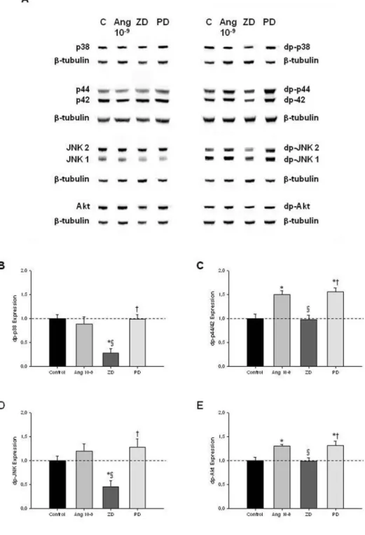
To clarify the intracellular signaling pathways that mediate ANGII actions on lung growth, lung explants treated with ANGII at 10–9mol/L (selected due to its maximal effect in explant growth), ZD-7155 or PD-123319 were evaluated for MAPK and Akt pathways activation
(Figure 3A). AT1receptor blockage in-duced a significant decrease of p-38 and JNK phosphorylation when compared with control explants (Figures 3B, D, re-spectively). On the other hand, the in-crease on lung branching, induced by ANGII at 10–9mol/L and AT
2receptor antagonist, significantly stimulated p44/42 and Akt phosphorylation (Fig-ures 3C, E, respectively).
In Vivo Antenatal PD-123319
Treatment Improves Fetal Lung Growth Left, right and total lung-to-body weight ratio (LW/BW) were analyzed for different experimental groups. Ac-cording to the nitrofen-induced CDH experimental model, pups of nitrofen-treated dams presented left and right lung hypoplasia, which was maximal in CDH + S group. Maternal PD-123319 treatment induced significant growth of both left and right lungs in control, nitrofen and CDH groups. In fact, LW/BW was significantly higher in the control, nitrofen and also CDH rats treated with PD-123319 when compared with the respective saline-treated groups (Figure 4A). Indeed, PD-123319 treatment stimulated partial recovery of lung hypoplasia in CDH neonates, in-ducing an increase of 11.4% in total lung weight (Figure 4B). Considering these results and to assess the potential of PD-123319 as a useful treatment for severe lung hypoplasia associated with CDH, we pursued our study focused in com-paring C + S, CDH + S and CDH + PD groups.
Biochemical analysis of lung protein and DNA content demonstrated that there was no significant difference in the protein/DNA ratio between C + S, CDH + S and CDH + PD groups (C + S 0.024 ± 0.005; CDH + S 0.030 ± 0.004; CDH + PD 0.026 ± 0.002).
The histological analysis of lung archi-tecture showed that CDH + S lungs ap-peared to have a thickened septal and saccular walls and an increased amount of interstitial tissue when compared with C + S. However, CDH + PD had a signif-icantly greater development of saccules
Figure 1.Protein expression pattern of RAS components during fetal lung development (from 15.5 until 21.5 dpc). (A) Renin was predominantly expressed in epithelium. (B) ACE expression. (C) Angiotensinogen expression in epithelial, endothelial (arrow) and vascular smooth muscle cells (arrowhead). (D) AT1receptor immunostaining. (E) AT2receptor
and airspaces when compared with CDH + S lungs (Figure 5A). In fact, RSC of CDH + S pups was significantly lower when compared with C + S group (Fig-ure 5B). However, PD-123319 treatment induced a significant increase on RSC of CDH pups, that is, promoted distal lung development (Figure 5B).
Regarding epithelial differentiation and lung maturity, CCSP (proximal ep-ithelial cell differentiation marker), SP-C (distal epithelial cell differentiation marker) and PAS+glycogen stores (an in-direct signal of immaturity) were evalu-ated (Figure 6). No significant differences in score of CCSP (Figure 6A), SP-C (Fig-ure 6B) and PAS-stained cells (Fig(Fig-ure 6C) between C + S and CDH groups were observed.
Concerning other organs than lungs, nitrofen treatment (nitrofen and CDH groups) induced decrease of heart-to-body weight (HW/BW) and kidney-to-body weight (KW/BW) ratios. However, antenatal PD-123319 treatment did not significantly change either HW/BW or KW/BW (see Supplemental Figure 2).
Antenatal PD-123319 Treatment Partially Reversed Arterial Structural Abnormality and Decreased Molecular Markers of PH on CDH Model
MA percentage was significantly in-creased in all arteries (independently of its size) in CDH + S group when com-pared with C + S and CDH + PD groups (Figure 7). CDH + PD neonates pre-sented a significant decrease in MA per-centage when compared with CDH + S for all arterial sizes, with maximal effect on smaller arteries. The effect was so evi-dent that CDH + PD arteries had no sig-nificant difference in MA percentage when compared with C + S group (Fig-ure 7B). Regarding adventitial layer, saline or PD-treated CDH groups pre-sented a significant increase in AA per-centage when compared with C + S for all arterial sizes (Figure 7C). Thus, PD-123319 treatment induced decrease of MA percentage and did not induce any change on AA percentage.
Regarding right ventricular hypertro-phy index and right ventricle thickness,
markers of right ventricular hypertrophy secondary to chronic PH, CDH neonates did not present differences when com-pared with control group. Moreover, PD-123319 treatment did not change these parameters (Table 2).
Additionally, expression levels of right ventricular molecular markers of PH (an-giotensinogen, BNP and ET-1) were as-sessed in pups allowed to be delivered at 5 min after birth. In Figure 7D, mRNA levels of these markers normalized to
β-actin are shown. CDH + S group presented a significant increase of angio -tensinogen and an increase (although not statistical significant) of BNP and ET-1 compared with C + S group. On its turn, PD-123319 treatment significantly de-creased these overload markers on CDH pups.
Antenatal PD-123319 Treatment Improves Lung Function and Survival
The adaptation of fetuses to extrauter-ine life was monitored within the first 5 min after delivery by determining an APGAR-like score (0-8). CDH + S neonates presented an APGAR score of 2.35 ± 0.17, 1.82 ± 0.12 and 2.28 ± 0.18, at 1, 3 and 5 min after birth, respectively. On the other hand, antenatal PD-123319 treatment improved APGAR score to 3.23 ± 0.26, 2.97 ± 0.09 and 2.71 ± 0.13, at 1, 3 and 5 min after birth (P< 0.05), respectively.
Five minutes after birth, a moment in which all neonates were alive, neonates of C + S, N + S, CDH + S, N + PD and CDH + PD groups were used for blood collection (collected after decapitation) and gasometric evaluation. When com-pared with control pups, neonates of nitrofen-treated dams (N + S and CDH + S) presented significant acidosis, decrease of PO2and SatO2, and increase of PCO2and lactate, which was maximal in CDH + S neonates (pups with maxi-mal degree of lung hypoplasia) (Table 3). On its turn, antenatal PD-123319 treat-ment allowed a statistical significant in-crease of pH, PO2and SatO2, and de-crease of PCO2and lactate concentration. Indeed, there was no difference in
gaso-Figure 2.Branching morphogenesis in a rat lung explant system. (A) Representative exam-ples of control and fetal lung explants treated with ANGII at 10–9mol/L, ZD-7155 (an AT
1
antagonist) or PD-123319 (an AT2-antagonist). Original magnification 25×. (B) Number of total airway buds of lung explants treated with increasing ANGII concentrations or (C) with ZD-7155 (ZD), PD-123319 (PD), ANG 10–9+ ZD or ANG 10–9+ PD. Results are expressed as
metric parameters between control, nitro-fen, and CDH groups treated with PD-123319 (Table 3).
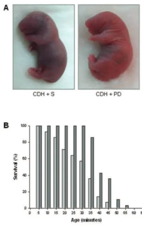
Concerning neonatal survival, the aver-age survival time was significantly longer in PD-123319-treated pups than in CDH + S pups (30.3 ± 3.2 min for CDH + S versus 42.4 ± 1.3 min for CDH + PD, P< 0.001). As a result, all CDH + PD pups, but only 57% of the CDH + S pups, survived for up to 30 min. At 45 min after birth, only 7% of CDH + S pups were alive, as op-posed to over a third of CDH + PD pups. In fact, PD-123319 treatment increased survival rates of CDH neonates for all time points evaluated (Figure 8).
Figure 3.MAPK and Akt kinase activities in control (C) lung explants and treated with ANGII at 10–9mol/L (Ang 10–9), ZD-7155 (ZD) or PD-123319 (PD). (A) Western blot analy-sis of p38, p44/42, JNK1/2 and Akt and to diphosphorylated forms of p38 (dp-p38), p44/42 (dp-p44/42), SAPK/JNK (dp-JNK1/2) and Akt (dp-Akt). Control loading was per-formed using β-tubulin (55 kDa). p38 corresponds to 38 kDa. p44/42 correspond to 44 and 42 kDa, respectively. JNK1 and 2 correspond to 46 and 54 kDa, respectively. Akt corresponds to 56 kDa. Semiquantitative analysis for dp-p38 (B), dp-p44/42 (C), dp-JNK1/2 (D), and dp-Akt (E). Results are presented as arbitrary units normalized for β-tubulin. P < 0.05: *versus ANGII at 0 mol/L (control, C), §versus ANGII at 10–9mol/L, †
versus ZD-7155 at 10–5mol/L.
Figure 4.In vivo antenatal PD-123319 treatment effects on lung growth. (A) Ratio of left, right and total lung-to-body weight in control (C), nitrofen (N) and CDH groups treated with saline (C + S, N + S, CDH + S) or PD-123319 (C + PD, N + PD, CDH + PD). Antenatal administration of PD-123319 en-hanced lung growth in all studied groups. (B) Effect of PD-123319 treatment on left, right and total lung hypoplasia. Prenatal administration of PD-123319 ameliorated both left and right lung hypoplasia. Results are expressed as %. P < 0.001: *versus C + S,
†versus N + S, ‡versus CDH + S, llversus C +
PD, §versus N + PD. For left lung, there is a
Regarding potential secondary effects of PD-123319 on maternal organs, their lungs, heart and kidneys were weighed. PD-123319 treatment did not signifi-cantly change maternal HW/BW, KW/BW or LW/BW ratios (see Supple-mental Figure 3).
DISCUSSION
This study demonstrated that all components of RAS (renin, ACE, angio -tensinogen, AT1and AT2receptors) were constitutively expressed in the lung dur-ing all studied gestational ages and that ANGII had a stimulatory effect on lung branching, mediated by AT1receptor, through p44/42 and Akt phosphoryla-tion. This stimulatory effect on lung growth was mimicked by treatment with AT2-antagonist. Therefore, AT2receptor antagonist was evaluated as a putative antenatal treatment for diseases charac-terized by fetal lung hypoplasia such as CDH. In an animal model of CDH, ante-natal PD-123319 treatment increased neo-natal lung growth, ameliorated indirect parameters of PH, improved lung
func-tion and survival, without maternal or fetal deleterious effects.
In past years, it has been demonstrated that local ANGII formation and its tissue-specific effects on growth and dif-ferentiation are thought to be extremely important for embryonic and fetal devel-opment (12). Regarding fetal lung, this study corroborated previous evidences concerning ACE, AT1and AT2expression (13–15,28). For the first time, the present study showed that renin and angio -tensinogen are also expressed during lung development. Interestingly, renin, ACE and AT2were expressed at very early stages (since 13.5 dpc), suggesting an important role for a local RAS since early stages of lung development.
The results of RAS components expres-sion prompted us to hypothesize that a local RAS is active in the developing lung. Therefore, the role of ANGII on lung morphogenesis was evaluated. ANGII supplementation induced an in-crease in lung explants growth, and it is necessary to stress that this enhancing ef-fect of ANGII on number of peripheral
airways buds of lung reached about 37%, whereas the stimulatory effect induced by fibroblast growth factor-10 (FGF-10), a classical and very important lung growth factor, in a similar model of murine lung explants, was around 20% (29). The pos-sible mechanism by which RAS inter-feres with the airway branching or pul-monary vascular development is still unclear, and further investigation is re-quired. However, it was already substan-tially demonstrated that reciprocal inter-actions between airways and blood vessels are critical for normal lung devel-opment. For instance, it was demon-strated that ablation of lung epithelium impair lung vascular cells development (30). Moreover, VEGF inhibition in neo-natal rats leads to arrested alveolar de-velopment, suggesting that inhibition of vascular growth itself may directly im-pair lung development (31–33). Thus, given that, in the present study, it was demonstrated that some components of RAS are expressed on epithelium and others on mesenchyme/vascular cells, it is possible that RAS is involved in both processes: airway and vasculature branching.
Interestingly, AT1receptor inhibitor de-creased, whereas AT2-antagonist signifi-cantly increased lung growth in explants. This opposite effect of AT1and AT2 re-ceptors, namely a stimulatory effect of AT1and inhibitory effect of AT2, is also described on other tissues (34–39).
Many of the effectors that modulate fetal lung branching seem to activate MAPK and/or PI3K/Akt cascades (40). Thus, MAPK and PI3K/Akt pathway ac-tivation by ANGII and AT1and AT2 an-tagonists in fetal lung development was investigated. ANGII and AT2receptor an-tagonist treatment induced an increase in lung branching by the stimulation of p44/42 and Akt phosphorylation. These intracellular mediators are also involved in AT1effects on proliferation and sur-vival of cells in other tissues (34,36). Re-garding lung growth inhibition induced by AT1antagonist, it was mediated by a decrease of p-38 and JNK phosphoryla-tion. These MAPK families were already
demonstrated to be involved on induc-tion of lung branching (22).
The in vitrostudies demonstrated that a local RAS is functional at early stages of lung morphogenesis. Moreover, the significant stimulatory effect on lung growth mediated by AT2receptor antag-onist led us to hypothesize that AT2 could be a new target for treatment of diseases characterized by fetal lung hy-poplasia, such as CDH. In fact, AT2 re-ceptor is described to be expressed virtu-ally only during fetal life (11,12,41), which would annul potential maternal adverse effects. Thus, treatment with AT2 antagonist (PD-123319) was selected for the in vivostudy, in which the nitrofen-induced CDH rat model (19,42) was used. The gestational age selected for maternal PD-123319 administration was based on the effect observed on lung ex-plants that were harvested at 13.5 dpc.
The nitrofen-induced CDH model is an experimental model of severe lung hy-poplasia, which reasonably replicates the major abnormalities and the pathophysi-ology described in human CDH (42–44). Although the mechanism by which nitro-fen induces the diaphragmatic defect and lung hypoplasia is not fully understood, recent evidences suggest the involve-ment of abnormalities linked with the retinoid signaling pathway in this model and also in human CDH etiology (42–45). Indeed, one clinical study demonstrated the presence of decreased levels of retinol and retinol-binding pro-tein in human CDH, suggesting a possi-ble deterioration of retinol transport across the placenta (46). Regarding the nitrofen-induced CDH model, it was al-ready demonstrated that nitrofen inhibits retinal dehydrogenase 2 (RALDH2), a key enzyme responsible for the
conver-sion of retinal to retinoic acid (47). More-over, the co-administration of retinoids (Vitamin A or retinoic acid) in nitrofen-induced CDH induces lung growth and reduces the incidence of CDH (9,10,48). Interestingly, some studies have de-scribed an inhibitory interaction between retinoid acid pathway and RAS, namely in adult cardiac remodeling (49,50). Thus, a possible interaction between retinoid acid pathway and RAS might be present during lung morphogenesis, but the underlying mechanisms remain unclear and further investigation is required.
Maternal PD-123319 subcutaneous ad-ministration significantly increased lung growth in control, nitrofen and CDH groups. In fact, in CDH neonates, despite the presence of mechanical forces that compress lungs, PD-123319 induced par-tial recovery of lung hypoplasia as as-sessed by LW/BW (an increase of 11.4%). In face of these results and to assess if the treated-lung is structural and func-tionally ameliorated, we focused further studies in comparing C + S, CDH + S and CDH + PD groups. In human and experimental CDH there is a reduction in peripheral lung development (1,10). PD-123319 treatment stimulated lung growth by promoting distal lung development as measured by the enhanced RSC. The po-tential clinical relevance of this effect should be emphasized, since such an in-crease in lung parenchyma can be deter-minant in providing a better adaptation of CDH fetus to extrauterine life.
Regarding epithelial differentiation and lung maturity, CCSP (a Clara cells marker, marker for proximal lung), SP-C (a type II pneumocytes marker, marker for distal lung) and PAS glycogen stores (a signal of immaturity) were assessed. No differences between groups were de-tected in CCSP, SP-C and PAS-stained cells score. Contradictory results about lung maturity and surfactant status in CDH animal models have been pub-lished, with studies demonstrating that the CDH lung is surfactant deficient (51–53) and others indicating no change in alveolar surfactant composition,
un-Figure 6.Antenatal PD-123319 treatment did not interfere with epithelial differentiation and lung maturation. (A) Representative CCSP-stained sections of C + S, CDH + S and CDH + PD lungs. Original magnification 100×. (B) Score of CCSP-stained cells in C + S, CDH + S and CDH + PD groups. (C) Representative SP-C stained sections in different groups. Original magnification 200×. (D) Score of SP-C-stained cells. (E) Representative PAS stained sections of C + S, CDH + S and CDH + PD lungs. Original magnification 400×. Ar-rows: PAS+(glycogen-rich) cells. (F) Score of PAS-stained cells. No significant difference
changed or even increased surfactant protein expression (54–57). However, one of the most recent studies per-formed with human CDH fetuses demonstrated that surfactant maturation is not delayed (54). The present study corroborates the idea that CDH does not appear to interfere or delay epithelial differentiation and surfactant accumula-tion (54). Moreover, PD-123319 treat-ment did not impair fetal lung matura-tion in CDH neonates.
Persistent PH accounts for significant mortality and morbidity in CDH (1,2,58). In severe human CDH and also in nitrofen-induced model of CDH, it was already demonstrated that PH re-sults from decreased number of arteries, increased thickness of media and ad-ventitia of pulmonary arterial walls and distal muscular extension to the non-muscular intraacinar arteries (58,59). In this study, the effect of PD-123319 treat-ment on PH was indirectly assessed by morphometric pulmonary vascular anal-ysis (59) and its cardiac repercussion by determination of right ventricular hy-pertrophy index and right ventricle thickness, and quantification of right ventricular overload molecular markers (Angiotensinogen, BNP and ET-1) (27). To better analyze the vessel morphol-ogy, areas were measured instead of thickness since this approach avoids the bias introduced by irregular shape of elastic laminae. Thus, PD-123319 treat-ment induced decrease of MA percent-age for all arterial sizes, with maximal effect on smaller arteries. Moreover, CDH + S neonates did not present dif-ferences in right ventricular hypertro-phy index and right ventricular wall thickness (morphological markers) when compared with C + S group, but they presented an increase of molecular markers of right ventricular overload. Thus, 5 min after birth, CDH + S neonates only presented molecular changes secondary to PH, but not yet morphological alterations (secondary to chronic PH). PD-123319-treated CDH pups presented a significantly decrease of right ventricular overload molecular
Figure 7.Indirect pulmonary hypertension assessment. (A) In upper panel are presented representative examples of pulmonary arteries > 50 µm stained with Weigert resorcin fuchsin solution of C + S, CDH + S and CDH + PD groups; in lower panel, representative examples of small pulmonary vessels (<30 µm) for each group are presented. Original magnification 400×. (B) Percentage medial area of pulmonary arteries of different nal diameters. (C) Percentage adventitial area of pulmonary arteries of different exter-nal diameters. (D) Right ventricular levels of angiotensinogen, BNP and ET-1 mRNA in C + S, CDH + S and CDH + PD neonates, expressed in arbitrary units normalized for β-actin. PD-123319 treatment significantly decreased arterial medial area and these overload markers on CDH pups. P < 0.001: *versus C + S, ‡
markers and did not influence morpho-logical markers. Therefore, PD-123319 maternal administration partially re-versed pulmonary arterial structural ab-normality that characterizes CDH and decreased molecular markers of PH, which suggest that PD-123319 might re-duce pulmonary vascular reactivity, and the risk of postnatal persistent PH ob-served in CDH neonates.
Regarding other antenatal pharmaco-logical strategies to decrease PH in CDH, Luong et al. demonstrated recently that antenatal sildenafil treatment (from 11.5 to 20.5 dpc, daily subcutaneous in-jection) attenuates PH in experimental CDH (3). In fact, that study demon-strated that antenatal sildenafil treat-ment improved lung structure, increased pulmonary vessel density and reduced right ventricular hypertrophy in CDH (3). However, in opposition to PD-123319 treatment, sildenafil had not pro-moted lung growth, as demonstrated by LW/BW, and consequently it did not in-duce a recover of lung hypoplasia (3). Moreover, it is necessary to stress that the idea that there is PH in CDH, since
fetal period, is underlying to Luong’s study (3). Nonetheless, it was already demonstrated that there is vascular hy-poplasia in nitrofen-induced CDH and neonatal PH, but it is not yet proven the presence of PH in CDH fetus (27,58,59). Indeed, immediately after the birth, Luong et al. demonstrated the presence of right ventricular hypertrophy (an in-direct signal of PH) in CDH neonates (3). In the present study, CDH neonates at 5 min after the birth did not present increase of right ventricular hypertrophy index or right ventricle thickness. How-ever, CDH + S neonates presented an in-crease of right ventricular overload markers which reveals a molecular car-diac repercussion. According to the liter-ature, it was already demonstrated that CDH vascular pulmonary alterations only affect neonatal and not fetal hemo-dynamics. Baptista et al. demonstrated that CDH is associated with significant molecular alterations secondary to PH, but only in the right ventricle and after birth (27). Moreover, also in fetal lamb CDH model, it was demonstrated that newborn CDH lambs had no differences
in right ventricular weight or right ven-tricular wall thicknesses compared with control lambs (60). Thus, despite the oc-currence of pulmonary vascular modifi-cations from early stages of prenatal de-velopment, the present study also shows that PH is only present after birth, likely occurring secondary to vascular hy-poplasia that characterizes CDH, and also consequently to pulmonary vaso-constriction, which is secondary to alve-olar hypoplasia, hypoxia and acidosis.
So, antenatal PD-123319 treatment in-terfered and improved the key determi-nants of mortality associated with CDH, namely lung hypoplasia and PH. Fur-thermore, maternal PD-123319 adminis-tration improved lung function, namely pulmonary gas exchange, as demon-strated by APGAR score, gasometric
Figure 8.Survival analysis. (A) Representa-tive examples of cyanotic color of CDH + S versus pink coloration of CDH + PD neonates at 5 min of life. (B) Survival rates (expressed as percentage of pups surviving at each 5 min) of CDH + S and CDH + PD neonates. Antenatal PD-123319 treatment improved survival time in CDH pups (mean survival time: CDH + S 30.3 ± 3.2 min; CDH + PD 42.4 ± 1.3 min, P < 0.001). , CDH + S;
, CDH + PD.
Table 2.Right ventricular hypertrophy evaluation 5 min after birth.a
Ratio of right/left ventricular weight Right ventricular wall thickness (µm)
C + S 0.49 ± 0.08 214.02 ± 9.29
CDH + S 0.44 ± 0.05 200.59 ± 4.27
CDH + PD 0.52 ± 0.05 220.31 ± 12.26
C, control; CDH, congenital diaphragmatic hernia; PD, PD-123319; S, saline.
aValues represent the mean ± SEM of measurements. P < .05: No significant difference
between experimental groups was observed.
Table 3.Neonatal blood gasometric evaluation 5 min after birth.a
pH PCO2(mmHg) PO2(mmHg) SatO2(%) Lactate (mmol/L)
C + S 7.30 ± 0.03 22.69 ± 2.33 102.80 ± 7.78 93.60 ± 2.50 6.83 ± 0.39 N + S 6.94 ± 0.03b 38.49 ± 3.21b 62.50 ± 5.36b 67.25 ± 5.58b 8.50 ± 0.51b
CDH + S 6.92 ± 0.02b 51.05 ± 1.71bc 52.00 ± 6.06b 58.50 ± 7.06b 9.56 ± 0.57b
N + PD 7.18 ± 0.08c 23.62 ± 3.39c 95.67 ± 6.49c 90.83 ± 2.44c 6.88 ± 0.82c
CDH + PD 7.03 ± 0.04de 24.63 ± 5.83d 95.00 ± 8.20d 87.80 ± 3.65d 6.73 ± 0.70d
C, control; CDH, congenital diaphragmatic hernia; N, nitrofen; PD, PD-123319; PCO2, CO2 partial pressure; PO2, O2partial pressure; S, saline; SatO2, O2saturation.
aValues represent the mean ± SEM of measurements.
bP < 0.05 versus C + S; cversus N + S; dversus CDH + S; eversus N + PD. There is a statistically
and survival evaluation. Regarding gas-ometry it is necessary to stress that due to low fetal blood volume, all blood possibly collected by decapitation was used for gasometric evaluation. So, gas-ometry evaluated a mix of arterial and venous blood. Nonetheless, antenatal PD-123319 treatment allowed a statisti-cal significant improvement of acidosis, hypercapnia, hypoxia and lactate con-centration that characterizes CDH fe-tuses. The results of these direct indica-tors of ventilation/perfusion matching quality suggest an obvious improve-ment of pulmonary gas exchange and peripheral O2delivery. Furthermore, this enhancement on lung function had important consequences on neonatal survival, namely PD-123319 treatment induced significantly longer average survival time. However, it is necessary to stress that the survival evaluation was performed without neonatal care or ventilatory support. This fact might be the explanation for the death of all neonates, despite the increase on lung function and survival time induced by antenatal PD-123319 treatment.
Regarding potential fetal adverse ef-fects, the nitrofen-exposed pups pre-sented decrease of HW/BW and KW/BW ratios as previously docu-mented (10,61–63). On the other hand, PD-123319 beneficial effect seemed lung-specific, since HW/BW and KW/BW ra-tios of the pups were not altered. Con-cerning potential maternal secondary effects induced by PD-123319, no differ-ences on heart, kidneys and lungs were observed. The absence of maternal dele-terious effects could be due to the fact of AT2receptor expression is dramatically decreased after birth, being restricted to a few organs (11,12,41). Indeed, an in-crease of AT2receptor expression during adult life has been only observed under pathological conditions (41).
CONCLUSION
In conclusion, this study demonstrated the existence of a functional local RAS in fetal lung. Moreover, it establishes AT2 receptor antagonist (PD-123319) as a
pu-tative antenatal therapy for pathologies characterized by fetal lung hypoplasia, such as CDH.
ACKNOWLEDGMENTS
This project was funded by Fundação para a Ciência e a Tecnologia
(PTDC/SAU-OBD/108051/2008) and by Secção de Neonatologia da Sociedade Portuguesa de Pediatria (Grant ZERU 2008). P Piairo was supported by Fun-dação para a Ciência e a Tecnologia (ref-erence SFRH/BD/33410/2008). RS Moura was supported by Fundação para a Ciência e a Tecnologia (reference SFRH/BPD/15408/2005). PD-123319 was kindly supplied by Medical Division of Pfizer Inc, Groton, Connecticut, USA.
We would like to thank to Luís Mar-tins for histological technical support and help on animal euthanasia and to Nuno M Pires for Weigert staining and vascular morphometric analysis support.
DISCLOSURE
The authors declare that they have no competing interests as defined by Molecu-lar Medicine, or other interests that might be perceived to influence the results and discussion reported in this paper.
REFERENCES
1. van den Hout L, et al. (2009) Can we improve outcome of congenital diaphragmatic hernia? Pe-diatr. Surg. Int. 25:733–43.
2. Keller RL, et al. (2010) Congenital diaphragmatic hernia: endothelin-1, pulmonary hypertension, and disease severity. Am. J. Respir. Crit. Care Med. 182:555–61.
3. Luong C, et al. (2011) Antenatal sildenafil treat-ment attenuates pulmonary hypertension in ex-perimental congenital diaphragmatic hernia. Cir-culation. 123:2120–31.
4. Puri P, Wester T. (1997) Historical aspects of con-genital diaphragmatic hernia. Pediatr. Surg. Int. 12:95–100.
5. Harrison MR, et al. (2003) A randomized trial of fetal endoscopic tracheal occlusion for severe fetal congenital diaphragmatic hernia. N. Engl. J. Med. 349:1916–24.
6. Jani JC, et al. (2009) Severe diaphragmatic hernia treated by fetal endoscopic tracheal occlusion. Ultrasound Obstet. Gynecol. 34:304–10. 7. Nogueira-Silva C, Moura RS, Esteves N,
Gon-zaga S, Correia-Pinto J. (2008) Intrinsic catch-up growth of hypoplastic fetal lungs is mediated by interleukin-6. Pediatr. Pulmonol. 43:680–9.
8. Santos M, et al. (2006) Ghrelin expression in human and rat fetal lungs and the effect of ghre-lin administration in nitrofen-induced congenital diaphragmatic hernia. Pediatr. Res. 59:531–7. 9. Baptista MJ, et al. (2005) Antenatal vitamin A
ad-ministration attenuates lung hypoplasia by inter-fering with early instead of late determinants of lung underdevelopment in congenital diaphrag-matic hernia. J. Pediatr. Surg. 40:658–65. 10. Thébaud B, et al. (1999) Vitamin A decreases the
incidence and severity of nitrofen-induced con-genital diaphragmatic hernia in rats. Am. J. Phys-iol. 277:L423–9.
11. Lavoie JL, Sigmund CD. (2003) Minireview: over-view of the renin-angiotensin system—an en-docrine and paracrine system. Endocrinology. 144:2179–83.
12. Paul M, Poyan Mehr A, Kreutz R. (2006) Physiol-ogy of local renin-angiotensin systems. Physiol. Rev. 86:747–803.
13. Goyal R, Leitzke A, Goyal D, Gheorghe CP, Longo LD. (2011) Antenatal maternal hypoxic stress: adaptations in fetal lung renin-angiotensin sys-tem. Reprod. Sci. 18:180–9.
14. Morrell NW, Grieshaber SS, Danilov SM, Majack RA, Stenmark KR. (1996) Developmental regula-tion of angiotensin converting enzyme and an-giotensin type 1 receptor in the rat pulmonary circulation. Am. J. Respir. Cell Mol. Biol. 14:526–37. 15. Shanmugam S, Corvol P, Gasc JM. (1996)
An-giotensin II type 2 receptor mRNA expression in the developing cardiopulmonary system of the rat. Hypertension. 28:91–7.
16. Diário da República [DR]. (1992) DR 245/92 SÉRIE I-B, Portaria n.º 1005/92 [of 1992 Oct 23]. [cited 2012 Mar 8]. Available from: http://www. dre. pt/ cgi/dr1s.exe?t=dr&cap=1-1200&doc= 1992343 0%20&v02=&v01=2&v03=1900-01-01& v04 = 3000-12-21&v05=&v06=&v07=&v08=&v09= &v10 =&v 11=Portaria&v12=&v13=&v14= &v15=& sort=0& submit=Pesquisar
17. World Medical Association [WMA]. (1964) WMA declaration of Helsinki - ethical principles for medical research involving humans. Last amended 2008 Oct. [cited 2012 Mar 8]. Available from: http://www.wma.net/en/ 30publications/ 10policies/b3/index.html
18. Institute of Laboratory Animal Resources; Com-mission on Life Sciences; National Research Council. (1996) Guide for the Care and Use of Labo-ratory Animals. Washington (DC): National Acad-emy Press. [cited 2012 Mar 8]. Available from: http://www.nap.edu/openbook.php?record_id= 5140
19. Tenbrinck R, et al. (1990) Experimentally induced congenital diaphragmatic hernia in rats. J. Pedi-atr. Surg. 25:426–9.
20. Eskildsen-Helmond YE, Mulvany MJ. (2003) Pressure-induced activation of extracellular signal-regulated kinase 1/2 in small arteries. Hy-pertension. 41:891–7.
an-giotensin II on the rat vascular structure. J. Clin. Invest. 98:418–25.
22. Nogueira-Silva C, Santos M, Baptista MJ, Moura RS, Correia-Pinto J. (2006) IL-6 is constitutively expressed during lung morphogenesis and en-hances fetal lung explant branching. Pediatr. Res. 60:530–6.
23. Dauger S, et al. (2001) MASH-1/RET pathway in-volvement in development of brain stem control of respiratory frequency in newborn mice. Phys-iol. Genomics. 7:149–57.
24. Bradford MM. (1976) A rapid and sensitive method for the quantitation of microgram quan-tities of protein utilizing the principle of protein-dye binding. Anal. Biochem. 72:248–54. 25. Cooney TP, Thurlbeck WM. (1982) The radial
alveolar count method of Emery and Mithal: a reappraisal 2 - intrauterine and early postnatal lung growth. Thorax. 37:580–3.
26. Pires NM, et al. (2007) Activation of nuclear recep-tor Nur77 by 6-mercaptopurine protects against neointima formation. Circulation. 115:493–500. 27. Baptista MJ, Nogueira-Silva C, Areias JC,
Cor-reia-Pinto J. (2008) Perinatal profile of ventricular overload markers in congenital diaphragmatic hernia. J. Pediatr. Surg. 43:627–33.
28. Shanmugam S, Monnot C, Corvol P, Gasc JM. (1994) Distribution of type 1 angiotensin II recep-tor subtype messenger RNAs in the rat fetus. Hy-pertension. 23:137–41.
29. Acosta JM, et al. (2001) Novel mechanisms in murine nitrofen-induced pulmonary hypoplasia: FGF-10 rescue in culture. Am. J. Physiol. Lung Cell Mol. Physiol. 281:L250–7.
30. Sarah A, Gebb B, Shannon JM. (2000) Tissue in-teractions mediate early events in pulmonary vasculogenesis. Dev. Dyn. 217:159–69. 31. Thébaud B, et al. (2005) Vascular endothelial
growth factor gene therapy increases survival, promotes lung angiogenesis, and prevents alveo-lar damage in hyperoxia-induced lung injury: ev-idence that angiogenesis participates in alveolar-ization. Circulation. 112:2477–86.
32. Jakkula M, et al. (2000) Inhibition of angiogenesis decreases alveolarization in the developing rat lung. Am. J. Physiol. Lung Cell Mol. Physiol. 279:L600–7.
33. Healy AM, Morgenthau L, Zhu X, Farber HW, Cardoso WV. (2000) VEGF is deposited in the subepithelial matrix at the leading edge of branching airways and stimulates neovascular-ization in the murine embryonic lung. Dev. Dyn. 21:341–52.
34. Yosypiv IV, El-Dahr SS. (2005) Role of the renin-angiotensin system in the development of the ureteric bud and renal collecting system. Pediatr. Nephrol. 20:1219–29.
35. Stoll M, et al. (1995) The angiotensin AT2-receptor mediates inhibition of cell proliferation in coro-nary endothelial cells. J. Clin. Invest. 95:651–7. 36. Nakajima M, et al. (1995) The angiotensin II type 2
(AT2) receptor antagonizes the growth effects of the AT1 receptor: gain-of-function study using
gene transfer. Proc. Natl. Acad. Sci. U. S. A. 92:10663–7.
37. Inagami T, Senbonmatsu T. (2001) Dual effects of angiotensin II type 2 receptor on cardiovascular hypertrophy. Trends Cardiovasc. Med. 11:324–8. 38. Gyurko R, Kimura B, Kurian P, Crews FT,
Phillips MI. (1992) Angiotensin II receptor sub-types play opposite roles in regulating phos-phatidylinositol hydrolysis in rat skin slices. Biochem. Biophys. Res. Commun. 186:285–92. 39. Maric C, Aldred GP, Harris PJ, Alcorn D. (1998)
Angiotensin II inhibits growth of cultured em-bryonic renomedullary interstitial cells through the AT2 receptor. Kidney Int. 53:92–9.
40. Kling DE, et al. (2002) Pre- and postnatal lung de-velopment, maturation, and plasticity: MEK-1/2 inhibition reduces branching morphogenesis and causes mesenchymal cell apoptosis in fetal rat lungs. Am. J. Physiol. Lung Cell Mol. Physiol. 282:L370–8.
41. Kaschina E, Unger T. (2003) Angiotensin AT1/AT2 receptors: regulation, signalling and function. Blood Press. 12:70–88.
42. Kling DE, Schnitzer JJ. (2007) Vitamin A defi-ciency (VAD), teratogenic, and surgical models of congenital diaphragmatic hernia (CDH). Am. J. Med. Genet. C Semin. Med. Genet. 145C:139–57. 43. Montedonico S, Nakazawa N, Puri P. (2008)
Con-genital diaphragmatic hernia and retinoids: searching for an etiology. Pediatr. Surg. Int. 24:755–61.
44. Gallot D, et al. (2005) Congenital diaphragmatic hernia: a retinoid-signaling pathway disruption during lung development? Birth Defects Res. A Clin. Mol. Teratol. 73:523–31.
45. Greer JJ, Babiuk RP, Thebaud B. (2003) Etiology of congenital diaphragmatic hernia: the retinoid hypothesis. Pediatr. Res. 53:726–30.
46. Major D, et al. (1998) Retinol status of newborn infants with congenital diaphragmatic hernia. Pediatr. Surg. Int. 13:547–9.
47. Mey J, Babiuk RP, Clugston R, Zhang W, Greer JJ. (2003) Retinal dehydrogenase-2 is inhibited by compounds that induce congenital diaphrag-matic hernias in rodents. Am. J. Pathol. 162:673–9. 48. Babiuk RP, Thebaud B, Greer JJ. (2004) Reductions
in the incidence of nitrofen-induced diaphrag-matic hernia by vitamin A and retinoic acid. Am. J. Physiol. Lung Cell Mol. Physiol. 286:L970–3. 49. Guleria RS, Choudhary R, Tanaka T, Baker KM,
Pan J. (2011) Retinoic acid receptor-mediated signaling protects cardiomyocytes from hyper-glycemia induced apoptosis: role of the angiotensin system. J. Cell Physiol. 226:1292–307. 50. Choudhary R, et al. (2008) All-trans retinoic acid prevents development of cardiac remodeling in aortic banded rats by inhibiting the angiotensin system. Am. J. Physiol. Heart Circ. Physiol. 294:H633–44.
51. Thébaud B, et al. (2001) Restoring effects of vita-min A on surfactant synthesis in nitrofen-induced congenital diaphragmatic hernia in rats. Am. J. Respir. Crit. Care Med. 164:1083–9.
52. Keijzer R, Liu J, Deimling J, Tibboel D, Post M. (2000) Dual-hit hypothesis explains pulmonary hypoplasia in the nitrofen model of congenital di-aphragmatic hernia. Am. J. Pathol. 156:1299–306. 53. Asabe K, Tsuji K, Handa N, Kajiwara M, Suita S.
(1998) Expression of clara cell 10-kDa protein (CC10) in congenital diaphragmatic hernia. Pedi-atr. Surg. Int. 14:36–9.
54. Boucherat O, et al. (2007) Surfactant maturation is not delayed in human fetuses with diaphrag-matic hernia. PLoS Med. 4:e237.
55. Van Tuyl M, et al. (2003) Pulmonary surfactant protein A, B, and C mRNA and protein expres-sion in the nitrofen-induced congenital diaphrag-matic hernia rat model. Pediatr. Res. 54:641–52. 56. Chapin CJ, et al. (2005) Congenital diaphragmatic
hernia, tracheal occlusion, thyroid transcription factor-1, and fetal pulmonary epithelial matura-tion. Am. J. Physiol. Lung Cell Mol. Physiol. 289:L44–52.
57. Santos M, et al. (2007) Pulmonary epithelial cell differentiation in the nitrofen-induced congenital diaphragmatic hernia. J. Pediatr. Surg.42:1231–7. 58. Mohseni-Bod H, Bohn D. (2007) Pulmonary hy-pertension in congenital diaphragmatic hernia. Semin. Pediatr. Surg. 16:126–33.
59. Kanai M, et al. (2001) Fetal tracheal occlusion in the rat model of nitrofen-induced congenital di-aphragmatic hernia: tracheal occlusion reverses the arterial structural abnormality. J. Pediatr. Surg. 36:839–45.
60. Karamanoukian HL, et al. (1995) Pathophysiol-ogy of congenital diaphragmatic hernia. XI: Anatomic and biochemical characterization of the heart in the fetal lamb CDH model. J. Pediatr. Surg. 30:925–8.
61. González-Reyes S, Martínez L, Tovar JA. (2005) Effects of prenatal vitamins A, E, and C on the hypoplastic hearts of fetal rats with diaphrag-matic hernia. J. Pediatr. Surg. 40:1269–74. 62. Montedonico S, Nakazawa N, Shinkai T,
Banni-gan J, Puri P. (2007) Kidney development in the nitrofen-induced pulmonary hypoplasia and congenital diaphragmatic hernia in rats. J. Pedi-atr. Surg. 42:239–43.