Longitudinal assessment of the lung mechanics of very
low birth weight preterm infants with and without
bronchopulmonary dysplasia
Avaliação longitudinal da mecânica pulmonar de crianças pré-termo de muito
baixo peso com e sem displasia broncopulmonar
Rosane Reis de Mello
I, Kátia Silveira da Silva
II, Anniele Medeiros Costa
III, José Roberto de Moraes Ramos
IVInstituto Nacional de Saúde da Mulher, da Criança e do Adolescente Fernandes Figueira/Fiocruz, Rio de Janeiro (RJ), Brazil
ABSTRACT
CONTEXT AND OBJECTIVE: Prematurity has been correlated with altered lung mechanics. Some infants develop lung injury as a consequence of lung immaturity, invasive mechanical ventilation and exposure to oxygen, thus resulting in bronchopulmonary dysplasia. The aim here was to compare the lung mechanics of preterm infants with and without bronchopulmonary dysplasia during the irst year of life.
DESIGN AND SETTING: Prospective cohort study in a tertiary-level hospital.
METHODS: This study included premature infants at a public hospital who underwent two pulmonary function tests: one at discharge and the other at the corrected age of 4 to 8 months. Tidal volume, lung compliance and lung resistance were measured. Statistical tests were used for comparisons between in-fants with and without bronchopulmonary dysplasia.
RESULTS: 102 children with mean gestational age of 29 ± 2.0 weeks were studied; 17 with bronchopul-monary dysplasia. Lung compliance (0.84 ± 0.29 versus 1.28 ± 0.46; P < 0.001) and tidal volume (6.1 ± 0.94 versus 7.2 ± 1.43; P < 0.01) at discharge were signiicant lower in children with bronchopulmonary dyspla-sia than in those without the disease, but no diferences were observed at the second test (compliance: 1.53 ± 0.77 versus 1.94 ± 1.01; P = 0.12; and tidal volume: 6.9 ± 1.4 versus 7.3 ± 1.6; P = 0.42).
CONCLUSION: Diferences in lung mechanics were observed between infants with and without bron-chopulmonary dysplasia at hospital discharge but these diferences were no longer detected at the inal follow-up. The lung mechanics of all the infants improved over this period of time.
RESUMO
CONTEXTO E OBJETIVO: Prematuridade tem sido associada com mecânica pulmonar alterada. Algumas crianças desenvolvem lesão pulmonar como consequência de imaturidade pulmonar, ventilação mecâ-nica invasiva e exposição a oxigênio, resultando em displasia broncopulmonar. O objetivo foi comparar a mecânica pulmonar de prematuros com e sem displasia broncopulmonar durante o primeiro ano de vida.
DESENHO DO ESTUDO E LOCAL: Estudo de coorte prospectivo em um hospital terciário.
MÉTODOS: O estudo incluiu prematuros, de hospital público, que realizaram duas provas de função pul-monar, uma na alta e outra entre quatro e oito meses de idade corrigida. Foram mensurados o volume corrente, a complacência e a resistência pulmonares. Testes estatísticos foram usados para comparações entre crianças com e sem displasia broncopulmonar.
RESULTADOS: Foram estudadas 102 crianças com idade gestacional média de 29 ± 2 semanas; 17 com displasia broncopulmonar. A complacência pulmonar (0,84 ± 0,29 versus 1,28 ± 0,46; P < 0.001) e o volume corrente (6,1 ± 0,94 versus 7,2 ± 1,43; P < 0.01) na alta foram signiicativamente inferiores nas crianças com displasia broncopulmonar comparadas às crianças sem a doença, mas não foram observadas diferenças signiicativas no segundo teste (complacência: 1.53 ± 0.77 versus 1.94 ± 1.01; P = 0.12; e volume corrente: 6.9 ± 1.4 versus 7.3 ± 1.6; P = 0.42).
CONCLUSÃO: Diferenças na mecânica pulmonar foram observadas entre crianças com e sem displasia broncopulmonar na alta hospitalar, mas essas diferenças não foram detectadas no seguimento inal. A mecânica pulmonar de todas as crianças melhorou no decorrer desse período de tempo.
IMD, PhD. Attending Physician, Department of
Neonatology, Instituto Nacional de Saúde da Mulher, da Criança e do Adolescente Fernandes Figueira/Fiocruz, Rio de Janeiro (RJ), Brazil.
IIMD, PhD. Epidemiologist, Clinical Research
Unit, Instituto Nacional de Saúde da Mulher, da Criança e do Adolescente Fernandes Figueira/ Fiocruz, Rio de Janeiro (RJ), Brazil.
IIIMSc. Physiotherapist, Department of Neonatology,
Instituto Nacional de Saúde da Mulher, da Criança e do Adolescente Fernandes Figueira/Fiocruz, Rio de Janeiro (RJ), Brazil.
IVMD, PhD. Head of Department of Neonatology,
Instituto Nacional de Saúde da Mulher, da Criança e do Adolescente Fernandes Figueira/ Fiocruz, Rio de Janeiro (RJ), Brazil.
KEY WORDS:
Respiratory mechanics. Respiratory function tests. Bronchopulmonary dysplasia. Infant, premature.
Lung compliance.
PALAVRAS-CHAVE:
Mecânica respiratória. Testes de função respiratória Displasia broncopulmonar. Prematuro.
INTRODUCTION
Bronchopulmonary dysplasia is a chronic lung disease that mostly afects prematurely born children. he disease is associated with use of mechanical ventilation and prolonged use of oxygen. Although these children are nowadays managed with “lung pro-tective strategies”, such as prenatal steroids, exogenous surfactants and minimally invasive ventilation strategies, along with other advances in neonatal care, bronchopulmonary dysplasia remains one of the most common complications of prematurity.1 he main
characteristic of children with bronchopulmonary dysplasia is late development of the lung acinus with abnormal alveolarization, abnormal deposition of elastin and abnormal vascularization.2
hese lung alterations may have signiicant consequences that go beyond childhood, and may result in obstruction and hyperreac-tivity of the airways.3 Infants with “new bronchopulmonary
dys-plasia” exhibit abnormalities in lung function ater birth, through-out childhood and into adolescence.4
Moreover, a few studies conducted during the last decade have evaluated the progression or regression of alterations to lung function detected before medical discharge in preterm infants.5 Extreme prematurity results in persistent alveolar
dam-age.6 Poor lung function during childhood has been reported in
both healthy preterm infants and in those with bronchopulmo-nary dysplasia, although it is unclear whether prematurity alone can explain the reduction in lung function observed in infants with bronchopulmonary dysplasia.7 Moreover, it has not yet been
established how the lung grows during bronchopulmonary dys-plasia and how the immature lung recovers from this disease.5
A longitudinal evaluation of lung function ater premature infants have been discharged may help to identify those at risk of incomplete recovery of respiratory function and hence of devel-opment of respiratory problems during childhood.5
OBJECTIVE
he purpose of this study was to compare the development of lung function at two times during the irst year of life of very low birth weight preterm infants with and without bronchopulmo-nary dysplasia.
METHODS
Study design and inclusion criteria
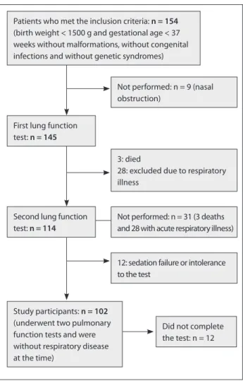
From 2005 to 2009, a prospective cohort study was conducted among very low birth weight premature infants (birth weight < 1500 g) who were admitted to and treated at a public neonatal intensive care unit. he protocol included two pulmonary func-tion tests, performed at the time of discharge from the unit and at the corrected age of 4 to 8 months (Figure 1). Infants with congenital malformations, genetic syndromes or congenital
infections and those who did not undergo the two lung function tests were excluded.
Evaluation of pulmonary function
Only infants who were clinically stable, were breathing room air and did not show any signs of respiratory infection over the three weeks prior to the tests were studied. Body weight and length were recorded prior to the tests. he pulmonary function tests were done at the lung function laboratory of our institution and were performed by a neonatologist and a physiotherapist who were unaware of the infants’ medical history. Recordings were made with the infant in an incubator during quiet sleep, i.e. non-rapid eye movement (non-REM) sleep. No sedation was used for the irst test, but chloral hydrate at a dosage of 50 mg/kg, 15 to 30 minutes beforehand, was given for the second pulmonary function test. he irst test was performed during the week scheduled for hospital discharge.
he pulmonary function evaluation included recordings of the following physiological signs: airlow, esophageal pressure and airways pressure. he PCLAB (Data Translation) sotware
Figure 1. Patient low chart.
Patients who met the inclusion criteria: n = 154
(birth weight < 1500 g and gestational age < 37 weeks without malformations, without congenital infections and without genetic syndromes)
Not performed: n = 9 (nasal obstruction)
3: died
28: excluded due to respiratory illness
12: sedation failure or intolerance to the test
Did not complete the test: n = 12 Second lung function
test: n = 114
Study participants: n = 102
(underwent two pulmonary function tests and were without respiratory disease at the time)
First lung function test: n = 145
was used for data acquisition. Airlow was measured using a resuscitation-type mask coupled to a pneumotachograph (Fleisch 00). Tidal volume was obtained through electronic inte-gration of the low signal. Esophageal pressure was measured with a water-illed catheter positioned in the distal third of the esophagus and connected to an ultra-sensitive pressure trans-ducer (Validyne MP45). Airway pressure was measured using an air-illed catheter placed at the resuscitation mask and connected to another pressure transducer (Validyne DP45).
The recordings were saved into a computer and the Anadat/Labdat software (Infodat, Montreal, Canada) was used for data analysis. Tidal volume, respiratory frequency, lung compliance and resistance were calculated. For each patient, 20 to 50 respiratory cycles were selected in order to calculate pulmonary resistance and compliance, using the linear regres-sion method (computed calculation/Abreath-Anadat).
Lung compliance and the tidal volume were adjusted by dividing their values by the body weight.8,9 Lung compliance
values < 1.2 ml/cm H2O/kg were considered abnormal.9,10
Lung resistance was considered abnormal if it was greater than 50 cm H2O/l/sec.11
All infants were clinically monitored through monthly visits to our hospital’s outpatient follow-up clinic for at-risk newborns.
Deinitions
Bronchopulmonary dysplasia was deined as a situation of use of oxygen therapy at 36 weeks of corrected age.12 he
classiica-tion of the adequacy of the infant’s weight for the gestaclassiica-tional age was based on Alexander’s intrauterine growth curve.13
Infants with birth weight below the 10th percentile were
classi-ied as small for the gestational age (SGA) and those with birth weight between the 10th and 90th percentiles were considered to
be appropriate for the gestational age (AGA). Septicemia was deined as the presence of a positive blood culture.
Sample size and statistical analysis
A sample size of 105 children (21 exposed and 84 unexposed) was estimated, taking into account the following parameters: expected frequency of functional impairment of 40% for infants with bronchopulmonary dysplasia (exposed group) and 10% for the unexposed group; relative risk of functional impairment at 4-8 months of 4.0; significance level of 0.05; and power of 80%.
he data were analyzed by using the Epi Info sotware (CDC, USA). Appropriate descriptive statistics were used for the cate-gorical variables. he t test was used for variables with normal distribution and Kruskal-Wallis for those without normal distri-bution. he chi-square or Fisher’s test was applied to determine ratio diferences. he statistical signiicance level was set at 5%.
his study was approved by the Research Ethics Committee of Instituto Fernandes Figueira and all the infants’ parents signed a consent statement.
RESULTS
Over the period between 2005 and 2009, 154 newborns met the inclusion criteria. Close to the time of medical discharge from the neonatal intensive care unit, 145 newborns (94.2%) underwent the pulmonary function test at a gestational age of 38 ± 3 weeks. For 9 patients, the irst test was not done because of nasal obstruc-tion, in conjunction with a positive family history of respiratory viral infection. Of the 145 newborns, 3 children died ater medi-cal discharge from the neonatal unit, and it was not possible to apply the second test to another 28 due to respiratory disease during the period established for the test. hree of these 28 chil-dren had bronchopulmonary dysplasia.
he second test was thus performed on 114 infants (74%), at an age of 5 ± 2.2 months. Twelve did not complete the sec-ond test due to technical problems such as sedation failure and intolerance to the esophageal catheter (Figure 1). No statistically signiicant diferences in birth weight, gestational age, duration of mechanical ventilation, duration of oxygen therapy or fre-quency of bronchopulmonary dysplasia were observed between the patients studied and those who were excluded.
he sample for this study therefore included 102 premature infants with birth weight under 1500 g, who underwent two pul-monary function tests and did not have any symptoms of respi-ratory disease. he characteristics of the population studied are described in Table 1. he mean gestational age of the sample was 29 weeks, and more than 80% of the mothers underwent ante-natal corticosteroid therapy to improve the lung condition of their infants. Almost 70% of the sample required mechanical
Maternal and neonatal characteristics
Bronchopulmonary dysplasia P
Yes (n = 17) No (n = 85)
Tobacco exposure during pregnancy 1 (5.8) 8 (9.4) 1.0 Antenatal corticosteroids 14 (82.4) 80 (94.1) 0.24 Birth weight (grams) 1001 [265] 1097 [236] 0.13
Gestational age (weeks) 28 [2] 29 [2] 0.15
Male 14 (82.4) 38 (44.7) < 0.01
Small for gestational age 7 (41.2) 32 (37.6) 0.78 Mechanical ventilation 16 (94.1) 52 (61.2) < 0.01
Oxygen therapy (hours) 1916 [1096] 375.3 [465] < 0.001
Patent ductus arteriosus 10 (58.8) 39 (45.9) 0.32 Intracranial hemorrhage 6 (35.3) 17 (20.0) 0.28
Septicemia 3 (17.6) 9 (10.6) 0.68
Postnatal corticosteroids 4 (23.5) 1 (1.2) < 0.01
Table 1. Characteristics of the sample of very low birth weight premature infants with and without bronchopulmonary dysplasia
ventilation and 95% of the infants who developed bronchopul-monary dysplasia underwent mechanical ventilation. he inci-dence of male babies was almost twice as high among infants with bronchopulmonary dysplasia as among those without bron-chopulmonary dysplasia.
he mean age at the time of the pulmonary function test, which was performed close to the time of medical discharge, was 37 ± 3.0 weeks for the children with bronchopulmonary dyspla-sia and 38 ± 3.0 weeks for those without the disease (P = 0.27). he lung compliance was abnormal in 88.2% (n = 15/17) of the children with bronchopulmonary dysplasia and in 48% (41/85) of those without bronchopulmonary dysplasia (P = 0.002). he lung resistance was abnormal in 94% of the children with bron-chopulmonary dysplasia and in 80% of those without broncho-pulmonary dysplasia. he second lung function test was applied at a mean age of 5.4 ± 2.6 months for the children with bron-chopulmonary dysplasia and 4.9 ± 2.1 months for those with-out the disease.
At 4-8 months of life, the children with bronchopulmonary dysplasia also had a higher rate of change in lung compliance (47%) than did the children without bronchopulmonary dysplasia (11.7%) (P = 0.001). Similar to what was seen at the time of medi-cal discharge, there was no statistimedi-cal diference in the frequency of impaired lung resistance between the children with bronchopul-monary dysplasia (29.4%) and those without the disease (22.4%). here were reductions in the frequencies of impaired pulmonary compliance and lung resistance, in both groups.
As shown in Table 2, the mean lung compliance close to the time of medical discharge from the neonatal unit among the children with bronchopulmonary dysplasia was consider-ably lower than the mean lung compliance among the children without bronchopulmonary dysplasia. he mean tidal volume of the children with bronchopulmonary dysplasia was consid-erably diferent from that to the children without bronchopul-monary dysplasia. here was no diference in anthropometric measurements in the irst test between those with and without bronchopulmonary dysplasia.
Table 2 also shows the values for lung mechanics among chil-dren with and without bronchopulmonary dysplasia at the cor-rected age of 4-8 months. It can be seen that the premature chil-dren with bronchopulmonary dysplasia had mean compliance that was lower than that of children without bronchopulmonary dysplasia, and had higher resistance. However, there were no sta-tistically signiicant diferences between infants with and without bronchopulmonary dysplasia over time.
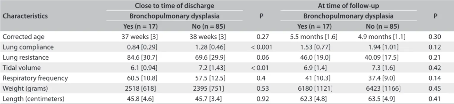
he increased in lung compliance was similar between the two groups as the children became older (Figure 2a). he lung resistance decreased over time for both groups. his decrease was greater among the children with bronchopulmonary dys-plasia (Figure 2b), but the diference was not statistically signii-cant. here was an increase in tidal volume among the children with bronchopulmonary dysplasia as their age increased, but the tidal volume among the children without bronchopulmonary dysplasia remained almost the same (Figure 2c). he increases in body weight and height/length were similar for the two groups over time.
DISCUSSION
he most important result from this study was that the diferences in lung function (compliance and tidal volume) that were observed at the time of hospital discharge were no longer observed at the follow-up assessment conducted at the age of 4-8 months.
his study showed that, at the time of medical discharge, the lung function damage among the premature infants with bronchopulmonary dysplasia was greater than that of the infants without the disease. Even considering the current lower sever-ity of the bronchopulmonary dysplasia, the lung compliance was signiicantly abnormal in these children, given that it was 35% lower than in the children without the disease. Other studies have reported lung compliance values at the age of 2-4 months of age that were 30-50% lower than those of control children born at term. Moreover, these studies reported that the lung resistance in children with bronchopulmonary dysplasia was twice the value found in children in the control group.14,15
Characteristics
Close to time of discharge
P
At time of follow-up
P
Bronchopulmonary dysplasia Bronchopulmonary dysplasia
Yes (n = 17) No (n = 85) Yes (n = 17) No (n = 85)
Corrected age 37 weeks [3] 38 weeks [3] 0.27 5.5 months [1.6] 4.9 months [1.1] 0.30
Lung compliance 0.84 [0.29] 1.28 [0.46] < 0.001 1.53 [0.77] 1.94 [1.01] 0.12
Lung resistance 84.6 [30.7] 69.6 [29.9] 0.06 46.0 [19.0] 40.09 [17.5] 0.21
Tidal volume 6.1 [0.94] 7.2 [1.43] < 0.01 6.9 [1.4] 7.3 [1.6] 0.42
Respiratory frequency 60.5 [10.8] 57.5 [12.5] 0.4 41 [10.3] 37.4 [9.0] 0.14
Weight (grams) 2518 [618] 2395 [751] 0.53 6180 [1121] 6423 [1166] 0.45
Length (centimeters) 45.8 [4.6] 45.7 [3.4] 0.92 62.3 [4.8] 63.5 [4.9] 0.41
he anatomical and physiological formation of the lungs dur-ing the prenatal and postnatal periods depends on a complex rela-tionship between factors that control vascular development and airway diferentiation. Prematurity, exposure of lung epithelial and endothelial cells, high oxygen level, use of ventilation support, presence of infection and presence of patent ductus arteriosus may compromise the process of lung vascular and bronchoalve-olar maturation, thus resulting in complications at an early stage of life (bronchopulmonary dysplasia) or later on.16 Hence, these
adverse conditions may cause abnormalities in the lung function of prematurely born children and these may be intensiied in chil-dren who develop bronchopulmonary dysplasia.8
Lower compliance results from the greater elastic limita-tion of the lungs of infants with bronchopulmonary dysplasia. Hjalmarson et al.8 reported that at the gestational age of 40 weeks,
the speciic compliance of the respiratory system of healthy pre-mature infants born at gestational ages between 25 and 33 weeks was 73% of the compliance of children born at term. Although our study used a method for assessing lung compliance and not respiratory system compliance (which would make measure-ments of lung elastic recoil and thoracic wall recoil combined), the results show the same impairment as seen in other studies.14,15
Although there was a diference in the pulmonary function test parameters at the time of medical discharge, both groups reached lung compliance, resistance and volume values close to normality in the tests at 4-8 months. Although abnormal lung mechanics have been described in infants with bronchopulmo-nary dysplasia at the time of discharge from the neonatal inten-sive care unit,4 the longitudinal follow-up of lung mechanics in
these patients is unclear.
he increases in lung and airway sizes over the irst year of life may result in normalization of lung compliance and lung resis-tance.17 his probably contributed towards the sharp increase in
tidal volume values seen in the children with bronchopulmonary dysplasia in the present study. hese values were close to nor-mality among the children without bronchopulmonary dyspla-sia at the time of medical discharge, and remained almost the same at 4-8 months.
Smaller airway caliber may be a result of anatomical difer-ences or subclinical inlammatory processes. Children with small airway caliber, relected in impairment of lung function, may be at higher risk for wheezing during the irst years of life. During childhood, a viral infection may induce reduction in the size of the peripheral airways, which will result in wheezing in children who already have a preexisting condition of small airway caliber,17
such as in the case of children with bronchopulmonary dysplasia. he results from the present study showed that lung function improved as the child grew older, but the compliance among chil-dren with bronchopulmonary dysplasia remained inferior to that of children without bronchopulmonary dysplasia and the resis-tance remained higher. his implies that even with growth, lung function seems to result from the prematurity and from any clini-cal complications that occurred soon ater birth.18 his is
concor-dant with the study by Gerhardt et al.,14 who reported that as
children with bronchopulmonary dysplasia grew, their pulmo-nary compliance improved. However, it was still only 80-90% of the values seen in the controls at the ages of 2-3 years.
he present study shows the importance of assessing the pulmonary mechanics of very low birth weight preterm infants. However, there is a need for further studies in order to indicate which method would be best for assessing lung function during 2.5
2.0
1.5
1.0
0.5
0.0
Lung c
omplianc
e (ml/cm H
2
O/kg)
Discharge
BPD
No BPD
BPD
No BPD Follow-up
100.0
80.0
60.0
40.0
20.0
0.0
Lung r
esistanc
e (cm H
2
O/ml)
Discharge Follow-up
9.6
8.0
6.4
4.8
3.2
1.6
0.0
T
idal v
olume (ml/kg)
Discharge Follow-up
BPD
No BPD
Figure 2. Lung mechanics in very low birth weight at discharge and at follow-up (4-8 months corrected age).
C. Tidal volume
A. Lung compliance
the various phases of the irst year of life. his need can be seen from our sample, given that although the lung compliance values increased over time in the population of children with broncho-pulmonary dysplasia, they still remained below those of the con-trols without the disease.
It has also been shown in the literature that children born prematurely but without respiratory disease also have abnormal expiratory lows and that at least up to the age of two years, these lows continue to present values lower than those of the controls born at term.19 Baraldi and Filipone showed that abnormal lows
continued up to adulthood.20
here is a need to carry out early analysis on several lung function parameters in order to identify these abnormalities earlier. It needs to be asked whether these infants with normal pulmonary compliance values at a corrected age of 4-8 months would also have normal values for forced expiratory low and vital capacity at this age. here is a need for functional biomark-ers for identifying children at risk of subsequent respiratory morbidity.21 he population of preterm infants and carriers of
bronchopulmonary dysplasia is more susceptible to respiratory morbidity, wheezing and development of asthma, according to the literature.22 Use of lung function tests among these children
as a follow-up measure could contribute towards early clinical intervention and improvement of survival.
Although the estimated sample size was 21, only 17 infants with bronchopulmonary dysplasia were enrolled in this study. This may be a limitation of the study, because this smaller number of children may have contributed towards lack of detection of any difference between the groups with regard to some features. Therefore, the present results should be inter-preted with caution.
Another of the limitations of this study was the varying ages at which the second lung function test was performed. Because respiratory infections occur frequently among children and it is recommended to wait for a minimum of three weeks thereater before the test is taken, this meant that we had to include a cor-rected age range from 4 to 8 months in the examination protocol. Another constraint was the high number of children excluded from the study because of respiratory infections presented at the time of the second test. his is a population susceptible to respi-ratory morbidity, which hinders availability of the entire popula-tion for the longitudinal tests.
One of the strengths of this study was that it enabled sequen-tial evaluation over the course of the irst year of life, using a less invasive technique to document the lung function of very low birth weight preterm infants. However, a single evaluation of lung function at a follow-up ater 4-8 months can be considered to be a limitation of the study.
here are few studies reporting serial measurements of lung function during childhood, thus showing the diiculties in making these measurements in this age group.17,23 In Brazil,
few studies on pulmonary mechanics close to the time of medi-cal discharge and over the course of the irst year of life of very low birth weight premature children have been conducted.24,25
hus, the present study provides an important contribution in this ield of knowledge.
CONCLUSION
Diferences in lung mechanics were observed between infants with and without bronchopulmonary dysplasia at the time of hospital discharge, but these diferences were no longer detected at a corrected gestational age of 4 to 8 months. he lung mechan-ics of all the infants improved over this period of time.
REFERENCES
1. Jobe AH. The new bronchopulmonary dysplasia. Curr Opin Pediatr. 2011;23(2):167-72
2. Mosca F, Colnaghi M, Fumagalli M. BPD: old and new problems. J Matern Fetal Neonatal Med. 2011;24 Suppl 1:80-2
3. Broström EB, Thunqvist P, Adenfelt G, Borling E, Katz-Salamon M. Obstructive lung disease in children with mild to severe BPD. Respir Med. 2010;104(3):362-70.
4. Doyle LW, Faber B, Callanan C, et al. Bronchopulmonary dysplasia in very low birth weight subjects and lung function in late adolescence. Pediatrics. 2006;118(1):108-13.
5. Schmalisch G, Wilitzki S, Roehr CC, Proquitté H, Bührer C. Development of lung function in very low birth weight infants with or without bronchopulmonary dysplasia: longitudinal assessment during the irst 15 months of corrected age. BMC Pediatr. 2012;12:37.
6. Narayanan M, Beardsmore CS, Owers-Bradley J, et al. Catch-up alveolarization in ex-preterm children: evidence from (3)He magnetic resonance. Am J Respir Crit Care Med. 2013;187(10):1104-9.
7. Sanchez-Solis M, Garcia-Marcos L, Bosch-Gimenez V, et al. Lung function among infants born preterm, with or without bronchopulmonary dysplasia. Pediatr Pulmonol. 2012;47(7):674-81. 8. Hjalmarson O, Sandberg K. Abnormal lung function in healthy
preterm infants. Am J Respir Crit Care Med. 2002;165(1):83-7. 9. Allen J, Zwerding R, Ehrenkranz R, et al. Statement on the care of the
child with chronic lung disease of infancy and childhood. Am J Respir Crit Care Med. 2003;168(3):356-96.
10. Respiratory mechanics in infants: physiologic evaluation in health and disease. American Thoracic Society/European Respiratory Society. Am Rev RespirDis. 1993;147(2):474-96.
12. Jobe AH, Bancalari E. Bronchopulmonary dysplasia. Am J Respir Crit Care Med. 2001;163(7):1723-9.
13. Alexander GR, Himes JH, Kaufman RB, Mor J, Kogan MA. A United States national reference for fetal growth. Obstet Gynecol. 1996;87(2):163-8.
14. Gerhardt T, Hehre D, Feller R, Reifenberg L, Bancalari E. Serial determination of pulmonary function in infants with chronic lung disease. J Pediatr.1987;110(3):448-56.
15. Morray JP, Fox WW, Kettrick RG, Downes JJ. Improvement in lung mechanics as a function of age in the infant with severe bronchopulmonary dysplasia. Pediatr Res.1982;16(4 Pt 1):290-4. 16. Carvalho CG, Silveira RC, Procianoy RS. Lesão pulmonar induzida pela
ventilação em recém-nascidos prematuros [Ventilator-induced lung injury in preterm infants]. Rev Bras Ter Intensiva. 2013;25(4):319-26. 17. Young S, Arnott J, O’Keefe PT, Le Souef PN, Landau LI. The association
between early life lung function and wheezing during the irst 2 years of life. Eur Respir J. 2000;15(1):151-7.
18. von Mutius E. Paediatric origins of adult lung disease. Thorax. 2001;56(2):153-7.
19. Jones M. Efect of preterm birth on airway function and lung growth. Paediatr Respir Rev. 2009; 10 Suppl 1:9-11.
20. Baraldi E, Filippone M. Chronic lung disease after premature birth. N Engl J Med. 2007;357(19):1946-55.
21. Proietti E, Riedel T, Fuchs O, et al. Can infant lung function predict respiratory morbidity during the irst year of life in preterm infants? Eur Respir J. 2014;43(6):1642-51.
22. McEvoy C, Venigalla S, Schilling D, et al. Respiratory function in healthy late preterm infants delivered at 33-36 weeks of gestation. J Pediatr. 2013;162(3):464-9.
23. Rosenfeld M, Allen J, Arets BH, et al. An oicial American Thoracic Society workshop report: optimal lung function tests for monitoring cystic ibrosis, bronchopulmonary dysplasia, and recurrent wheezing in children less than 6 years of age. Ann Am Thorac Soc. 2013;10(2):S1-S11. 24. de Mello RR, Dutra MV, Ramos JR, et al. Lung mechanics and high-resolution computed tomography of the chest in very low birth weight premature infants. Sao Paulo Med J. 2003;121(4):167-72. 25. de Mello RR, Dutra MV, Ramos JR, et al. Neonatal risk factors for
respiratory morbidity during the irst year of life among premature infants. Sao Paulo Med J. 2006;124(2):77-84.
Acknowledgements: Conselho Nacional de Desenvolvimento Cientíico e Tecnológico (CNPq) and Fundação Oswaldo Cruz, Rio de Janeiro (Papes IV Project), for inancial support.
Sources of funding: Conselho Nacional de Desenvolvimento Cientíico e Tecnológico (CNPq) and Fundação Oswaldo Cruz, Rio de Janeiro (Papes IV Project). Project number 400115 /2006-2009
Conlicts of interest: None
Date of irst submission: December 17, 2014
Last received: December 17, 2014
Accepted: December 18, 2014
Address for correspondence:
Rosane Reis de Mello
Departamento de Neonatologia Av. Rui Barbosa, 716 — 3o andar
Flamengo — Rio de Janeiro (RJ) — Brasil CEP 22250-020