• Nenhum resultado encontrado

Braz. Dent. J. vol.21 número3

N/A
N/A
Protected

Academic year: 2018

Share "Braz. Dent. J. vol.21 número3"

Copied!
6
0
0

Texto

(1)

INTRODUCTION

Osteochondroma (OC) or osteocartilagenous exostosis, a cartilage-capped exophytic lesion that arises from the bone cortex, is one of the most common benign bone tumors. It usually occurs in the axial skeleton, especially long bones, such as the distal metaphisis of the femur or the proximal metaphisis of the tibia (1,2). The oral and maxillofacial regions are not common sites of OCs, but the embryonic development of the temporomandibular joint (TMJ), by the endochondral ossification, makes this area the most frequent facial site of this type of tumor (2-7). Differently from long bones, craniofacial OCs occur at older ages with a slow growth, even at the end of puberty, most frequently affecting women in their second decade of life (1,8,9). The etiology and pathogenesis of the lesion is

Osteochondroma of the Temporomandibular Joint:

A Case Report

Estevam Rubens UTUMI1 Irineu Gregnanin PEDRON1

Andréia PERRELLA1 Camila Eduarda ZAMBON2 Marcelo Minharro CECCHETI3 Marcelo Gusmão Paraíso CAVALCANTI4

1Department of Stomatology, Dental School, University of São Paulo, São Paulo, SP, Brazil

2Department of Oral and Maxillofacial Surgery, Clinics Hospital, University of São Paulo, São Paulo, SP, Brazil 3Department of Oral and Maxillofacial Surgery, Dental School, University of São Paulo, São Paulo, SP, Brazil 4Department of Radiology, Dental School, University of São Paulo, São Paulo, SP, Brazil; Department of Radiology,

College of Medicine, University of Iowa, Iowa City, IA, USA

Osteochondroma of the mandibular condyle has been found in the oral and maxillofacial region rarely. This paper describes a case of osteochondroma of the mandibular condyle in a 20-year-old woman, who was referred to our service with facial asymmetry, prognathic deviation of chin, cross-bite to the contralateral side, changes in condylar morphology, limited mouth opening, and malocclusion. Computed tomography (CT) was performed for better evaluation to the pathological conditions on the temporomandibular joint. Based on the clinical examination, patient history, and complementary exams, the hypothesis of osteochondroma was established. Condylectomy was performed using a preauricular approach with total removal of the lesion. After 3 years of postoperative follow up and orthodontic therapy, the patient is symptom-free, and has normal mouth opening with no deviation in the opening pattern.

Key Words: osteochondroma, mandibular condyle, condylectomy, oral surgery.

not fully understood and neither is its development and neoplastic or reparative nature. The histological appearance reveals an endochondral ossification capped by a proliferative hyaline cartilage. This feature is similar to those seen in epiphysal plates before closure, supporting the theory of pluripotential periosteal cells as precursors of this lesion (1,2,7,10,11). Porter and Simpson (12) suggested that a genetic component might also be involved in the neoplastic pathogenesis due to somatic mutations found in chromosomes 8 and 11. However, the cellular origin of this process is controversial.

Although condyle OC can present several different clinical characteristics, facial asymmetry, malocclusion, prognathic deviation of chin, crossbite in the contralateral side, and mouth opening disturbance are the most commonly seen manifestations of the

(2)

disease. These conditions may also be found in unilateral condylar hyperplasia and other differential diagnoses including osteoma, chondroma, giant cell tumor, myxoma, fibro-osteoma, fibrous dysplasia, fibrosarcoma and chondrosarcoma. In spite of the common clinical features, the definitive diagnosis should always be based on clinical, radiological and histological criteria (2,10,11). Complementary examinations such as panoramic radiography and computed tomography (CT) can be useful to visualize the existing relationships among anatomic structures (13). Scintigraphy may also be used to detect the presence of intense uptake in the lesion (2,14). In addition, growing bone surrounded by cartilage is expected to be observed under histological evaluation (2,13).

Condylectomy with complete lesion removal is the most common treatment of condylar OC. However, treatment could also be performed without condylectomy (2,3,9), and an association with orthodontic treatment should also be considered.

This paper presents an uncommon case of OC on the left mandibular condyle treated surgically and followed up for 3 years postoperatively.

CASE REPORT

A 20-year-old female patient was referred to our hospital complaining of eating difficulty, facial asymmetry, and pain in the left TMJ. She had noted a slowly progressive facial asymmetry and tooth crowding for at least 4 years. No history of trauma was reported.

Clinical examination revealed severe malocclusion, facial asymmetry with approximately 10 mm deviation of the midline to the left side, posterior crossbite on the right side, and negative overjet resulting in eating difficulty. Her maximum mouth opening was 34 mm, and Class I occlusion without open bite was present (Fig. 1).

The panoramic radiograph showed a slight, well-defined radiopacity in the left condyle head, causing deviation of the midline (Fig. 2). Coronal and axial CT scans showed a large hyperdense bone growth on the surface of the left condylar head (Fig. 3A-B). The lesion extended from the medial surface of the condyle towards the glenoid fossa. Scintigraphy showed an increased activity in the left TMJ (Fig. 4).

Based on clinical examination, patient history, and complementary tests, a diagnosis of OC was hypothesized.

Orthodontic treatment was performed along with the surgical treatment for correction of tooth crowding. Under general anesthesia, we performed total excision and condylectomy with a modified temporal incision. After skin incision, the underlying subcutaneous tissue, muscle, and fascia were carefully dissected from the condyle. The facial nerve was located at its normal anatomic position and it was carefully moved. After exposing the condyle, it was observed that the tumor

Figure 1. Clinical preoperative facial appearance. A= Chin deviation to the right side; B= Lateral view showing mandibular prognathism; C= Midline deviation and malocclusion; D= Negative overjet.

(3)

could not be separated from the condyle (Fig. 5A-B). Then, the lesion excised and the condylar neck was reshaped and repositioned underneath the preserved TMJ disk. The excision site of the condylar region was curetted sufficiently to remove any remaining tumoral

cells. Finally, a drain was placed and all tissues were sutured with 3.0 catgut and 5.0 nylon (skin).

The histopathological examination revealed a nodular lesion with cartilaginous cap and immature bone tissue with presence of fiber, neoformed bone and cartilaginous hyaline tissue, confirming the diagnosis of osteochondroma of the condyle (Fig. 5C).

In order to guide the correct position of the mandible, the patient used guiding elastics for 2 weeks. Jaw exercises were undertaken for 3 weeks and repeated 5 times a day after removal of the elastics. Mandibular movements could be easily performed without pain. Orthodontic therapy was continued after surgery to improve occlusion. Complete correction

Figure 3. CT scans. A = Sequences of coronal CT images in bone window showing irregular mass of mineralization in the condyle. B = Sequences of axial CT images in bone window showing a large nodular mass around the left TMJ.

Figure 4. Scintigraphy with 99mTc-HMDP show an intense uptake

of radiopharmaceutical in the left condyle (arrows).

(4)

of the malocclusion and midline deviation could not be achieved. However, after 3 years of postoperative follow up and orthodontic therapy, neither recurrence nor complications were observed (Fig. 6). The patient was satisfied with the postoperative results and remission of pain.

DISCUSSION

OC is not a common disease (13). Its causes are still unclear and symptoms vary depending on the location of the tumor. Trauma and inflammation have been suggested as contributory factors. There have been controversies if such lesions should be considered of developmental, neoplastic or reparative nature (2,11,14). Common clinical manifestations of the OC of the mandibular condyle include facial asymmetry, swelling at the TMJ region, disturbance of mouth opening, and joint pain (7). Our patient had facial asymmetry, pain in the left TMJ and malocclusion.

The growth of an OC is usually slow, causing

gradual displacement and elongation of the mandible (10). Seki et al. (11) reported a case of condylar OC with complete hearing loss. In the present case, the patient had not experienced trauma at the TMJ region or ear infection before the onset of symptoms, which included pain in the TMJ region, severe facial asymmetry, midline deviation, but no obstructive hearing sensations. Based on the fact that the tumor arose from the posteromedial surface of the condyle and little limitation of mouth opening was observed, we believed that the patient had suffered a minor trauma to the condyle.

A careful assessment of the patient’s history might provide valuable information for the diagnosis and treatment of facial asymmetry (16).

The diagnosis of OC was proposed based on clinical and radiographic findings. Imaging techniques can be valuable tools for accurately diagnosing and determining treatment for a variety diseases and are supportive to clinical examination (10,13). CT scans can easily demonstrate the continuity of cortex and medulla of the parent bone tumor. In the present case, they were useful to determine the margins of the OC causing facial asymmetry.

Sales et al. (17) reported that CT imaging brings to radiologists and clinicians the possibility of evaluating complex cases in the maxillofacial field and giving information that leads to more accurate and specific diagnosis of some TMJ pathological conditions. Preoperative CT assessment can be of great important role in the treatment planning of these tumors (1,18,19). Although CT scans have not been considered the best tool to evaluate non-calcified cartilage caps, they have a recognizably high accuracy to demonstrate calcified cartilage, and to delineate soft-tissue alterations secondary to tumor growth and atrophy of the masticatory muscles for complementary surgical indications. CT images are also of great value for differential diagnoses, especially in differentiating condylar OC from unilateral condylar hyperplasia. OC is usually seen as a growth of the morphologically normal condyle, while condylar hyperplasia is seen as an enlargement of the condylar process (1).

According to Villanueva et al. (13), the main goal of OC treatment, regardless of the lesion etiology, should be the achievement of acceptable mouth opening ranges. Our main objective in treating our patient was to recover her facial symmetry and reestablish facial harmony and occlusion after surgery associated with orthodontic treatment. Haag et al. (16) emphasized that treatment Figure 6. Postoperative views. Front (A) and lateral (B) views of

(5)

goals should be specified according to the diagnosis of facial asymmetry. In cases of acceptable facial esthetics, orthodontic camouflage treatment could be done to correct dental asymmetries. It has been recommended that correction of asymmetric occlusion should be done at the early stages (16).

Several surgical approaches have been suggested for the treatment of condylar OCs, including complete resection of the tumor using condylectomy, condylectomy with reconstruction, or selected tumor removal without condylectomy (5,17). The treatment of choice in this case was resection through condylectomy after orthodontic planning. Orthodontic evaluation was performed before surgery and used as a guidance to evaluate occlusion. Condylectomy cannot be recommended as a routine procedure for all cases. If the tumor involves only a limited area of the condylar surface, preservation of the remaining part of the condyle, and reshaping should be done.

A conservative condylectomy with articular disc repositioning combined with orthognathic surgery is an acceptable option for treatment of condylar OC (2). The patient was satisfied with the postoperative results and continued the orthodontic treatment.

Some authors have proposed reconstruction using vertical sliding osteotomy of the mandibular ramus and two miniplates for osteosynthesis (20). This technique can be an alternative for the reconstruction of small and medium defects resulting from condylectomy, as well as small vertical dimension losses derived from posttraumatic avascular necrosis of the condyle and idiopathic condylar resorption.

Histologically, the diagnosis of an OC includes chondrocytes of the cartilaginous cap arranged in clusters parallel to lacunar spaces. Differential diagnoses of OC include osteoma, benign osteoblastoma, chondroma, and chodroblastoma (2). It is very important to differentiate OC from these previous lesions.

Condylar OC should be considered in the differential diagnoses of tumors of the TMJ region. Orthopantomograph at best can be considered as a screening tool in the detection of these lesions. CT scans should be performed in all cases of suspected condyle OC (1).

Most cases reported in the literature did not associate surgery with orthodontic treatment (2,3,5,8,10,14,20). In the present case, the orthodontic treatment corrected the tooth crowding, but it did not correct the facial asymmetry. Therefore, we considered

surgery associated with orthodontic treatment a valid approach to minimize facial asymmetry, contributing to the recovery of occlusion and facial harmony. When facial asymmetry persists after surgery, orthognathic surgery is recommend for correction.

RESUMO

Osteocondroma de côndilo mandibular é raro na região craniofacial. Este artigo descreve um caso de osteocondroma de côndilo mandibular em uma mulher de 20 anos que foi encaminhada ao nosso serviço apresentando assimetria facial, desvio de mento, mordida cruzada para o lado contralateral, alterações na morfologia condilar, limitação de abertura bucal e maloclusão. Tomografia computadorizada foi realizada para melhor avaliação da condição patológica da ATM. Devido à base no exame clínico, histórico do paciente e exames complementares, foi estabelecida uma hipótese de osteocondroma. Um procedimento de condilectomia utilizando abordagem preauricular com uma total remoção da lesão foi executado. Após três anos de acompanhamento pós-operatório e ortodôntico, o paciente está livre dos sintomas e tem uma abertura normal sem desvio de padrão durante a abertura.

ACKNOWLEDGEMENTS

To The São Paulo State Research Foundation (FAPESP) for

granting a Masterʼs degree scholarship to Dr. Estevam Rubens

Utumi (Process#2006/05328-0). The authors would like to thank Dr. Jan Peter Ilg for helping with the case, and Dr. Flavio E. Hirai for reviewing the manuscript.

REFERENCES

1. Avinash KR, Rajagopal KV, Ramakrishnaiah RH, Carnelio S, Mahmood NS. Computed tomographic features of mandibular osteochondroma. Dentomaxillofac Radiol 2007;36:434-436. 2. Ortakoglu K, Akcam T, Sencimen M, Karakoc O, Ozyigit HA,

Bengi O. Osteochondroma of the mandible causing severe facial asymmetry: a case report. Oral Surg Oral Med Oral Pathol Oral Radiol Endod 2007;103:e21-28.

3. Lizuka T, Schroth G, Laeng RH, Ladrach K. Osteochondroma of the mandibular condyle: report of a case. J Oral Maxillofac Surg 1996;54:495-501.

4. Tanaka E, Lida S, Tsuji H, Kogo M, Morita M. Solitary osteochondroma of the mandibular of the mandibular symphysis. Int J Oral Maxillofac Surg 2004;33:625-626.

5. Karras SC, Wolford LM, Cottrell DA. Concurrent osteochondroma of the mandibular condyle and ipsilateral cranial base resulting in temporomandibular joint ankylosis: report of a case and review of the literature. Oral Maxillofac Surg 1996;54:640-646.

6. Koga K, Toyama M, Kurita K. Osteochondroma of the mandibular angle: report of a case. J Oral Maxillofac Surg 1996;54:510-513. 7. Wu W, Hu X, Lei D. Giant osteochondroma derived from pterygoid

(6)

9. Peroz I, Scholman HJ, Hell B. Osteochondroma of the mandibular condyle: a case report. Int J Oral Maxillofac Surg 2002;31:455-456.

10. Holmlund AB, Gynther GW, Reinholt FP. Surgical treatment of osteochondroma of the mandibular condyle in the adult. A 5-year follow-up. Int J Oral Maxillofac Surg 2004;33:549-553. 11. Seki H, Fukuda M, Takahashi T, Iino M. Condylar osteochondroma

with complete hearing loss: report of a case.J Oral Maxillofac Surg 2003;61:131-133.

12. Porter DE, Simpson AH. The neoplastic pathogenesis of solitary and multiple osteochondromas. J Pathol 1999;188:119-125. 13. Villanueva J, González A, Cornejo M, Núñez C, Encina S.

Osteochondroma of the coronoid process. Med Oral Patol Oral Cir Bucal 2006;11:E289-291.

14. Vezeau PJ, Fridrich KL, Vincent SD. Osteochondroma of the mandibular condyle: literature review and report of two atypical cases. J Oral Maxillofac Surg 1995;53:954-963.

15. Sakai H, Minemura T, Ito N, Miyazawa H, Kurashina K. Isolated osteochondroma near the mandibular angle. Int J Oral Maxillofac Surg 2007;36:274-275.

16. Haag U, Wong R, Ng AFS. Facial asymmetry The Orthodontist’s viewpoint. Dental Asia. 2007; 19-24.

17. Sales MA, Oliveira JX, Cavalcanti MG. Computed tomography imaging findings of simultaneous bifid mandibular condyle and temporomandibular joint ankylosis: case report. Braz Dent J 2007;18:74-77.

18. Iizuka T, Schroth G, Laeng RH, Lädrach K. Osteochondroma of the mandibular condyle: report of a case. J Oral Maxillofac Surg 1996;54:495-501.

19. Koole R, Steenks MH, Witkamp TD, Slootweg PJ, Shaefer J. Osteochondroma of the mandibular condyle. A case report. Int J Oral Maxillofac Surg 1996;25:203-205.

20. González-Otero S, Navarro-Cuéllar C, Escrig-de Teigeiro M, Fernández-Alba-Luengo J, Navarro-Vila C. Osteochondroma of the mandibular condyle: Resection and reconstruction using vertical sliding osteotomy of the mandibular ramus. Med Oral Patol Oral Cir Bucal 2009;14:E194-197.

Imagem

Figure  1.  Clinical  preoperative  facial  appearance.  A=  Chin  deviation to the right side; B= Lateral view showing mandibular  prognathism;  C=  Midline  deviation  and  malocclusion;  D=
Figure 3. CT scans. A = Sequences of coronal CT images in bone  window showing irregular mass of mineralization in the condyle

Referências

Documentos relacionados

Dos objectivos traçados no nosso trabalho, verificamos que da análise quantitativa da transição das Lux GAAP para as IFRS nas diferentes rubricas, bem como nos

Esta pesquisa pode ser descrita como um estudo de caso, com abordagem qualitativa e quantitativa, que utilizou a técnica de entrevista (mapeamento cognitivo) para coletar dados

Ousasse apontar algumas hipóteses para a solução desse problema público a partir do exposto dos autores usados como base para fundamentação teórica, da análise dos dados

The probability of attending school four our group of interest in this region increased by 6.5 percentage points after the expansion of the Bolsa Família program in 2007 and

In the former Chapter, I have given you a short Account of that Part of the Laws of England which is called Lex Scripta, namely, Statutes or Acts of Parliament, which in their

[r]

Figura 20: Utilização do cluster pelos experimentos com 90% para limite superior, 40% para limite inferior e 45% para média exponencial. tempo para a finalização dos

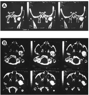
The clinical features of the fibrous-ankylosis are severely limited mouth-opening capacity (limited range of motion during the opening), usually no pain and no joint sounds,