Analysis of the
Agger nasi cell
and frontal sinus ostium sizes using
computed tomography of the paranasal sinuses
Abstract
Fernando Veiga Angélico Junior1, Priscila Bogar Rapoport2
1 MSc in Health Sciences; Assistant Professor and Coordinator of the Medical Residency Program in Otorhinolaryngology of the Medical School of the ABC. 2 PhD in Otorhinolaryngology; Full Professor of Otorhinolaryngology - Medical School of the ABC.
Medical School of the ABC (FMABC).
Send correspondence to: Fernando Veiga Angélico Junior. Rua Mediterrâneo, nº 290. Cj. 127/128. São Bernardo do Campo - SP. Brazil. CEP: 09750-420. veiga.fernando@yahoo.com.br
Paper submited to the BJORL-SGP (Publishing Management System - Brazilian Journal of Otorhinolaryngology) on November 16, 2012;
and accepted on March 14, 2013. cod. 10599.
T
he Agger nasi cell (ANC) and the frontal sinus ostium (FO) are important structures that can influence the anatomy and physiology of the frontal recess. The aim of this study was to evaluate the presence and size of ANC and the FO and correlate them according to gender, race and among themselves.Method: A prospective study with 40 patients who underwent CT of the paranasal sinuses with sagittal reconstruction. Measurements: ANC (APAN) anteroposterior diameter, ANC (CCAN) cranio-caudal diameter, ANC (LLAN) side-to-side diameter, anteroposterior diameter of the FO (APFO) and side-to-side diameter of the FO (LLFO).
Results: Twenty-two patients were male and 18 females, mean age 33.7 years. Most patients were white (45%), followed by browns (32.5%), blacks (20%) and asians (2.5%). The ANC was present in 98.7% of patients. There was statistical difference for APAN on females and LLAN on females and on the total sample. There were no differences for all measurements regarding gender, as well as the race. ANC and FO measurements showed positive correlation, but poor or very poor.
Conclusion: The prevalence of ANC in our sample was high and did not show a statistically significant difference for most measurements. The correlation between measurements of ANC and the FO was poor or very poor.
ORIGINAL ARTICLE Braz J Otorhinolaryngol.
2013;79(3):285-92.
BJORL
Keywords:
anatomy; ethmoid sinus; frontal sinus; measures; tomography.
INTRODUCTION
The frontonasal region anatomy acquired much importance, mainly after the development of techniques for approaching this region through nasal endoscopy.
In recent years, endoscopic endonasal surgery (EES) has been widely used for the treatment of the most varied sinonasal disorders, especially of the frontal sinus (FS)1,2.
However, the frontal endoscopic sinusotomy remains a challenge for most otorhinolaryngologists, due to the com-plexity and anatomical variability of the three-dimensional spaces called ethmoidal infundibulum and frontal recess (FR)3-5.
The FR is the anterosuperior border of this complex, being the embryological origin of the FS. Its medial border is the lateral surface of the anterior portion of the middle concha, all the way to its insertion in the skull base. Should the unciform process curve medially and inserts in the mi-ddle concha, it will also be part of the FR medial border. The lamina papyracea is the lateral recess border, and the unciform process may be part of such border when it is inserted superiorly or laterally to it. The posterior boundary is created from the anterior surface of the ethmoid bulla, which is typically inserted at the skull base, which may be an incomplete insertion. The anterior border includes the Agger nasi (AN), which can be pneumatized and of varied size. When the ANC is pneumatized, we have the
Agger nasi cell (ANC) formation6-8.
In the sagittal plane, the frontonasal communication takes the form of an hourglass, which more closely corres-ponds to the frontal sinus ostium (FO)9,10. Below the FO
the FR dimensions are determined by several structures which also contribute to its physiological functioning.
This anatomical pattern may be influenced by anterior ethmoidal cells that develop embryolo-gically around the FR, which could have profound anatomical and functional repercussions on the frontonasal communication11.
Therefore, any malformation or existing anatomical variation in this area can affect FS drainage and ventilation, causing difficult-to-treat rhinosinusitis. Several factors can narrow the FR, especially the ANC, the ethmoidal bulla, the unciform process and even the middle concha head9,12,13.
Thus, surgery aiming at eliminating a FR disorder or to correct an anatomical alteration or malformation will lead to restoration of normal FS function, due to improved ventilation and drainage11.
With the development of computed tomography (CT) and fiber optic systems, more data can be obtained in the preoperative period, aiming at a better treatment choice. The critical analysis of these data, associated with intraoperative findings are important in the decision to do a more extensive approach on the ethmoidal infundibulum
and FR, sometimes leading to a resection of the thick bone adjacent to the FO.
The paranasal sinuses computed tomography scan (PSCT) is the method of choice for image evaluation of nasal and sinonasal diseases and to study the ostiomeatal complex14,15.
Usually, there are two views: coronal and axial. These views may bring detailed information about FS size, ANC size and ethmoidal infundibulum structures that can compromise and alter the FR.
The sagittal reconstruction, with fine slices used in recent years, allows for a better analysis of nasal structures and brings a fresh push to understanding the complex anatomy, allowing us to measure the sizes of structures that make up or contribute to the FR formation9,16,17. It also
allows the identification of the anteroposterior relationship between the FR, the unciform process, ethmoidal bulla, basal lamella of the middle concha and the upper concha, which can be difficult to assess only under nasal endoscopy or conventional CT scan.
Thus, the study of the FR region is of fundamental importance in the management of frontal rhinosinusitis.
Thus, the objectives of this study are: to evaluate the prevalence of the Agger nasi cell (ANC), measure the dimensions of the ANC and the frontal sinus ostium (FO), compare the dimensions of the ANC and FO in both gen-ders and between the races, and check the relationship of the ANC and FO dimensions.
METHOD
We consecutively included volunteer adults, of both genders, coming from the general outpatient clinic of a tertiary hospital, who required PNSCT to investigate frontal headache with or without other nasal symptoms.
The study protocol was reviewed and approved by the Ethics in Research Committee of the institution under number 102/2003, and all patients signed an informed consent form.
All patients were evaluated, obtaining data from clinical history, general physical examination and ENT physical exam. They were also submitted to nasofibros-copy with a 4 mm 0 degree hard endoscope (Fiegert Endotech brand).
of the maxilla to the apex of the frontal sinus, parallel to the hard palate. From the acquired images, we made a reconstruction of the coronal and sagittal planes on the workstation installed on the scanner and the recording was made in discs for later analysis.
The exclusion criteria were: patients under the age of 18 years; absence of frontal sinus; presence of nasal-related headache, chronic rhinosinusitis, sinonasal polyposis, bone erosions, sinonasal tumors, anatomical malformations of the paranasal sinuses or any other con-dition that prevents the visualization of the bony structures of the region studied; presence of ethmoidofrontal cells, frontal intersinusal cells, supraorbital ethmoidal cells, supra bullae cells, fontal bulla or any other anatomical variation close to or obstructing the FR or FO, altering its dimen-sions; any CT alteration which prevents measurements of the ANC and/or the FO, such as, for example, artifacts caused by dental restorations; and those who refused to sign the Informed Consent Form.
We analyzed 80 nasal cavities, corresponding to 40 patients, evaluating the axial, coronal and sagittal vies.
For FO identification we initially used the sagittal views, where it is easier to pinpoint the internal nasal spine, the FR and the FO. On the CT scanner worksta-tion we could see, at the same time, the axial, coronal and sagittal views in one same screen and, moving the marking cursor in order to measure the structures on the image in one determined type of view, the cursor moves automatically in the others two views, simultaneously. So, we were able to identify and more accurately measure the dimensions of the structures under consideration. Similarly, we pinpointed the ANC and measured it.
The following measures were taken:
• Larger anteroposterior diameter of the frontal sinus ostium (OFAP) (Figure 1);
• Larger anteroposterior diameter of the Agger nasi cell (AGAP) (Figure 3);
Figure 1. Anteroposterior measure of the frontal sinus ostium (OFAP).
• Side-to-side diameter of the frontal sinus ostium (OFLL) (Figure 2);
Figure 2. Side-to-side measure of the frontal sinus ostium (OFLL).
Figure 3. Anteroposterior measure of the Agger nasi cell (AGAP). • Larger side-to-side diameter of the Agger nasi
cell (AGLL) (Figure 4);
Figure 4. Side-to-side measure of the Agger nasi cell (AGLL). • Larger craniocaudal diameter of the Agger nasi
For the statistical analysis, we used the following programs: SPSS V16 (Statistical Package for Social Sciences - version 16.0) 15, Minitab and Excel OfficeTM to obtain
the results. We used the following tests: ANOVA (Variance Analysis), Student’s paired t-test and the test for equality of two ratios to assess FO and ANC measures and the Pearson’s correlation coefficient in order to establish possible relationships between the variables above.
For all analyses, we considered the 95% confidence interval and 5% significance interval (p < 0.05).
RESULTS
Our series comprises 40 patients, 18 (45%) females and 22 (55%) males, with no statistical difference between them (p = 0.371).
The patient’s age range varied between 19 and 54 years, averaging 33.7 years and with a standard deviation of 9.7 years. There was no statistically significant difference between the genders vis-à-vis age (p = 0.922).
Most patients were caucasian (45%), followed by browns (32.5%) and blacks (20%). We found only one patient (2.5%) of the yellow race.
Presence of theAgger nasicell
The ANC was present in 98.7% of the sides, not being seen in only one side of a patient (right side).
Agger nasi cell measures
Anteroposterior diameter of the Agger nasi cell (AGAP)
There was no significant difference in the measures vis-à-vis the side among men and in the total sample, only among women (Table 1).
Side-to-side diameter of the Agger nasi cell (AGLL)
Table 2 shows the mean values for females, males and total sample. There was a significant difference in the measures vis-à-vis the right and left sides among women
Agger nasi cell Craniocaudal Diameter (AGCC)
Evaluating the largest cranial-caudal diameter of the Agger nasi cell (AGCC), no statistically significant differences were found between the genders and between the right and left sides (Table 3).
Figure 5. Craniocaudal measure of the Agger nasi cell (AGCC).
Table 1. Measure in millimeters of the largest anteroposterior diameter of the Agger nasi cell (AGAP).
AGAP Females Males All
Right Left Right Left Right Left
Mean 7.28 5.67 6.48 6.86 6.85 6.31
Standard
deviation 2.97 2.47 2.68 2.85 2.81 2.72
Minimum
value 3 2 4 3 3 2
Maximum
value 12 10 13 14 13 14
p < 0.001 0.428 0.116
Table 2. Measure in millimeters of the largest anteroposterior diameter of the Agger nasi cell (AGLL).
AGLL Females Males All
Right Left Right Left Right Left
Mean 5.72 4.78 6.14 5.71 5.95 5.28
Standard
deviation 2.40 2.05 1.77 2.03 2.06 2.06
Minimum
value 2 2 3 2 2 2
Maximum
value 11 9 9 10 11 10
p 0.025 0.369 0.036
Table 3. Measure in millimeters of the largest craniocaudal diameter of the Agger nasi cell (AGCC).
AGCC Females Males All
Right Left Right Left Right Left
Mean 6.67 7.39 7.29 6.90 7.00 7.13
Standard
deviation 3.20 3.84 1.79 2.32 2.52 3.08
Minimum
value 2 2 4 3 2 2
Maximum
value 13 14 12 11 13 14
p 0.195 0.390 0.714
Frontal sinus ostium measures
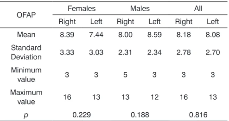
Anteroposterior diameter of the frontal sinus ostium (OFAP)
Side-to-side diameter of the frontal sinus ostium (OFLL)
The mean values of the frontal sinus ostium side-to-side diameter (OFLL) in the total sample was 6.50 mm (SD = 2.17 mm) to the right and of 6.45 mm (SD = 2.05 mm) to the left. Statistically significant difference was not observed between the sides and the genders (Table 5). Considering the 80 sides, the mean value was 6.48 mm.
Relation between the Agger nasicell and frontal sinus ostium measures
Analyzing the relationships between the various measures of the ANC and FO through the Pearson cor-relation, we concluded that they all have a statistically significant positive correlation, but with a bad or very bad correlation rate (Table 7).
DISCUSSION
The FR is one of the most complex areas of the paranasal sinuses anatomy, causing difficulties even for the rhinologists who are frequently working in this region. This if justified by the large number of structures that can participate and influence the formation of this communi-cation with the FS, the FO and the FS itself.
Any study that brings some new contribution to the knowledge of this region helps us better understand its anatomy and provides more data for decision-making aimed at the management of patients with sinonasal diseases.
In our study, the first question to be argued is the use of PSCT scan to assess the FR, the FO and the ANC. Most studies in the literature use CT scans to assess these structu-res, but there is no uniform methodology for such analysis. The type of cross-sectional view (axial, sagittal or coronal) and the slice thickness vary among the studies, making data comparison much too variable. Some authors used only the axial and coronal views for their conclusions18-23. Although
Landsberg et al.10 reported that the ANC identification is very
difficult even with the use of the sagittal reconstruction, we chose this technique to complement the analysis of the axial and coronal views, since it helped in the identification of the FR, the FO and the ANC, as numerous authors have reported in the literature7,9,17,24,25, besides facilitating the measuring of
the structures of interest in our study.
One important data of our analysis was the presence or absence of the ANC. Several studies have evaluated the presence of ANC using PSCT scan. We found this cell in 98.7% of the sides studied - such structure was seen only on the left side in one patient. Disregarding the sides, the ANC was found in 100% of our patients. It is worth stressing that the literature data is presented differently, sometimes considering the individual, sometimes the sides to calculate the ANC prevalence. We chose to use the individual-related prevalence, as most of the studies did. In general, the ANC presence varied between 7.77% and 100%. Our study corroborates data from various authors who found the ANC in 95% or more of the patients studied26,27. Others
found the ANC in about 90% of the patients - very close to the values reported in our study22,28. On the other hand,
our data is different from that of some authors who lower percentages in their respective studies20,29,30.
Table 4. Measure in millimeters of the largest anteroposterior diameter of the frontal sinus ostium (OFAP).
OFAP Females Males All
Right Left Right Left Right Left
Mean 8.39 7.44 8.00 8.59 8.18 8.08
Standard
Deviation 3.33 3.03 2.31 2.34 2.78 2.70
Minimum
value 3 3 5 3 3 3
Maximum
value 16 13 13 12 16 13
p 0.229 0.188 0.816
Table 5. Measure in millimeters of the largest side-to-side diameter of the frontal sinus ostium (OFLL).
OFLL Females Males All
Right Left Right Left Right Left
Mean 6.17 5.94 6.77 6.86 6.50 6.45
Standard Deviation 2.53 1.70 1.85 2.25 2.17 2.05
Minimum value 3 3 4 4 3 3
Maximum value 12 8 12 13 12 13
p 0.675 0.835 0.880
Gender comparison
As far as gender is concerned, there was no signifi-cant difference between the measures evaluated (Table 6).
Comparison between the races
In order to assess the race influence on the above measures, we made a paired analysis of each one of the measures, comparing the races (white versus black, white versus brown, brown versus black). All measures (AGAPE, AGLLD, AGLLE, AGCCD, AGCCE, OFAPD, OFAPE, OFLLD and OFLLE) showed no statistically significant differences with the exception of AGAPD between the black and brown races, in which we observe a significant difference (p = 0.037).
Analyzing the reason behind such differences, we concluded that the technique used can be an important explanation for the discrepancy found. Some authors used only the CT scan with thicker axial and coronal slices (thi-ckness equal to or larger than 3 mm), getting lower ANC percentages. Since we choose the PSCT with sagittal re-construction and thin slices (1 mm), this technique proved to be better for ANC identification when compared to the other studies, and we believe it should be the choice of approach to analyze this region. Finally, we found other authors who, even using the sagittal CT reconstruction with thin slices, similar technique to the one used in this study, did not achieve our results16,31,32.
Perhaps the size of our sample has been the factor that led to this difference, since the sample of the studies from Landsberg et al.10 and Mazza et al.31 were larger.
Hilger et al.16 assessed only ten cases, and this has certainly
compromised their final results. The only data that called our attention in the literature review, was the one from Kayalioglu et al.29, who found the ANC in only 7.77% of
the studied cases. Revising their paper, we noticed that this low prevalence occurred due to the ANC definition they chose, which differs from the regularly used nomenclature6
and it is closer to the definition of an ethmoidofrontal cell and not the ANC itself.
The ANC measures, on the other hand, are not fre-quently reported in the literature: only one study evaluated the anteroposterior measure of the ANC9. In our results
we found a slightly lower mean value (6.85 mm on the right and 6.31 mm on the left) in relation to the results from this author (9.1 mm on the right and 8.7 mm on the left). We believe that this difference is due to the type of population assessed (Brazilian versus Americans), not being able to discard the influence of the race and physical biotype, as per already demonstrated in another compa-rative study between different peoples, which assessed, for instance, the ANC presence20. We did not find in the
literature data on ANC measurements in the side-to-side and craniocaudal directions and our data only points to
Table 6. Gender comparison (measures in mm).
Mean Median SD p
OFAP
Right Females 8.39 9 3.33 0.666
Males 8.00 8 2.31
Left Females 7.44 7 3.03 0.185
Males 8.59 9 2.34
OFLL
Right Females 6.17 6 2.53 0.387
Males 6.77 7 1.85
Left Females 5.94 6 1.70 0.161
Males 6.86 7 2.25
AGAP
Right Females 7.28 8 2.97 0.381
Males 6.48 6 2.68
Left Females 5.67 5 2.47 0.180
Males 6.82 6 2.79
AGCC
Right Females 6.67 6 3.20 0.452
Males 7.29 7 1.79
Left Females 7.39 7 3.84 0.510
Males 6.73 6 2.41
AGLL
Right Females 5.72 6 2.40 0.533
Males 6.14 6 1.77
Left Females 4.78 5 2.05 0.101
Males 5.91 6 2.18
Table 7. Correlation (R) between the Agger nasi cell measures and those from the frontal sinus ostium.
General OFAP OFLL AGAP AGCC
OFLL R 44.3% - -
-p < 0.001 - -
-AGAP R 31.2% 42.0% -
-p 0.005 < 0.001 -
-AGCC R 38.7% 22.2% 44.8%
-p < 0.001 0.050 < 0.001
-AGLL R 29.7% 27.5% 36.3% 36.9%
measures associated to the small population sample and one from a given region, but it can be an incentive and a basis for future studies.
FO size is the object of investigation of some authors and the measures found vary in the literature. All the evaluated studies describe the FO dimensions considering the mean values of the measures of the right and left sides together and not individualizing the nasal cavities. In our study, we differentiated the measures for each side and we also calculated the mean value of the measures for the 80 studied sides in order to better compare our findings and those from the other researchers. In our sample, the OFAP measured 8.13 mm, considering the 80 sides, slightly larger than those found by Landsberg et al.10 (mean = 7.22
mm) and DelGaudio et al.33, who found the mean value
of 7.4 mm. On the other hand, our result was closer to the one found by Farhat et al.34 (mean = 7.9 mm). The
OFAP measures, both for the right and for the left sides, found in our series were lower (mean = 8.08 mm to left, and 8.18 mm to the right) in relation to those reported by Jacobs et al.9 (mean = 10.5 mm in the left and 10.3 mm
in the right), with the caveat that the sample used by that author was half of ours (20 patients), and this may have influenced their results. For the OFLL, we found a mean value of 6.48 mm, which was different from that of Landsberg al.10 who found a mean value of 8.92 mm for
the same measure, but they assessed more patients (144 patients). Although the technique used in the PSCT was the same in our study and in all the other studies cited, once again the sample size and the population diversity could have influenced the values found.
As far as the races are concerned, our sample’s characteristic is very similar to the one of the Brazilian population in general (IBGE - Census 2010)35. The only
studies in which race was considered, involve the ANC prevalence. In our sample, the ANC was present in 100% of the sides for whites, blacks and yellows; and in 92.3% of the sides in browns. Most of the studies involve patients of the yellow race, and corroborate our findings (92.1%21;
94%36; 94.1%37; 95.3%23). Only Badia et al.20 found a lower
prevalence (47%), but their sample was also larger (100 patients). This same author found a 44% prevalence of ANC among Caucasians (100 patients).
In the literature we found no race distinctions in the numerous studies published concerning ANC and FO me-asures. In our study we did not find statistically significant differences between the measures (AGAP, AGCC, AGLL, OFAP and OFLL) among whites, browns and blacks. We did no statistical assessment of patients from the yellow race, since we had only one patient in that category. The only measure with relevant statistical difference was the AGAPD among blacks and browns. When we compared the measures of the ANC and FO among the races, without
considering gender and size, we found no statistically significant differences among them.
Numerous authors report that the ANC block the FR, and such anatomical alteration can be one of the possible causes of frontal sinus disease6,13,19,26,38,39. Daniels et al.40
stress that the ANC is an important structure that influences the dimensions of the FO and the size of the internal nasal spine, data corroborated by Wormald4, who stresses that
the ANC pneumatization influences the size of the inter-nal nasal spine and, consequently, the FO size. They all report only qualitative findings derived from the analysis of the anatomical structures in the PSCT scan and their spatial positioning. However, we did not find quantitative studies measuring how much the ANC influences the FO dimensions. In our study, we used the Pearson’s correlation in an attempt to quantify how much the ANC measures variation would influence the FO dimensions. We found that all the variables assessed (AGAP, AGLL, AGCC, OFAP e OFLL) had positive correlation, that is, when one increases or decreases, the others also increases or decreases, in a direct ratio, but this is a poor correlation. These findings goes hand-in-hand with Wormald’s qualitative findings13,
who concluded that great pneumatization of the ANC cause smaller internal nasal spines and, consequently, larger frontal ostia. Thus, in our sample we cannot state that the ANC strongly influenced the anteroposterior and side-to-side dimensions of the FO.
CONCLUSIONS
The Agger nasi cell (ANC) prevalence was 98.7% in the nasal cavities and in 100% of the patients studied. There was no statistically significant difference among the mean values of the ANC and FO measures vis-à-vis the total sample. There was no statistically significant difference between the genders and among the races for all the measures. The ANC measures had a poor or very poor correlation with the FO measures.
REFERENCES
1. Lucente FE, Schoenfeld PS. Calibrated approach to endoscopic sinus surgery. Ann Otol Rhinol Laryngol. 1990;99(1):1-4. PMid:2294828 2. Loury MC. Endoscopic frontal recess and frontal sinus ostium
dissec-tion. Laryngoscope.1993;103(4 Pt 1):455-8. http://dx.doi.org/10.1002/ lary.5541030417 PMid:8459757
3. Cohen NA, Kennedy DW. Endoscopic sinus surgery: where we are-and where we’re going. Curr Opin Otolaryngol Head Neck Surg. 2005;13(1):32-8. http://dx.doi.org/10.1097/00020840-200502000-00009 4. Wormald PJ. Surgery of the frontal recess and frontal sinus. Rhinology.
2005;43(2):82-5. PMid:16008060
5. Danielsen A, Reitan E, Olofsson J. The role of computed tomogra-phy in endoscopic sinus surgery: a review of 10 years’ practice. Eur Arch Otorhinolaryngol. 2006;263(4):381-9. http://dx.doi.org/10.1007/ s00405-005-1032-0 PMid:16456692
7. Lee D, Brody R, Har-El G. Frontal sinus outflow anatomy. Am J Rhi-nol. 1997;11(4):283-5. http://dx.doi.org/10.2500/105065897781446720 PMid:9292179
8. Coates MH, Whyte AM, Earwaker JW. Frontal recess air cells: spectrum of CT appearances. Australas Radiol. 2003;47(1):4-10. http://dx.doi. org/10.1046/j.1440-1673.2003.t01-1-01124.x PMid:12581047 9. Jacobs JB, Lebowitz RA, Sorin A, Hariri S, Holliday R. Preoperative
sa-gittal CT evaluation of the frontal recess. Am J Rhinol. 2000;14(1):33-7. http://dx.doi.org/10.2500/105065800781602948 PMid:10711330 10. Landsberg R, Friedman M. A computer-assisted anatomical study of
the nasofrontal region. Laryngoscope. 2001;111(12):2125-30. http:// dx.doi.org/10.1097/00005537-200112000-00008 PMid:11802009 11. Jacobs JB. 100 years of frontal sinus surgery. Laryngoscope.
1997;107(11 Pt 2):1-36. http://dx.doi.org/10.1097/00005537-199711001-00001 PMid:9365106
12. McLaughlin RB Jr, Rehl RM, Lanza DC. Clinically relevant frontal sinus anatomy and physiology. Otolaryngol Clin North Am. 2001;34(1):1-22. http://dx.doi.org/10.1016/S0030-6665(05)70291-7
13. Wormald PJ. The agger nasi cell: the key to understanding the anatomy of the frontal recess. Otolaryngol Head Neck Surg. 2003;129(5):497-507. http://dx.doi.org/10.1016/S0194-5998(03)01581-X
14. Zinreich J. Imaging of inflammatory sinus disease. Otolaryngol Clin North Am. 1993;26(4):535-47. PMid:8414526
15. Jones NS. CT of the paranasal sinuses: a review of the correlation with clinical, surgical and histopathological findings. Clin Otola-ryngol Allied Sci. 2002;27(1):11-7. http://dx.doi.org/10.1046/j.0307-7772.2001.00525.x PMid:11903365
16. Hilger AW, Ingels K, Joosten F. Sagittal computerized tomography reconstruction of the lateral nasal wall for functional endoscopic sinus surgery. Clin Otolaryngol Allied Sci. 1999;24(6):527-30. http:// dx.doi.org/10.1046/j.1365-2273.1999.00309.x PMid:10607001 17. Kew J, Rees GL, Close D, Sdralis T, Sebben RA, Wormald PJ.
Multipla-nar reconstructed computed tomography images improves depiction and understanding of the anatomy of the frontal sinus and recess. Am J Rhinol. 2002;16(2):119-23. PMid:12030358
18. Bradley DT, Kountakis SE. The role of agger nasi air cells in patients requiring revision endoscopic frontal sinus surgery. Otolaryngol Head Neck Surg. 2004;131(4):525-7. http://dx.doi.org/10.1016/j. otohns.2004.03.038 PMid:15467630
19. Kantarci M, Karasen RM, Alper F, Onbas O, Okur A, Karaman A. Remarkable anatomic variations in paranasal sinus region and their clinical importance. Eur J Radiol. 2004;50(3):296-302. http://dx.doi. org/10.1016/j.ejrad.2003.08.012 PMid:15145491
20. Badia L, Lund VJ, Wei W, Ho WK. Ethnic variation in sinonasal ana-tomy on CT-scanning. Rhinology. 2005;43(3):210-4. PMid:16218515 21. Nitinavakarn B, Thanaviratananich S, Sangsilp N. Anatomical
varia-tions of the lateral nasal wall and paranasal sinuses: A CT study for endoscopic sinus surgery (ESS) in Thai patients. J Med Assoc Thai. 2005;88(6):763-8. PMid:16083216
22. Zhang L, Han D, Ge W, Xian J, Zhou B, Fan E, et al. Anatomical and computed tomographic analysis of the interaction between the uncinate process and the agger nasi cell. Acta Otolaryngol. 2006;126(8):845-52. http://dx.doi.org/10.1080/00016480500527284 PMid:16846928
23. Liu SC, Wang CH, Wang HW. Prevalence of the uncinate process, agger nasi cell and their relationship in a Taiwanese population. Rhinology. 2010;48(2):239-44. PMid:20502768
24. Kanowitz SJ, Shatzkes DR, Pramanik BK, Babb JS, Jacobs JB, Lebowitz RA. Utility of sagittal reformatted computerized tomographic images in the evaluation of the frontal sinus outflow tract. Am J Rhinol. 2005;19(2):159-65. PMid:15921215
25. Leunig A, Sommer B, Betz CS, Sommer F. Surgical anatomy of the frontal recess--is there a benefit in multiplanar CT-reconstruction? Rhinology. 2008;46(3):188-94. PMid:18853869
26. Jones NS, Strobl A, Holland I. A study of the CT findings in 100 pa-tients with rhinosinusitis and 100 controls. Clin Otolaryngol Allied Sci. 1997;22(1):47-51. http://dx.doi.org/10.1046/j.1365-2273.1997.00862.x PMid:9088680
27. Pérez-Pi-as, Sabaté J, Carmona A, Catalina-Herrera CJ, Jiménez-Castellanos J. Anatomical variations in the human paranasal sinus region studied by CT. J Anat. 2000;197 (Pt 2):221-7. PMid:11005714 PMCid:1468121 28. Lee WT, Kuhn FA, Citardi MJ. 3D computed tomographic analysis of
frontal recess anatomy in patients without frontal sinusitis. Otolaryn-gol Head Neck Surg. 2004;131(3):164-73. http://dx.doi.org/10.1016/j. otohns.2004.04.012 PMid:15365531
29. Kayalioglu G, Oyar O, Govsa F. Nasal cavity and paranasal sinus bony variations: a computed tomographic study. Rhinology. 2000;38(3):108-13. PMid:11072655
30. Chaiyasate S, Baron I, Clement P. Analysis of paranasal sinus de-velopment and anatomical variations: a CT genetic study in twins. Clin Otolaryngol. 2007;32(2):93-7. http://dx.doi.org/10.1111/j.1365-2273.2007.01404.x PMid:17403223
31. Mazza D, Bontempi E, Guerrisi A, Del Monte S, Cipolla G, Perrone A, et al. Paranasal sinuses anatomic variants: 64-slice CT evaluation. Minerva Stomatol. 2007;56(6):311-8. PMid:17625488
32. Robinson M, Donlon D, Harrison H, Houang M, Stammberger H, Wolf G. Variations of the paranasal sinuses in Melanesians as observed by CT. Rhinology. 2010;48(1):11-7. PMid:20502729
33. DelGaudio JM, Hudgins PA, Venkatraman G, Beningfield A. Multipla-nar computed tomographic analysis of frontal recess cells: effect on frontal isthmus size and frontal sinusitis. Arch Otolaryngol Head Neck Surg. 2005;131(3):230-5. http://dx.doi.org/10.1001/archotol.131.3.230 PMid:15781763
34. Farhat FT, Figueroa RE, Kountakis SE. Anatomic measurements for the endoscopic modified Lothrop procedure. Am J Rhinol. 2005;19(3):293-6. PMid:16011137
35. Instituto Brasileiro de Geografia e Estatística (IBGE). Censo Demo-gráfico 2010-Características da população e dos domicílios. Rio de Janeiro 2010. Disponível em: http://www.ibge.gov.br/home/estatis-tica/populacao/censo2010.
36. Cho JH, Citardi MJ, Lee WT, Sautter NB, Lee HM, Yoon JH, et al. Comparison of frontal pneumatization patterns between Koreans and Caucasians. Otolaryngol Head Neck Surg. 2006;135(5):780-6. http:// dx.doi.org/10.1016/j.otohns.2006.05.750 PMid:17071312
37. Han D, Zhang L, Ge W, Tao J, Xian J, Zhou B. Multiplanar computed tomographic analysis of the frontal recess region in Chinese subjects without frontal sinus disease symptoms. ORL J Otorrhinolaryngol Relat Spec. 2008;70(2):104-12. http://dx.doi.org/10.1159/000114533 PMid:18408408
38. Brunner E, Jacobs JB, Shpizner BA, Lebowitz RA, Holliday RA. Role of the agger nasi cell in chronic frontal sinusitis. Ann Otol Rhinol Laryngol. 1996;105(9):694-700. PMid:8800055
39. Kennedy DW, Senior BA. Endoscopic sinus surgery. A review. Oto-laryngol Clin North Am. 1997;30(3):313-30. PMid:9162119