Original Article
Artigo Original
Daniele Tugumia1 Alessandra Giannella Samelli1 Carla Gentile Matas1 Fernanda Cristina Leite Magliaro1 Camila Maia Rabelo1
Keywords
Tinnitus Evoked Potentials, Auditory Audiometry Neuronal Plasticity Questionnaires
Descritores
Zumbido Potenciais Evocados Auditivos Audiometria Plasticidade Neuronal Questionários
Correspondence address:
Alessandra Giannella Samelli Rua Cipotânea, 51 – Cidade Universitária São Paulo (SP), Brazil, CEP: 05360-160. E-mail: [email protected]
Received: 09/12/2014
Accepted: 05/27/2015
Study carried out at the Department of Physical Therapy, Speech-Language Pathology and Audiology, and Occupational Therapy, School of Medicine at Universidade de São Paulo – USP – São Paulo (SP), Brazil. (1) Department of Speech-Language Pathology and Audiology, School of Medicine at Universidade de São Paulo – USP – São Paulo (SP), Brazil.
Financial support: Fundação de Amparo à Pesquisa do Estado de São Paulo – Fapesp (10/06699-8).
Conlict of interests: nothing to declare.
Auditory training program
in subjects with tinnitus
Programa de treinamento
auditivo em portadores de zumbido
ABSTRACT
Tinnitus may be deined as the conscious perception of sound or noise, without the presence of external acoustic stimulation. Given the damage caused by tinnitus and the lack of effective treatment, alternatives are necessary to rehabilitate subjects with tinnitus. There is an assumption that auditory training (AT) could lead to a plastic reorganization of this system, thus promoting an improvement of the symptom. Objective: To verify the effect of an AT program in subjects with tinnitus in the perception of this symptom. Methods: Twelve subjects with tinnitus were included in the study and divided into two groups: Study Group (SG) and Control Group (CG). All of them underwent audiometric and electrophysiological assessments; acuphenometry; Tinnitus Handicap Inventory and auditory processing assessment (GIN – Gaps in Noise, Frequency Pattern Test and Speech-in-Noise). Afterward, trainings began and the SG was submitted to the formal AT, and the CG to the visual training. Results: In the characterization of the groups, no statistically signiicant difference was found for the variables such as age, pitch, and loudness of tinnitus, or for hearing thresholds in conventional audiometry and high frequencies. Comparisons were performed between the groups, before and after the trainings, regarding electrophysiological, behavioral, and Tinnitus Handicap Inventory tests, and no statistically signiicant differences were found between them for any of the evaluations. Conclusion: Findings showed no statistically signiicant differences between groups in the comparison between the moments pre- and post-trainings (auditory or visual), nor for the electrophysiological indings or behavioral assessment of the auditory processing and for the Tinnitus Handicap Inventory, although some speciic differences in the individual analysis have occurred.
RESUMO
O zumbido pode ser deinido como a percepção consciente de um som, sem a presença de estimulação acústica externa. Considerando o prejuízo causado pelo zumbido e a falta de tratamentos deinitivos, ica evidente a necessidade de alternativas para reabilitação de pessoas com zumbido. A hipótese é que o treinamento auditivo pode causar uma reorganização plástica desse sistema, promovendo uma melhora do sintoma. Objetivo: Veriicar o efeito de um programa de treinamento auditivo em portadores de zumbido na percepção desse sintoma. Métodos: Participaram 12 indivíduos portadores de zumbido, divididos em dois grupos: Grupo Estudo (GE) e Grupo Controle (GC). Todos os indivíduos realizaram: audiometria; avaliação eletroisiológica; acufenometria; aplicação do THI e avaliação do processamento auditivo (GIN – Gaps In Noise, Teste de Padrão de Frequência e Fala com Ruído). Após a avaliação, foram iniciados os treinamentos e o GE foi submetido ao treinamento auditivo formal enquanto o GC ao treinamento visual. Resultados: Na caracterização dos grupos não foram observadas diferenças estatisticamente signiicantes para as variáveis: idade, pitch e loudness do zumbido, nem para os limiares auditivos na audiometria convencional e altas frequências. Na comparação entre os grupos, pré e pós-treinamento, dos testes eletroisiológicos, comportamentais e THI não houve diferenças estatisticamente signiicantes entre eles para nenhuma das avaliações realizadas.
INTRODUCTION
Several investigators without a consistent conirmation have suggested many hypotheses on tinnitus development. Thus, the heterogeneity of outcomes in many studies does not allow the determination of a single, speciic, and deinite treatment for all cases.
There is a consensus among investigators that tinnitus might be extremely damaging to the quality of life of people who have this symptom(1). Some have proposed that tinnitus could be the consequence of a normal spontaneous activity in some parts or in the entire auditory pathway, which would cause a plastic reorganization of the central nervous system (CNS)(2).
Based on the concepts of neuroplasticity, the Auditory Training (AT) is a largely used technique in the rehabilitation therapies of auditory processing and in the process of adaptation of hearing aids. The AT is deined as a combination of condi-tions and/or activities directed to the activation of the auditory system and of associated systems so that beneicial changes in the auditory behavior and CNS might exist. The modiica-tions caused in the Central Auditory Nervous System (CANS) through the AT are frequently associated with behavioral and electrophysiological changes, which might be measured through speciic evaluation(3,4).
There are only a few studies associating AT with tinnitus; however, literature indings suggest that tinnitus might be a negative consequence of plasticity in the CNS after a peripheral aggression; therefore, training of auditory skills could partially reverse these changes and, in consequence, improve tinnitus(5,6).
Based on the above mentioned aspects, as well as taking into consideration the damage caused by tinnitus and lack of effective and viable deinite treatments for most of the cases, the need of proposing some alternatives to rehabilitate these patients is clear.
Therefore, this study hypothesis is that the use of AT could reduce the inconvenience caused by tinnitus, possibly due to the strengthening of synapses of the auditory pathway, as well as the triggering synchrony of neurons involved in the audi-tory processing. This improvement created by the AT could be seen through the behavioral assessment (tests that assess audi-tory skills of speech discrimination, frequency, and temporal skills(5,7,8,9)), the electrophysiological assessment (long-latency potential(10)) and through the improvement in the quality of life of these subjects (identiied by means of a speciic question-naire – the Tinnitus Handicap Inventory – THI(11)).
The objective of this study was to verify the effect of a formal AT program in subjects with tinnitus in the perception of this symptom and in the inconvenience caused by it.
METHODS
The Ethics Committee of the Institution approved the research under registration protocol number 1.068/10. All subjects signed the free informed consent before the beginning of evaluations.
First, a survey in the medical records from patients in the Audiology Department of a public hospital in São Paulo,
Brazil, was conducted. One hundred seventy-ive subjects in compliance with the inclusion criteria were chosen, including: age above 18 years; constant presence of tinnitus for more than two years (unilateral or bilateral); absence of metabolic altera-tions; absence of neurological alteraaltera-tions; presence of hearing thresholds in the normality standards or with sensorineural hearing loss of up to 55 dBHL in any assessed frequency so that no interferences of the tonal hearing thresholds appeared in the auditory evoked potentials (AEPs); imitanciometry in-dicating absence of alterations in the middle ear; and normal brainstem AEP.
After contact and reevaluation of these subjects, 162 were excluded because they did not present tinnitus anymore (after medical treatment) or because they did not follow the inclusion criteria after reevaluation.
Thus, 12 subjects with tinnitus effectively participated in the investigation and were randomly divided into two groups: Study Group (SG) (mean age 34.83 and SD 16.28 years); and Control Group (CG) (mean age 34.5 and SD 17.17 years), with four women and two men in each group.
The study was divided into three stages: evaluation, inter-vention (training), and reevaluation.
First stage: Evaluation
Subjects performed the following procedures:
• Clinical anamnesis and questionnaire of tinnitus severity (THI – validated to Portuguese), including 25 questions, and the result was categorized in ive levels of tinnitus severity (slight to catastrophic)(11).
• Imitanciometry (with the Interacoustics middle ear ana-lyzer, model AT235h).
• Pure-Tone Threshold Audiometry (250–20,000 Hz) in cabin, with an audiometer model GSI-61, Grason-Stadler brand, supra-auricular headphones, TDH-50P model for audiometry in the frequencies of 0.25–8 kHz and Sennheiser HDA-200 phones for higher frequencies. • Acuphenometry for the identiication of tinnitus loudness
and pitch.
• Brief evaluation of auditory processing that is composed of the tests: Gaps in Noise – GIN(7), Speech-in-Noise(8), and Frequency Pattern(9).
2.000 stimuli, analysis window of 10.24 ms, low-pass (3000 Hz), and high-pass (100 Hz) ilters.
The P300 was registered with the electrodes positioned in the vertex (Cz), in the left (A1), in the right (A2), and in the front (Fpz) mastoids. Tone-burst stimuli were used in 1,000 Hz (frequent) and 1,500 Hz (rare), which were mono-aurally presented at 75 dBnHL with speed of 1.1 stimuli per second, totaling 300 stimuli (20% rare and 80% frequent), analysis window of 512 ms, low-pass ilters of 30 Hz and high-pass ilter of 1 Hz.
Subjects were instructed to mentally count the rare stimuli, and by the end of presentations, they reported the number of stimuli. The criterion for normality used was the one proposed in a previous study(12).
Second stage: Intervention (training)
After evaluation, the subjects were divided into two groups: • Study Group (SG), which was submitted to eight weekly
sessions of formal AT in an acoustic cabin, for around 40 minutes, whose activities focused mainly on temporal hearing and attention skills (temporal ordination, temporal resolution, hearing closure, igure-background, integration, and binaural separation). Activities were planned so they would not be the same in all sessions, and the level of dif-iculty increased in each session; thus the same activities were used for all subjects(13).
• Control Group (CG), which underwent eight visual training (VT) sessions, once a week, for around 40 min-utes. The visual activities (7-error game, word search, cross words, and Sudoku) were printed and were the same for all subjects, by beginning through the less complex activities and as the positive answers were becoming more consistent, the activities became more dificult. It is worth emphasizing that subjects from this group did not receive any kind of hearing stimulation during the VT session.
At the end of eight training sessions (AT or VT), there was a two-month interval, and then the reevaluation was performed.
Third stage: Reevaluation
Once again, the following procedures were performed: brief evaluation of the Auditory Processing; Auditory Electrophysiological Assessment; use of THI.
With regard to the statistical analysis, a descriptive analy-sis of variables was initially conducted. Variables presenting different measurements for each ear, like the Speech-in-Noise test with a result for each ear, were compared in each group and since they did not show statistically signiicant differences, they were grouped among themselves. In the case of interven-tion analysis before and after training and comparison between groups, we used the ANOVA test. Therefore, in the comparison
before and after training, the following rule was created: post-result minus the pre-post-result equals the difference. This variation was compared between the groups for each performed test. A 5% signiicance level was adopted for all analyses.
RESULTS
Characterization of study population
With regard to tinnitus, among the six subjects in the SG, one presented unilateral tinnitus in the right ear and the other ive presented bilateral tinnitus. The CG included two subjects with unilateral tinnitus (one in the right ear and one in the left ear) and four with bilateral tinnitus.
The tinnitus pitch analysis for the CG showed a mean for both ears of 7,675 Hz (SD: 6,593) and the SG had a mean of 7,606 Hz (SD: 6,041), without a statistically signiicant differ-ence between groups (p=0.984).
As to loudness, the CG had a mean for both ears of 19 dBSL (SD: 18.67) and the SG had a mean of 7.27 dBSL (SD: 7.19), without a statistically signiicant difference between groups (p=0.146).
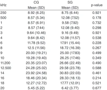
With regard to the hearing thresholds for the frequen-cies from 0.25 to 20 kHz (Table 1), there was no statistically signiicant difference between the two groups for any of the tested frequencies.
It is worth mentioning that, although the hearing thresh-olds means for the groups were within the normality limits for the conventional audiometry, some subjects presented hearing loss. For the SG, two of them had a mild sensorineu-ral hearing loss and one presented a moderate sensorineusensorineu-ral hearing loss. For the CG, two subjects had a mild sensori-neural hearing loss.
kHz CG
Mean (SD)
SG
Mean (SD) p-value 250 8.92 (6.25) 8.75 (6.44) 0.921 500 8.57 (5.34) 12.08 (7.52) 0.178 1 8.57 (6.91) 9.58 (7.92) 0.732 2 8.57 (7.44) 12.08 (9.40) 0.298 3 9.64 (10.46) 9.16 (9.49) 0.921 4 9.64 (8.42) 12.08 (11.57) 0.538 6 11.78 (9.52) 17.91 (15.44) 0.228 8 12.5 (11.56) 18.72 (16.39) 0.267 9 20.00 (19.21) 25.00 (17.83) 0.499 10 19.28 (19.40) 26.25 (17.46) 0.349 11.200 20.35 (23.07) 26.66 (22.49) 0.490 12.500 24.28 (25.02) 29.58 (23.78) 0.588 14 23.92 (24.58) 30.83 (22.03) 0.461 16 18.46 (20.34) 28.33 (18.13) 0.214 18 12.27 (12.72) 17.77 (12.01) 0.521 20 5.45 (5.22) 6.42 (3.77) 0.677 Table 1. Hearing thresholds (in dBHL) considering the right and left ears together, according to the mean and standard deviation per group and p-value
Behavioral Assessment
Table 2 shows that although the results show a remarkable difference for the SG, no statistically signiicant difference was found in all the performed behavioral tests.
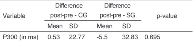
Electrophysiological assessment
Table 3 shows that P300 latencies after training for the SG were decreased when compared to pre-training latencies. However, this difference was not statistically signiicant.
Questionnaire of tinnitus severity
In the comparison of the THI score between the moments before and after training, no statistically signiicant difference was seen between the groups (Table 4). Although there was no signiicant difference, we observed an improvement in the scoring for the SG, while there was a worsening for the CG. It is worth mentioning that, according to the classiication of the THI score, the SG changed from mild to slight level, whereas the CG remained in the mild level.
In the individual classiication of participants for the THI, CG subjects presented before intervention included the follow-ing: three, mild tinnitus; three, slight tinnitus. As to the SG, two presented mild tinnitus and four, slight tinnitus. For the post-intervention score, one of the SG subjects changed the classiication from mild to slight and one of the CG changed from mild to moderate. The other subjects from both groups maintained their initial classiications.
DISCUSSION
With regard to the characteristics of tinnitus that were as-sessed through the acuphenometry, the pitch, for both groups, obtained a mean of around 7 kHz. These indings are in agree-ment with another study(15) that also veriied values regarding the pitch of tinnitus in higher frequencies.
Tinnitus has also been associated with pitch frequency with the presence of hearing loss in a certain frequency or with the frequency in which the hearing thresholds presented higher in the audiogram(14). Other authors disagree with this statement and report that tinnitus pitch in cases of sensorineural hearing loss tend to be more acute and that the symptom might be present in any area of frequency where there is a signiicant hearing loss(15).
As to the mean of loudness, the CG obtained a mean of 19 dBSL and the SG had a mean of 7.27 dBSL. Our results are similar to the loudness values obtained by other authors(14), who veriied values that varied from 5 to 15 dBSL.
With regard to tinnitus laterality, we observed a higher prevalence of bilateral tinnitus in both groups, which is in agreement with previous studies(16,17).
As to the conventional audiometry, we noticed that the means of hearing thresholds from both ears are within the normality standards and no statistically signiicant differences were found between the groups, although some subjects from the mentioned study showed a sensorineural hearing loss of mild to moderate degree, both in the SG and in the CG. Among the ive subjects with sensorineural hearing loss, four of them are aged 50 years or older.
Some investigators observed that the tinnitus incidence is higher in the population of older adults, thus associating this fact to hearing losses, brain traumas, noise exposure, hyperten-sion, and anxiety, among other factors(18).
A previous study(19) emphasized that the increase of tinnitus prevalence in older patients does not mean necessarily that tinnitus, a single symptom, will increase with advancement of age. It afirmed, similarly, that the audiometric measurements in subjects with tinnitus might be more correlated with the age factor and that in older subjects, tinnitus presence is associated, in most cases, with hearing loss.
In the comparison between groups for high frequency audiometry, the results did not show statistically signiicant differences; however, the values of hearing thresholds for high frequencies were more increased in older subjects, follow-ing the same standard seen for the conventional audiometry. These indings are in agreement with another study(17), which emphasized the relation between increase of age and increase of hearing thresholds in high frequencies.
Variable
Difference post-pre - CG
Difference
post-pre - SG p-value Mean SD Mean SD
RE/LE SN
together (in %) 2 3.51 5.33 6.89 0.149 GIN (in %) 3.61 8.39 7.77 5.44 0.331 GIN (in ms) -0.33 0.81 -0.66 1.03 0.551 FST (in %) 1.39 1.25 2.22 7.57 0.796 Table 2. Post- and pre-intervention difference for behavioral assessment tests of the auditory processing per group, according to mean, standard deviation, and p-value
Caption: CG = Control Group; SG = Study Group; RE = right ear; LE = left ear; ms = millisecond; SN = Speech with Noise; GIN = Gaps in noise; FST = Frequency Pattern Test; SD = standard deviation
Variable
Difference post-pre - CG
Difference
post-pre - SG p-value Mean SD Mean SD
P300 (in ms) 0.53 22.77 -5.5 32.83 0.695
Table 3. Post- and pre-intervention difference for the P300 latency per group, according to mean, standard deviation, and p-value
Caption: CG = Control Group; SG = Study Group; ms = millisecond; SD = standard deviation.
Variable
Difference after and before - CG
Difference after
and before - SG p–Value Mean SD Mean SD
THI 5.33 8.26 -3 13.43 0.187 Table 4. Post- and pre-intervention difference for the Tinnitus Handicap Inventory score per group, according to mean, standard deviation, and p–value
Some authors that compared the hearing thresholds of high frequencies among subjects with and without tinnitus(20) veriied a signiicant worsening in subjects with tinnitus when compared to subjects without it. This fact suggests that the co-chlear function might be compromised in subjects with tinnitus, even in the absence of hearing loss found through conventional audiometry(14,21,22).
Behavioral assessment
In a previous study that compared subjects with and without tinnitus complaint, it was identiied that tinnitus interferes in the skill of auditory closure. However, due to the high prevalence of hearing loss in that population, it is hard to estimate the exact effect of tinnitus, after exclusion of the hearing loss(23).
In the comparison of results for the Speech-in-Noise test between before and after moments, we observed that no sta-tistically signiicant difference was found between the groups, thus suggesting that the used training (auditory or visual) did not interfere with the auditory closure skill.
In an individual analysis, all subjects showed the results of the Speech-in-Noise test within the normality at pre-interven-tion evaluapre-interven-tion. This fact might have collaborated to the lack of a signiicant improvement in the percentage of correct answers, even after AT. The presence of ceiling effect in auditory activi-ties was already reported(24). Because of the ceiling effect, the investigation of magnitude effects in auditory tasks for subjects without alteration of the auditory processing is limited(24), which can be applied to the present study, since participants did not present altered results in this test.
As to the case of temporal resolution, there is no consensus in literature about the alteration of this skill in subjects with tin-nitus. In a previous investigation(25), for example, no statistically signiicant differences were found between the groups with and without tinnitus regarding the GIN. But, in another study(26), it was seen that subjects with tinnitus had some impairment in the auditory skills for temporal resolution.
In the present study, among the 12 participants, two showed alterations in the results of GIN (one of the SG and one of the CG). Thus, although there was no agreement in literature regarding the alteration of temporal resolution in patients with tinnitus, and although the present investigation popula-tion is small, we suggest this auditory skill could be further investigated in future studies, because it might collaborate to elucidating existing doubts on tinnitus.
In the comparison of the moments before and after training between the two groups for the GIN, no statistically signiicant differences were seen. As to the two subjects that presented alterations in this test, the SG participant showed an evolution comparing the moments before and after training (from 58 to 70% of correct answers), while the CG participant maintained his/her performance. These results were already expected, since there was a training of the temporal resolution skill for the SG(3).
For the Frequency Pattern Test, the means obtained for both groups are within the normality standards for adults. However, if we analyze the results individually, we verify that two sub-jects showed altered results for this test, both from the CG.
The results remained altered after intervention, as expected, since this group was not submitted to AT.
As to the comparison of pre- and post-moments between the studied groups for the Frequency Pattern test, no statistically signiicant differences were found.
Previous studies(5,6) used different paradigms of training for auditory discrimination, seeking to obtain an improvement in the tinnitus of patients with this symptom. However, these studies did not use behavioral tests to evaluate the auditory processing before and after intervention. Other studies cor-relating the auditory skill of discrimination with tinnitus and, especially, evaluating this auditory skill in the population were not found in literature.
Therefore, in a general analysis, we can assume that for the behavioral tests, no statistically signiicant differences were found before and after intervention when the SG and CG were compared. Thus, the AT used in the present study may not have caused a signiicant modiication in the CANS, which is measurable through behavioral assessment.
Electrophysiological assessment
The neural generators of late- or long-latency auditory evoked potentials, despite not being accurately established, involve the supratemporal auditory cortex, frontal cortex, and hippocampus(12), as well as the thalamus(27).
Some investigations used late AEP to assess subjects with tin-nitus, for example, and they veriied in subjects with tinnitus: normal latencies and reduction of amplitudes for N1, P2, and P300(28); in-creased latencies for the N1 component(29); and increase of N1, N2, and P3 latencies(20). It was suggested(30) that some of the alterations seen in the late potentials in patients with tinnitus could relect an abnormal central auditory processing associated with tinnitus sen-sation, which could be the relex of neural adaptive processes that happened after damage in the peripheral auditory system.
In the present investigation, the means obtained for P3 wave latencies, for both groups, are found within the normality stan-dards. The analyses of the records, individually, revealed that all CG and SG subjects presented P300 latencies within the normality standards, which is not in agreement with one of the previously mentioned studies(16), but it is in agreement with another(28).
With regard to the comparison of P300 results before and after training between the SG and CG, no statistically signiicant differences were found.
Some studies used the AEP before and after training to verify modiications in the auditory pathway from the intervention(4,10). However, there were no studies in literature that compared the AEP before and after training in patients with tinnitus.
Thus, for the P300, there was an improvement in latency time in the difference before and after intervention for the SG, but this change was not signiicant if compared to the difference before and after intervention for the CG.
Questionnaire of tinnitus severity
aspects: emotional, functional, and catastrophic aspects of subjects with tinnitus.
In the present study, no statistically signiicant differences were found for the comparison of the situations before and after training between the groups regarding the THI. However, the classiication of tinnitus for the SG changed from mild to slight, while it remained mild for the CG classiication.
Other studies used this instrument to measure the severity of tinnitus before and after intervention. For example, one of them(1) found a statistically signiicant difference regarding the THI, in the comparison before and after nutritional intervention in subjects with tinnitus and metabolic alterations.
The THI was also applied in patients submitted to auditory discrimination training, obtaining a statistically signiicant improvement in the score of the questionnaire if compared to the situations before and after training. The authors excluded from the investigation subjects that presented tinnitus with a severe or catastrophic classiication(6).
The mentioned studies were able to verify differences seen in the tinnitus severity between pre- and post-intervention situ-ations. The present research did not ind a signiicant difference between the moments before and after training between the groups for the THI, but there was a decrease in the level of discom-fort for the SG. This fact might suggest that the AT had an effect, even if it is a small one, on the sensation of tinnitus for the SG.
It is important to comment on the change of mild degree to moderate degree of one of the CG subjects that passed in the VT. This could be related to the increased awareness of this CG participant on the symptom, since he/she passed in the VT, in the absence of any speciic hearing stimulus, favoring the perception of the tinnitus that might have increased the discomfort caused by the symptom.
Another fact to be considered is the CG loudness. The loud-ness mean was higher than that referred by the SG, and it might be related to the presence of a higher number of subjects in the CG with more remarked complaint of tinnitus discomfort. This relation between loudness and tinnitus severity had already been previously described in a speciic study(30).
In summary, we can state that the AT used in the present investigation did not cause any modiication that could be iden-tiiable by the already used instruments, since no differences between the groups that passed in the auditory training (SG) and VT (CG) were found.
Isolatedly, for some SG subjects, we observed small modiications in the results of tests. However, these modiica-tions could not be observed for the group. In future studies, a qualitative analysis of results could help in the observation of the improvements obtained individually.
Some factors might have contributed so these results could happen. The small number of subjects limits the visualization of possible modiications before and after intervention, given the variability of possible answers intra- and inter-subjects. Hence, some evident modiications would result in statistically signiicant differences.
Another worthy factor is the difference between groups with regard to the severity of tinnitus. Even randomly, the CG comprised subjects with higher severity of tinnitus, which can be seen both through loudness and the THI score. Future studies using the same methodology should try to form more homogenous groups regarding this aspect.
As to the AT itself, a possible limitation is the periodicity of sessions. In this study, the training was carried out following the standard for studies with patients that have an alteration in the central auditory processing(13). It is possible that for subjects with tinnitus, this periodicity is not ideal, since these people live with the symptom 24 hours a day, every day, for several years. However, the increase of periodicity develops an increase of cost for the volunteers.
Other studies(5,6) that investigated the auditory discrimi-nation training in subjects with tinnitus used more frequent periodicity of AT and obtained better results regarding the tinnitus improvement.
In order to solve these two problems (increase of AT pe-riodicity and decrease of costs), the suggestion would be the development of a training program which could be used at home, more days during the week.
In summary, although the indings of the present study did not conirm our initial hypothesis, we believe tinnitus happens due to plastic modiications involving the CANS, therefore the AT could be a viable alternative for the improvement of this symptom. Nevertheless, some gaps such as training time, periodicity, form of training conduction, and monitoring of intervention should be studied in order to achieve an eficient training for this symptom.
CONCLUSION
Our results did not show statistically signiicant differences between the groups for the comparison between the moments before and after training (auditory or visual), both for the electrophysiological tests and for the behavioral assessment of the auditory processing and for the THI, although some punctual differences in the individual analysis have occurred. Thus, future studies on the subject involving a larger sample might obtain better results.
*DT elaborated the research and schedule, investigated literature, performed data collection and analysis, and wrote the article; AGS guided the paper, participated in the elaboration of investigation and schedule, analyzed data, corrected article writing, and approved the inal version; CGM, FCLM and CMR analyzed data, corrected article writing, and approved the inal version.
REFERENCES
1. Almeida TAS, Samelli AG, Mecca FDN, Martino E de, Paulino AM. Sensação subjetiva do zumbido pré e pós intervenção nutricional em alterações metabólicas. Pró Fono. 2009;21:291-6.
3. Musiek FE, Shinn J, Hare C. Plasticity, Auditory Training, and Auditory Processing Disorders. Semin Hear. 2002;23(4):263-75.
4. Tallal P, Gaab N. Dynamic auditory processing, musical experience and language development. Trends Neurosci. 2006;29(7):382-90.
5. Flor H; Hoffmann D; Struve M; Diesch E. Auditory discrimination training for the treatment of tinnitus. appl psychophysiol biofeedback. 2004;29(2):113-20.
6. Herraiz C; Diges I; Cobo P; Aparicio JM; Toledano A. Auditory discrimination training for tinnitus treatment: the effect of different paradigms. Eur Arch Otorhinolaryngol. 2010;267(7):1067-74.
7. Musiek FE, Shinn J, Jirsa R, Bamiou DE, Baran JA, Zaidan E. GIN (Gaps in Noise) test performance in subjects with conirmed central auditory nervous system involvement. Ear Hear. 2005;26(6):608-18. 8. Pereira LD, Schochat E. Testes auditivos e comportamentais para
avaliação do processamento auditivo central. São Paulo: Pró Fono; 2011. 9. Musiek FE. Frequency (pitch) and duration patterns tests. J Am
Audiol.1994; 5(4):265-8.
10. Jirsa RE. Clinical efficacy of electrophysiologic measures in APD management programs. Semin Hear. 2002;23(4):349-56.
11. Newman CW, Jacobson GP, Spitzer JB. The development of tinnitus handicap inventory. Arch Otolaryngol Head Neck Surg. 1996;122(2):143-8. 12. McPherson DL. Late potentials of the auditory system (evoked
potentials). San Diego: Singular Publishing Group; 1996.
13. Chermak GD, Musiek FE. Auditory Training: Principles and Approaches for Remediating and Managing Auditory Processing Disorders. Semin Hear. 2002;23(4):297-308.
14. Buzo BC, Carvallo RMM. Psychoacoustic analyses of cochlear mechanisms in tinnitus patients with normal auditory thresholds. Int J Audiol. 2014;53(1):40-7.
15. Shekhawat GS, Stinear CM, Searchield GD. Transcranial direct current stimulation intensity and duration effects on tinnitus suppression. Neuro Neural Repair. 2013;27(2):164-72.
16. Attias J, Pratt H, Haran ID, Bresloff I, Horowitz G, Polyakov A, et al. Detailed analysis of auditory brainstem responses in patients with noise induced tinnitus. Audiology. 1996;35(5):259-70.
17. Nageris BI, Attias J, Raveh E. Test-retest tinnitus characteristics in patients with noise-induced hearing loss. A J of Otol – Head Neck Med and Surg. 2010;31(3):181-4.
18. Nondahl DM, Cruickshanks KJ, Wiley TL, Klein R, Klein BE, Chappell R, et al. The ten-year incidence of tinnitus among older adults. Int J Audiol. 2010;49(8):580-5.
19. Savastano M. Tinnitus with or without hearing loss: are its characteristics different? Eur Arch Otorhinolaryngol. 2008;265(11):1295-300. 20. Burguetti FAR, Peloggia AG, Carvallo RMM. Limiares de audibilidade
em altas frequências em indivíduos com queixa de zumbido. Arq Int de Otorrinolaryngol. 2004;8(4):277-83.
21. Paglialonga A, BO LD, Ravazzani P, Tognola G. Quantitative analysis of cochlear active mechanisms in tinnitus subjects with normal hearing sensitivity: multiparametric recording of evoked otoacoustic emissions and contralateral suppression. Auris Nasus Larynx. 2010;37(3):291-8. 22. Weisz N, Hartmann T, Dohrmann K, Schlee W, Norema A.
High-frequency tinnitus without hearing loss does not mean absence of deaffrentation. Hear Res. 2006;222(1-2):108-14.
23. Huang CY, Lee HH, Chung KC, Chen HC, Shen Y, WU Jl. Relationships among speech perception, self-rated tinnitus loudness and disability in tinnitus patients with normal pure-tone thresholds of hearing. Orl J Otorhinolaryngol Relat Spec. 2007;69(1):25-9.
24. Bellis TJ, Ross J. Performance of normal adults and children on central auditory diagnostic tests and their corresponding visual analogs. J Am Acad Audiol. 2011;22(8):491-500.
25. Acrani IO, Pereira LD. Resolução temporal e atenção seletiva de indivíduos com zumbido. Pró Fono. 2010;22(3):233-8.
26. Sanches SG, Sanchez TG, Carvallo RM. Inluence of cochlear function on auditory temporal resolution in tinnitus patients. Audiol Neurotol. 2010;15(5):273-81.
27. Kraus N, McGee T. Potenciais Auditivos Evocados de Longa Latência. In: Katz J. Tratado de Audiologia Clínica. São Paulo: Manole; 1999. 28. Attias J, Urbach D, Gold S, Shemesh Z. Auditory event related potentials
in chronic tinnitus patients with noise induced hearing loss. Hear Res, 1993;71(1-2):106-13.
29. Jacobson GP, Calder JA, Newman CW, Peterson EL, Wharton J, Ahmad BK. Electrophysiological indices of selective auditory attention in subjects with and without tinnitus. Hear Res. 1996;97(1-2):66-74.