Division of Neuro s u rg e ry, Hospital da Restauração, University of Pernambuco and Division of Neuro s u rg e ry, Hospital Memorial São José, Recife PE, Brazil:1MD, MSc, PhD Neuro s u rgeon;2MD, Neuroradiologist;3MD, MSc, PhD, FRCS, Chairman of Neuro s u rg e ry
Department University of Pernambuco.
Received 2 December 2005, received in final form 1 March 2006. Accepted 17 April 2006.
Dr. Paulo Thadeu Brainer-Lima - Rua das Fronteiras 175 / 208 - 50070-170 Recife PE. E-mail: [email protected]
GANGLIOGLIOMA
Comparison with other low-grade brain tumors
Paulo Thadeu Brainer-Lima
1, Alessandra Mertens Brainer-Lima
2,
Hildo Rocha Azevedo-Filho
3ABSTRACT -Method:F o rty-two patients with low-grade brain tumor and re f r a c t o ry epilepsy were stud-ied. The mean age was 22.3 years. They were divided into two groups: Group A, patients with gangli-oglioma (n=19) and group B, patients with other low-grade tumors (n=23) (14 astrocytoma, 6 oligoden-d roglioma, 2 oligoden-dysembryoplastic neuroepithelial tumor, anoligoden-d 1 xanthoastro c y t o m a ) . Results:Age at seizure ’s onset was 7 years or less in 73% of the patients in group A and in 30.4% of the patients in group B (p=0.045). Complex partial occurred frequently in group A and B (94.7% versus 82%, respectively). Seizure ’s fre q u e n-cy was higher in group B (p=0.002).Computerized tomography (CT) was normal in 36.8% of group A patients and abnormal in all group B patients. Magnetic resonance imaging (MRI) was abnormal in all patients. S u rgical removal was complete in 89.5% of the patients in group A and in 78.2% of the patients in gro u p B . Conclusion:The association of re f r a c t o ry epilepsy and complex partial seizures, at a relatively low fre-q u e n c y, in young patients potentially normal CT and a MRI hypointense temporal lobe lesion in T1-weighed slices were habitual image findings in ganglioglioma, rather than other low-grade tumor.
KEY WORDS: ganglioglioma, refractory epilepsy, low-grade brain tumors.
Ganglioglioma: estudo comparativo com outros tumores cerebrais primarios de baixo grau
RESUMO - Método:Foram estudados 42 pacientes com tumor cerebral primário de baixo grau e epilepsia refratária. A idade média foi 22,3 anos. Eles foram divididos em dois grupos: no grupo A os pacientes com ganglioglioma (n=19) e no grupo B os pacientes com outros tumores primários de crescimento lento (n=23) (14 astrocitomas, 6 oligodendrogliomas, 2 tumores desembrioblástico neuroepitelial e um xantoastro c i t o-m a ) . Resultados:A idade de início das crises convulsivas foi 7 anos ou menos em 73% dos pacientes no g rupo A e 30,4% dos pacientes no grupo B (p=0,045). A crise convulsiva do tipo parcial complexa foi a mais identificada nos grupos A e B (94,7% versus 82%, respectivamente). A freqüência de crise foi mais alta no g rupo B (p=0,002). A tomografia computadorizada foi normal em 36,8% dos pacientes no grupo A e anor-mal em todos no grupo B. A ressonância magnética foi anoranor-mal em todos os pacientes. A remoção cirúr-gica foi completa em 89.5% dos pacientes no grupo A e 78,2% no grupo B. Conclusão:A associação de epilepsia refratária e crise parcial complexa, principalmente quando a freqüência não é muito alta, em pacientes jovens, mesmo com tomografia computadorizada normal e alteração hipointensa na seqüência de T1 da ressonância magnética é sugestiva de ganglioglioma mais que outros tipos de tumor cere b r a l primário de baixo grau.
PALAVRAS-CHAVE: ganglioglioma, epilepsia refratária, tumor cerebral de baixo grau.
Ganglioglioma (GG) is one of the commonest caus-es of tumor- related re f r a c t o ryepilepsy in young pa-t i e n pa-t s1, and together with other low-grade brain
neo-p l a s m ’s, comneo-prises 10-30% of the neo-pathological sub-strate in patients with chronic intractable partial epi-l e p s y2. GG is frequently found in patients younger
than 30 years (80%) with re f r a c t o ry epilepsy. It re p re-sents 0.5 to 1.7% of all neuroepithelial tumors3and
constitutes 1.7% to 7.6% of all tumors of the central n e rvous system in the pediatric population4 , 5. The
pos-sibility that such lesions are of embryonal type6. GGs
are included in the category of primary cerebral tu-mors in which mature ganglion cells and dysplastic neurons may be present7.
GG contains neoplastic glial cells, mainly astro c y-tes in varying stay-tes of diff e re n t i a t i o n8 - 1 0. These glial
cells directly affect the biological behavior of the tumor; they are usually benign and are related to his-tological features typical of low-grade pilocytic astro-c y t o m a8. The presence of an increased number of
NMDA (N-methyl-D-aspartate) receptors and the a b n o rmal production of neurotransmitters found in the cortex around GG, unlike what is seen in other low-grade tumors, might explain the increased ten-dency towards seizure ’s generation before an after tumor resection in some patients1 1. GG occurs
main-ly in the temporal lobe, it is well defined and intra-c o rtiintra-cal, firm intra-consistenintra-cy and shows intra-calintra-cifiintra-cations and cystic components in about 50% of the patients. When surgical removal is incomplete, radiotherapy is warranted if anaplastic histological findings or tu-mor pro g ression are documented. These occur in less than 20% of the patients9,12.
Most previous studies on brain tumor and epilep-sy have analysed patients with diff e rent low-grade tumors as a single group, without specifically delin-eating the findings in those with ganglioglioma. In this study, we performed a retrospective analysis of patients with intractable epilepsy and histologically verified ganglioglioma and other low-grade brain tumors that underwent tumor resection. We com-p a red clinical, neurocom-physiologic and neuro i m a g i n g findings in these two groups in order to better define the patients with GG.
METHOD
The presence of medically intractable epilepsy (at least 1 complex partial seizure per month over the last 2 years), low-grade primary cerebral tum or and at least 2 years of postoperative follow-up were the clinical inclusion criteria in this series of consecutive patients (n=42). Patient’s post-operative outcome in relation to seizures was rated
accord-ing to Engel’s classification1 3. All patients whose tumors have
shown any sign of malignancy were submitted to compli-m e n t a ry treatcompli-ment with radiotherapy and checompli-motherapy. In all patients, at least two pre and postoperative inter-ictal EEG re c o rdings were obtained during sleep and wake-fulness, using the 10-20 system, with at least 1 hour of dura-tion. Both computadorized tomography (CT) and magnet-ic ressonance image (MRI) (1.5T) were acquired using high resolution scanners and thin slices.
Operative techniques included stereotactic tumor
localiza-t i o n1 4 , 1 5, electro c o rticographic (ECoG) monitoring and brain
mapping to identify eloquent nonresectable rolandic and
language cortex. Two operations were perf o rmed with the patient under local anesthesia and neuroleptoanalgesia.
Intraoperative ECoG w as perf o rmed in all patients. It consisted of placement of carbon-tipped electrodes for sur-face re c o rdings over the lateral cortex and multicontact subdural strips and grids to sample sub temporal cortex.
C o rtical mapping of the motor cortex under general anesthesia was obtained with bipolar square pulses with 2 to 10 mA, at 100Hz and with 0,1 msec of duration.
T h i rty men and 12 women with ages between 6 and 57 years (mean 22.3 years) were studied.
Patients were divided in two groups, according to his-tological findings: in Group A (n=19), patients had GG and in group B (n= 23), other low-grade tumors were pre s e n t ( a s t rocytoma, oligodendroglioma, dysembryoplastic neu-roepithelial tumor and xanthoastrocytoma).
The following variables were studied in groups A and B: age at surg e ry, age at onset of epilepsy, time before diag-nosis, types of seizure, number of seizures per month, re s u l t s of neurological examination, electroencephalogram, CT and MRI findings, surgical technique, and outcome re s u l t s in relation to seizures.
Statistical analysis was carried out using techniques of descriptive statistics, through tables, including absolute or p e rcentile distributions and statistical measures. Chi-square or Fisher exact tests were used whenever necessary. The significance level was p<0.05.
RESULTS
The pathological findings can be seen in Table 1. A summary of clinical data can be seen in Table 2 and 3. Group A consisted of 14 men and 5 women and group B comprised 16 men and 7 women. The c e rebral tumors were located within the temporal lobe in 14 (73.6%) of the patients in group A and in 15 (65.2%) in group B. There were 3 frontal lobe, 2 parietal lobe and 1 occipital lobe tumors in group A. In group B, 4 frontal, 2 parietal and 2 occipital lobe tumors were found.
Epilepsy began at a mean age of 7.8 years (range 2.4-48 years) in group A and 12,0 years (range 4.1-34 years) in group B. The number of patients eight years
Table 1. Etiology in patients with primary brain tumors and refractory epilepsy.
Histology Frequency %
Ganglioglioma 19 45.2
Astrocytoma 14 33.3
Oligodendroglioma 6 14.3
Xanthoastrocytoma 1 2.4
Dysembryoplastic neuroepithelial tumor 2 4.8
or over at the beginning of their seizures was signif-icantly higher (p=0,045) in group B (n=16) than in group A (n=7).
At the time of surg e ry, patients in group A had experienced an average of 11.2 seizures per month and those in group B 18.1 seizures per month. An average number of seizures higher than 17 per
month was significantly more common in group B (n=18) than in the group A patients (n=3). Complex partial seizures, alone or in combination with other s e i z u re ’s type were observed in 20 patients (87%) in g roup B and 18 patients (94.7%) in group A, with-out statistical diff e rence. All patients with temporal lobe tumors had complex partial seizures, as did 78%
Table 3. Distribution of the patients according to seizures, neuro p h y s i o l o g y, neuroimaging and the groups of cerebral tumor. Group
A B p value
N % N %
Age at seizure onset (yrs)
7 or less 12 63.1 7 30.4 2=4.2
From 8 to 30 7 36.9 16 69.6 p=0.0045
Age at operation (yrs)
6 to 20 11 57.9 10 43.5 2=0.477
21 to 62 8 42.1 13 56.5 p=0,490
Duration of epilepsy (yrs)
2 to 9 9 47.4 13 56.5 2=0.093
10 to 30 10 52.6 10 43.5 p=0.760
Mean preoperative seizure frequency (per mo)
1 to 16 16 84.2 5 21.7 2=9.445
17 to 30 3 15.8 18 78.3 p=0.002
Interictal scalp EEG epileptiform discharges 13 68.4 19 82.6 P=0.370
Postoperative EEG epileptiform discharges 11 57.9 15 65.2 P=0.480
CT with lesion demonstrated 12 63.2 23 100 p=0.010
MRI gadolinium enhancement 11 57.9 17 74.0 p=0.240
MRI mass effect 8 42.1 15 65.2 p=0.170
MRI and CT calcifications 12 63.1 8 34.8 p=0.084
Temporal lobe location 14 73.7 15 65.2 p=1.000
Complete resection 17 89.5 18 78.2 p=0.455
Seizure outcome 18 94.7 20 87.0 p=1.000
p value based on Fischer’s Exact Test.
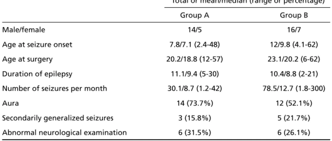
Table 2. Summary of clinical findings.
Total or mean/median (range or percentage)
Group A Group B
Male/female 14/5 16/7
Age at seizure onset 7.8/7.1 (2.4-48) 12/9.8 (4.1-62)
Age at surgery 20.2/18.8 (12-57) 23.1/20.2 (6-62)
Duration of epilepsy 11.1/9.4 (5-30) 10.4/8.8 (2-21) Number of seizures per month 30.1/8.7 (1.2-42) 78.5/12.7 (1.8-300)
Aura 14 (73.7%) 12 (52.1%)
of the patients in group A and 64% in group B. An aura, described by 14 patients in group A (73.7%) and 12 patients in group B (52.1%), was present in all those with temporal lobe epilepsy. Secondarily generalized seizures were present in 5 patients in g roup B and 3 patients in group A, all with temporal lobe epilepsy, and predominantly in the first second years of their history of epilepsy.
N e u rological status was normal in 13 (68.4%) pa-tients of group A and in 17 (73.9%) of group B (p> 0.05). The neurological examination abnorm a l i t i e s found in these patients were related to the localiza-tion of the tumor, with no statistical difference bet-ween the groups (visual field defects in 8, dysart h r i a in 2, and hemiparesis in 5).
Interictal epileptiform discharges occurred in 13 (68.4%) of the patients in group A, while in group B these abnormalities were detected in 19 patients (82.6%). The EEG findings were most often focal in both groups A and B (62% and 54%, re s p e c t i v e l y ) ; they were multifocal in 24% and 35%, and general-ized in 5% and 8%, respectively, in groups A and B.
Postoperative EEG re c o rdings were available in all patients. Epileptiform discharges were present in 11 (57.9%) patients in group A and 15 (65.2%) in group B (no statistical difference).
Computed tomography was abnormal in all pa-tients in group B and normal in 7 (36.8%) papa-tients of group A (p<0.05).
MRI detected the tumor in all 42 patients. MRI findings in group A and B patients included, re s p e c-t i v e l y, conc-trasc-t enhancemenc-t (11 and 17), mass eff e c c-t (8 and 15), cystic component (10 and 7), peritumoral edema (1 and 7) and calcifications (13 and 8; con-firmed by CT).
P re and post resection ECoG monitoring was per-f o rmed in 32 patients (15 in group A). In 12 patients in group A and 13 in group B resection of the epilep-tic zone was complete and all were seizure free post-o p e r a t i v e l y. In 3 patients (1 in grpost-oup A and 2 in grpost-o u p B), there was a subtotal (90%-95%) tumor re s e c t i o n and a complete resection of the ECoG spiking zone: all of them were rendered seizure’s free postopera-t i v e l y. In all papostopera-tienpostopera-ts, adequapostopera-te resecpostopera-tion of postopera-the epilep-togenic zone re q u i redtissue removal that extended beyond that which would have been necessary for tumor resection alone. In 7 patients, resection of the epileptic zone was incomplete, due to overlap of elo-quent (motor or language) cortex and the epileptic zone. Postoperatively, these patients continued to experience seizures, but a 90-95% improvement in seizure’s frequency was noted.
S u rgical resection was complete in 17 (89.5%) patients in group A and in 18 (78.2%) patients in group B.
Mean postoperative follow-up time was 33.4 months. Postoperatively, 18 (94.7%) patients in gro u p A and 20 (87%) in group B were classified as Engel’s grade I and the others as Engel’s grade II.
DISCUSSION
Haddad et al.1 2found that GG was an uncommon
finding among the etiologies of epilepsy. That might be true if we considered epilepsy in all its pre s e n t-ing forms; however, if we considered only patients with re f r a c t o ry seizures, the situation might be diff e-rent. The Cleveland Clinic re p o rt e d5that 12% of the
patients operated on for re f r a c t o ryepilepsy over a 10-years period had low-grade tumors; all patients have had less than 30 seizures per month and a long history of epilepsy (more than 6 years).
The mean duration of epilepsy before surgery in patients with low-grade tumors was 10.8 years in our series, in agreement with others1 6 , 1 7. This behavior
d i ffers from that seen in patients with high-grade brain tumors. In the latter, the high frequency of sei-z u res usually leads to earlier investigation and diag-n o s i s1 6. In group A patients, seizures began earlier
than in group B. That diff e rence reached statistical significance and is in agreement with findings in oth-er centoth-ers6 , 8 , 1 8. GG seems to be related to neural
mat-uration and is frequently found in young children. The higher prevalence of GG in this young popu-lation could have important clinical implications. Early s u rgical intervention might offer the best chance of relief of intractable epilepsy and might reduce neu-ropsychological and social disability.
In patients with low-grade brain tumor and re f r a c-t o ry epilepsy, seizure ’s frequency can pro g re s s i v e l y d e c rease over time1 6 , 1 9. This was not noted in our
se-ries. There was a high prevalence of partial seizures in group A and B. This was also noted in other stud-ies of patients with re f r a c t o ryepilepsy and brain tu-mors, which found that 85% to 92% of them suff e r-ed partial seizure s2 0 - 2 2. Partial epilepsy might occur
in patients with tumors in the temporal lobe and out-side it as well2 3. Extracranial EEG, although lacking
adequate spatial resolution may be used as a scre e n-ing tool24. The presence of epileptic discharges
post-operative EEG data did not correlate with seizure outcome. According to some authors2 5 , 2 6, 30% of
re f r a c t o ry temporal lobe epilepsy have surface EEG re c o rdings showing independent bitemporal spik-ing. This prevalence of bitemporal abnorm a l i t i e s i n c reases as the duration of the EEG re c o rdings in-creased. Other studies have also shown that surface EEG findings consisting of bilateral independent tem-poral foci, did not correlate with the effect of sur-gery in seizure’s control26,27.
MRI was abnormal in all patients in this series. Ha-bitual findings consisted of hypointensity in the T1-weighted and hyperintensity in T2-T1-weighted slices. On the other hand, CT was normal in seven patients, all of whom had GG (group A) within the temporal lobe. MRI was more sensitive than CT in the detec-tion of structural lesions in the temporal lobe2 8. MRI
was also more effective in suggesting specific tumor types and is presently considered the gold-standard in imaging evaluation of patients with epilepsy2 9.
MRI findings such as the presence or absence of gado-linium enhancement, mass effect, and cystic compo-nents had no measurable influence on the seizure outcome following tumor resection in our series and in others7,24.
The temporal lobe is the favorite location for GG (40% to 77% of the patients). It is often associated with neuronal migration disorders12,16,20.
Ninety-four percent of our patients with GG have been re n d e red seizure - f ree after surg e ry. Morris et al. found that 74% of the patients operated on for re f r a c t o ry temporal lobe epilepsy and GG had excel-lent results in relation to seizure ’s control. This is espe-cially true for young patients, with short duration of the epileptic syndrome and with absence of epilep-tic activity in postoperative EEG5. Secondary
autono-mous mirror foci may develop in patients with GG due to the presence of a prolonged re f r a c t o ry epilep-tic syndro m e3 0. Lesionectomy alone achieved seizure ’s
c o n t rol in 9 (64.2%) of 14 patients with an extratem-poral lesion but in only 2 (22.2%) of 9 patients with a temporal lobe lesion1 3, suggesting that at least in
temporal lobe lesions, resection of a cortical marg i n guided by ECoG might be useful. It is possible that s e c o n d a ry epileptic foci might be more pre v a l e n t within the temporal lobe where they tend to become autonomous more quickly than in other areas of the brain. Other studies2 5 , 3 1identified incomplete tumor
resection or tumor re c u rrence as causes of poor post-operative seizure control. Despite the small number of patients who had had incomplete resection in our
series, we believe that the presence of residual tumor is an important cause of postoperative seizures.
GG seems to be more indolent than other primary c e rebral tumors, with longer history of epileptic s e i z u res. In young patients, it could easily be confus-ed with neuronal migration disorders if the lesion is located in the cortical surf a c e1 1 , 3 2. Our patients had
a long history of seizures before diagnosis and re c e i v-ed appropriate surgical treatment late. It would be n e c e s s a ry to develop a clinical paradigm to early iden-tify patients with GG. Early diagnosis might provide a better chance of total tumor removal and re m i s-sion of epilepsy1,33,34.
Postoperative psychosis was found to be more common in patients bearing temporal lobe GG than in other temporal lobe pathology3 5. No
postopera-tive psychosis was noted in our series.
In our series, patients with GG were young (usual-ly under 8 years old) and presented characteristical-ly with complex partial seizures (94.7% of the pati-ents), at a frequency of less than 16 seizures per month (usually less then 6), normal CT in one third of the patients, and MRI-defined tumor in all (hypointense in T1 and hyperintense in T2 slices). These feature s might be considered as suggestive of GG and could aid in the preoperative diff e rential diagnosis fro m other low-grade tumors. Lesionectomy with cort i c a l m a rgins defined by intraoperative ECoG seems to be the best operative approach, especially in extra tem-poral lobe lesions.
REFERENCES
1. Rousseau A, Kujas M, Berg e m e r-Fouquet AM, van Eff e n t e r re R, Hauw JJ. Survivin expression in ganglioglioma. J Neurooncol 2005;15:1-17. 2. Babb TL, Brown WJ. Pathological findings in epilepsy. In Engel J Jr (ed).
S u rgical treatment of epilepsies. New York: Raven Press, 1987:511 - 5 4 0 . 3. Lote K, Stenwing AE, Skullerud K. Prevalence and prognostic signifi-cance of epilepsy in patients with gliomas. Eur J Cancer 1998;34:98-102. 4. Giulioni M, Galassi E, Zucchelli M, Volpi L. Seizure outcome of lesionec-tomy in glioneuronal tumors associated with epilepsy in children. J Neurosurg 2005;102(Suppl 3):S288-S293.
5. Morris HH, Estes ML, Gilmore R, et al. Chronic intractable epilepsy as the only symptom of primary brain tumor. Epilepsia 1993;34:1038-1043. 6. Prayson RA. Composite ganglioglioma and dysembryoplastic
neu-roepithelial tumor. Arch Pathol Lab Med 1999;123:247-250. 7. Tatter SB, Wilson CB, Hars GR. Neuroepithelial tumors of the adult
brain. In Youmans JR (ed). Neurological surg e r y, 4t hed. Philadelphia:
Saunders, 1996:2612-2684.
8. Dash RC, Provenzale JM, Mccomb RD, et al. Malignant supratentori-al ganglioglioma (ganglioncell-giant cell glioblastoma): a case re p o r t and review of the literature. Arch Pathol Lab Med 1999;123:342-345. 9. Luyken C, Blumcke I, Fimmers R, Urbach H, Wiestler OD, Schramm
J. Supratentorial gangliogliomas: histopathologic grading and tumor re c u r rence in 184 patients with a median follow-up of 8 years. Cancer 2004;101:146-155.
10. DeA r r i b a - Villamor CM, Martinez-Mata A, Espinosa-Mogro H, et al. Ganglion cell tumors. Rev Neurol 1998;27:1008-1011.
N-methyl-D-aspartati c acid re c e p t o r. Acta Neuropathol (Berl) 2001; 101:383-392.
12. Haddad SF, Moore S A, Menezes AH, et al. Ganglioglioma: 13 years of experience. Neurosurgery 1992;31:171-178.
13. Engel J, Van Ness PC, Rasmussen TB, et al. Outcome with respect to epileptic seizures. In Engel J Jr (ed). Surgical treatment of epilepsies. New York: Raven Press, 1993:609-621.
14. Cascino GD, Kelly PJ, Hirschorn KA, et al. Stereotactic resection of intraxial cerebral lesions in partial epilepsy. Mayo Clin Proc 1990;65: 1053-1060.
15. Kelly PJ. Stereotactic resection: general principles. In Kelly PJ (ed). Tumor stereotaxis. Philadelphia: Saunders, 1991:268-295.
16. Altman DG. Practical statistics for medical re s e a rch. Great Britain: Chapman & Hall, 1991.
17. C a v a l i e reR, Lopes MB, Schiff D. Low-grade gliomas: an update on pathology and therapy. Lancet Neurol 2005;4:760-770.
18. Vi l e m u re JG, Tribolet, N. Epilepsy in patients with central nervous sys-tem tumors. Curr Opin Neurol 1996;9:424-428.
19. A ronica E, Leenstra S, van Veelen CW, et al. Glioneural tumors and medically intractable epilepsy: a clinical study with long-term follow up of seizures outcome after surgery. Epilepsy Res 2001;43:179-191. 20. Weber P, Silbergeld D L, Winn HR. Surgical resection of epileptogenic
cortex associated with structural lesions. Neuro s u rg Clin N Am 1993;2: 327-337.
21. Siegel AM, Cascino GD, Meyer FB, et al. Resective reoperation for failed epilepsy surgery: seizure outcome in 64 patients. Neurology 2004;63: 2298-2302.
22. B r a i n e r-Lima PT, Rao S, Cukiert A, et al. Surgical treatment of re f r a c-tory epilepsy associated with space occupying lesions: experience and review. Arq Neuropsiquiatr 1996;54:384-392.
23. Pace A , Bove L, Innocenti P, et al. Epilepsy and gliomas: incidence and treatment in 119 patients. J Exp Clin Cancer Res 1998;17:479-482. 24. Gil-Nagel A, Risinger MW. Ictal semiology in hippocampal versus
extrahippocampal temporal lobe epilepsy. Brain 1997;120:183-192.
25. Choi JY, Chang JW, Park YG, Kim TS, Lee BI, Chung SSA re t ro s p e c t i v e study of the clinical outcomes and significant variables in the surg i c a l t reatment of temporal lobe tumor associated with intractable seizure s . Stereotact Funct Neurosurg 2004;82:35-42.
26. Aw a rd IA, Rosenfeld J, Ahal J, et al. Intractable epilepsy and stru c t u r-al lesions of the brain: mapping, resection strategies, and seizure out-come. Epilepsia 1991;32:179-186.
27. Cukiert A, Puglia P, Scapolan HB, et al. Congruence of the topography of intracranial calcifications and epileptic foci. A rq Neuro p s i q u i a t r 1994;52:289-294.
28. G u e r re i ro MM, Andermann F, Andermann E, et al. Surgical tre a t m e n t of epilepsy in tuberous sclerosis: strategies and results in 18 patients. Neurology 1998;51:1263-1269.
29. Lee DH, Gao FU, Rogers JM, et al. MR in temporal lobe epilepsy: analy-sis with pathologic confirmation. Am J Neuroradiol 1998;19:19-27. 30. Chan A, McAbee G, Queenan J, et al. Ganglioneurocytoma mimicking
a malignant tumor: case report with literature review of the MRI appear-ance of neurocytomas and gangliogliomas. J Neuroimaging 2001;11 : 47-50.
31. Miller SP, Li LM, Cendes F, et al. Medial temporal lobe neuronal dam-age in temporal and extratemporal lesional epilepsy. Neuro l o g y 2000;54:1465-1470.
32. Fried I. Management of low-grade gliomas: results of resections with-out electrocorticography. Clin Neurosurg 1995;42:453-463.
33. Klein M, Engelberts NH, van der Ploeg HM, et al. Epilepsy in low-grade gliomas: the impact on cognitive function and quality of life. Ann Neurol 2003;54:514-520.
34. Piepmeier J, Baehring JM. Surgical resection for patients with benign primary brain tumors and low grade gliomas. J Neurooncol 2004;69: 55-65.