Phototherapy has been indicated as an adjunctive treatment for tissue repair, including the pulp tissue. However, there are no defined irradiation parameters, which is a great challenge to the clinical use of phototherapy. The aim of this study was to evaluate the effect of phototherapy with red LED on odontoblast-like MDPC-23 cells, using different parameter settings. Cells were seeded (104 cells/cm²), incubated for 12 h in complete
DMEM and then the culture medium was replaced by DMEM supplemented with 0.5% FBS. After 12 h incubation, irradiations were performed (630±10 nm) using a LEDTable device with a 20 or 40 mW/cm² power density and 2 J/cm² energy dose. The cells were irradiated 1 or 3 times, at 1 min intervals. Non-irradiated cells served as control. The cells were evaluated for viability (MTT assay), total protein dosage (Lowry method) and number of viable cells (Trypan blue). The data (n=12 per group) were submitted to Kruskal-Wallis
and Mann-Whitney tests (p=0.05). A single irradiation with 20 or 40 mW/cm² enhanced cell viability, which was negatively affected after 3 consecutive irradiations. Cells irradiated only once with 20 mW/cm² produced more proteins compared with those irradiated with 40 mW/cm². Reduction in the number of viable cells occurred only after 3 consecutive irradiations with 40 mW/cm². In conclusion, red LED was capable of biomodulating the metabolic activities of cultured MDPC-23 odontoblast-like cells. The best cell biostimulation was obtained when a single irradiation with 2 J/cm2 energy dose and 20 mW/cm2 power
density was delivered to the pulp cells.
R e d L E D P h o t o b i o m o d u l a t e s
t h e M e t a b o l i c A c t i v i t y o f
O d o n t o b l a s t - L i k e C e l l s
Leopoldina de Fátima Dantas de Almeida1, Ana Paula Silveira Turrioni², Fernanda Gonçalves Basso², Liege Aldrovandi Montoro², Carlos Alberto de Souza-Costa3, Josimeri Hebling²
1Department of Restorative
Dentistry, Araraquara Dental School, UNESP - Universidade Estadual Paulista, Araraquara, SP, Brazil
2Department of Orthodontics
and Pediatric Dentistry, Araraquara Dental School, UNESP - Universidade Estadual Paulista, Araraquara, SP, Brazil
3Department of Physiology and
Pathology, Araraquara Dental School, UNESP - Universidade Estadual Paulista, Araraquara, SP, Brazil
Correspondence: Profa. Dra. Josimeri Hebling, Rua Humaitá, 1680, 14801-903 Araraquara, SP, Brasil. Tel: +55-16-3301-6334. e-mail: [email protected]
Key Words: phototherapy, odontoblasts, cell metabolism, cell proliferation.
Introduction
The effects of low-level phototherapy with wavelengths in the red and infrared light spectrum have been investigated in different areas of health (1) including dentistry (2,3). The main overall mechanism of action of this therapy is the stimulation of cell metabolism and tissue repair, thus favoring a more effective and rapid response of the tissue or organ exposed to an aggression (4). It has been demonstrated that irradiation with red and infrared lights improves the ATP synthesis and stimulates cell proliferation (5,6). However, the mechanism of action of this therapy is still not completely elucidated (3).
Wavelengths in the red region of the spectrum have been considered the best stimulatory source in the studies regarding the application of light as a therapy. Compared with other wavelengths, such as blue or green (1,7), the red light induces more penetration than spreading (7). This behavior suggests the application of red light as an adjuvant therapy to the pulp repair after pulp capping or restorative treatments performed in deep cavities. Although some in vitro and in vivo studies evaluated the effect of
light sources on this process, the mechanism of action of light on the pulp tissues is not clear (3,8). It is known that light exerts positive effects on primary cultures derived
from rodent pulp, inducing an increase in cell metabolism and proliferation, followed by greater production of mineralized tissue (3).
The positive effect of low-level phototherapy has been observed in human teeth using red laser (670 nm) in reactionary dentinogenesis on the increase in type III collagen, tenascin and fibronectin expression (9). In the same study, a less intense inflammatory reaction was also observed in the teeth subjected to phototherapy, when compared to the non-irradiated teeth. Although studies observed encouraging results from the use of phototherapy with LED (3,8,10,11), finding reliable and reproducible parameters for the application of this technique in different therapeutic situations is still a challenge (12).
L. F
. D. de Almeida et al.
Given the lack of standardization of the parameters used for low-intensity light therapy, laboratorial studies are important to screen the best set of physical parameters. Thus, in vitro studies are necessary to determine both
energy dose and power density parameters for successful stimulation of cells, resulting in clinically optimal application.
In the present study, odontoblast-like cells (MDPC-23) were investigated with regards to the effect of red LED (630 nm) irradiation. Cells with such phenotype are found within the dental pulp of mammalian teeth, forming a single layer that underlies the dentin, which is able to promote pulp tissue repair by deposition and mineralization of dentin matrix (9,14). Therefore, MDPC-23 cells were subjected to red LED irradiation (630 nm), using different power densities and irradiation frequencies, in order to determine adequate parameters capable of promoting photobiostimulation of these cells. The establishment of specific irradiation parameters is important to develop an alternative therapy to preserve the viability of teeth, stimulate dental pulp cells and consequently accelerate the pulp repair process. The tested null hypothesis was that the irradiation with a red LED does not affect the metabolism of odontoblast-like cells.
Material and Methods
Cell Culture and Experimental Protocol
MDPC-23 odontoblast-like cells were seeded (10,000 cells/cm2) in 24-well acrylic plates (n=12) in Dulbecco’s Modified Eagle’s Medium (DMEM; Sigma Chemical Co., St. Louis, MO, USA) supplemented with 10% fetal bovine serum (FBS), for 12 h. Then the medium was replaced by a new DMEM containing 0.5% FBS, in order to induce a cell metabolic stress, due to the lower concentration of growth factors. This contributed to reduce the metabolism of cells and therefore determine the possible adjuvant effects of phototherapy. After 12 hours of incubation, the irradiation procedures were performed and the samples were maintained in DMEM medium with 0.5% FBS. Cell number and viability as well as their potential to produce proteins were assessed 24 h after exposure to the red LED.
Irradiation Device and Parameters
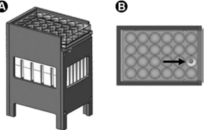
The irradiation procedures were performed with a device called LEDTable (ʎ 630±10 nm), developed by the Optical Group (OG) of the Optics and Photonics Research Center (Centro de Pesquisa em Óptica e Fotônica - CePOF) of the São Carlos Institute of Physics (IFSC) at the University of São Paulo (USP). This device contains 24 indium gallium nitrate (InGaN) diodes positioned underneath each compartment of the 24-well plate, in such a way that they could be individually and homogeneously exposed to the red light
. To ensure uniform distribution of light, there was also an 11 mm distance between the plate base plate and LED. In order to standardize the correct direction of LED, a collimator was included in the lamps, allowing proper individualization of irradiation for each sample
The energy doses were selected by pilot studies, the chosen power densities were those most frequently found in the literature (15). The time needed to deliver 2 J/cm2 using 20 mW/cm2 power density was 1 min and 40 s, while the time using 40 mW/cm2 was 50 s. The evaluated exposure frequencies were 1 or 3 consecutive irradiations, with an interval of 1 min between each. Thus, the samples subjected to the consecutive exposures received three energy doses of 2 J/cm² each, according to the used power densities (20 and 40 mW/cm²). During the interval, the samples were kept in an incubator at 37 °C with 5% CO2.
Non-irradiated MDPC-23 cells served as control. The 24-well culture plate containing the cells was placed in the LEDTable for the same time, except that the LEDs were not turned on.
Cell Viability
Cell viability was analyzed by the methyl tetrazolium assay (MTT Assay) (16), which evaluates the activity of the succinate dehydrogenase enzyme (SDH). Therefore, 24 h after the last irradiation, the culture medium was aspirated and replaced by a solution containing 900 µL of DMEM and 100 µL of MTT solution (Sigma Chemical Co.) at 5 mg/mL concentration. The cells were incubated for 4 h in contact with this solution, which was aspirated and replaced by 600 μL acidified isopropanol (HCl 0.04 N) for solubilization of the formazan crystals. Three 100 μL aliquots of this solution were individually transferred to the compartments of 96-well acrylic plates (Costar Corp., Cambridge, MA, USA) and the absorbance was determined at 570 nm in an Elisa reader (TP - Reader, ThermoPlate, China). The mean of the absorbance values of the three aliquots was calculated and the values presented as percentage, considering the median of the control group as being 100% metabolism.
Total Protein Production
Red LED photobiomodulates odontoblast cells
way as described for cell viability, the data were presented as percentage of the control.
Number of Viable Cells
The number of viable cells was evaluated by the Trypan Blue test, in which the dye added to the culture medium penetrates into the cells that had rupture of the membrane, thus staining them blue (17). For this test, the culture medium of each compartment was aspirated and replaced by 300 µL 0.25% trypsin (Invitrogen, Carlsbad, CA, USA), which had contact with the cells for 10 min. Aliquots of 50 µL of this cell suspension and 50 µL of 0.04% Trypan blue solution (Sigma-Aldrich,) were placed in the wells of a 96-well acrylic plate, where they remained for 2 min at ambient temperature. To count the viable cells, 10 µL of this final solution was taken to the hemocytometer and evaluated under an inverted light microscope (Nikon Eclipse TS 100, Nikon Corporation, Tokyo, Japan). The number of viable cells was determined by subtracting from the total number of cells, the cells determined as non-viable (stained blue). Thus, the number of viable cells obtained in the count corresponded to nx104 cells per mL of the suspension. The
values were converted into percentage of the control.
Data Analysis
No set of data (cell viability, total protein production and number of viable cells) presented normal distribution. Therefore, the nonparametric Kruskal-Wallis complemented by Mann-Whitney tests for comparison of the groups two by two. Statistically significant difference between the groups was inferred when p<0.05, i.e., at the level of significance of 5%.
Results
Cell Viability
Data from MDPC-23 cell viability are shown in Figure
2A.When a single irradiation was performed, the viability of MDPC-23 cells was significantly higher after using both power densities in comparison with non-irradiated cells. However, the best results were observed for 20 mW/cm². After 3 irradiations, a significant reduction in cell viability was observed in comparison with the control, for both power densities. Considering the same power density, a single irradiation was always better than a triple irradiation.
Total Protein Production
An increase in total protein production was observed in the group with a single irradiation using the 20 mW/ cm² power density, but this was not observed for the 40 mW/cm² power density. When the cells were exposed to 3 irradiations with a 40 mW/cm² power density, there was a statistically significant reduction in total protein production in comparison with the non-irradiated group, while no effect was observed when the 20 mW/cm² power density was used. A single irradiation, irrespective of the power density, was superior to multiple irradiations (Fig. 2B).
Number of Viable Cells
Significant decrease in the number of viable MDPC-23 cells was observed when they were irradiated with a 40 mW/cm² power density, compared with 20 mW/cm² power density and no irradiation, with no difference between them. No effect on the number of cells was detected for the groups irradiated 3 times, by both energy densities. Similarly, no effect was observed with regards to the number of irradiations when the same power density was compared (Figure 2C).
Discussion
This study demonstrated that the odontoblast-like cell metabolism was affected in different ways depending on the combination of the parameter settings. The best biostimulatory response was observed after a single irradiation with red LED at 630 nm wavelength, 2 J/cm² energy dose and 20 mW/cm² power density. Therefore, the null hypothesis that the LED irradiation would have no effect on odontoblast-like cells was rejected.
The biostimulative effect induced by low level phototherapy on the repair capacity of the dentin-pulp complex has been demonstrated in vitro and in vivo
(3,9,18,19). This effect is characterized by the increased cell proliferation and mineralized matrix production. Although low-level phototherapy has demonstrated good results in in vivo studies, there is still little research in the
basic area, aiming to determine the optimal parameters for light application among various therapeutic activities (1,12). Additionally, the effect of low-level phototherapy on a specific cell population derived from dental pulp tissue
L. F
. D. de Almeida et al.
has not yet been described.
In this study, red LED irradiation was performed in MDPC-23 cell culture. The fixed dose of 2 J/cm² was selected based on pilot studies (data not shown) using wavelengths selected from previous reports in the literature (8,16), while cell irradiation was performed one or three times consecutively to simulate a clinical protocol where the phototherapy would be applied before the insertion of the restorative materials. The results demonstrated that the increase in the frequency of light application had a detrimental effect on cell metabolic activity for both power densities.
The photoeffect on cell proliferation may vary according to the energy dose, power density and frequency. These parameters applied at higher values may cause adverse effects on cells such as structural damage to the DNA and exacerbated increase in reactive oxygen species (20). From the results obtained in the present study and based on those from Oliveira et al. (16), it was evident that the increase in frequency from one to three consecutive
irradiations reduced the viability of odontoblast-like cells. The combination of a single irradiation of the cells with red LED at 20 mW/cm² power density was efficient in promoting an increase in cell viability.
It has been reported that the power density has a direct influence on the effect of light by the generation of a two-stage dose-response effect, in which inhibition of stimulatory effects is achieved when the threshold of energy received by the cell is exceeded (15,20). Therefore, since the dose-response effect varies according to the set of irradiation parameters (21), it may be suggested that a higher frequency of exposure of MDPC-23 cells to LED exceeded the threshold of biological tolerance of these cells, leading to a reduction in their metabolism. Previous studies that evaluated the bio-stimulatory effects of light sources have observed such biphasic effect (1,15,20). These studies have shown that the effect of light is not linear, being dependent on the frequency of application and energy doses. In other words, light stimulus can both cause benefits and damage, depending on the total energy
Red LED photobiomodulates odontoblast cells
applied to the cell.
Previous studies have evaluated the effects of red LED applied on different cell lines (3,9-11). Irradiation in the red wavelength range, energy dose of 9.5 J/cm² caused an increase in type I collagen and TGB-β expression in a fibroblast culture (11). Increase in cell proliferation and angiogenesis in rodents was reported by Nishioka et al. (10), when the energy dose of 2.49 J/cm² was delivered by a device with a cross-sectional area of 0.5 cm² and 150 mW output power. In the present research, the increase in cell viability and total protein production occurred when the MDPC-23 cells were subjected to a single irradiation with red LED at 20 mW/cm² power density and energy dose of 2 J/cm². It seems evident that different types of cells need a specific energy dose in order to be bio-stimulated. When evaluating the effect of red LED (632 nm) on a culture of pulp cells obtained from rodents, Holder et al. (3) demonstrated that the cells irradiated for 40 or 60 s with a 3.73 mW/cm² power density had increased viability and production of mineralized nodules. These positive cell effects have been obtained with the use of red LED at a power density almost five times lower than the one used in the present study. Biostimulation was observed using 20 mW/cm². Holder et al. (3) used a primary culture of rodent dental pulp, in which fibroblasts, stem cells, endothelial and other cells are present, whereas in the present study, an immortalized culture of odontoblast-like cells were used. The sensitivity of these cells is different from cells in a primary pulp culture (21). The difference in sensitivity between cell cultures may be related to the intrinsic characteristics of each cell, as well as to the process of immortalization of the cell culture (22). Therefore, a higher power density was required to biostimulate MDPC-23 cells in culture than the one used by Holder et al. (3) to irradiate the primary culture of rodent pulp.
In spite of phototherapy presenting positive cellular bio-stimulation results, the light mechanism of action, specifically its participation in the tissue repair process, has still not been completely elucidated (3,20). It is believed that the light may be absorbed by chromophores in the cells (23), providing an increase in the activation of the cytochrome c oxidase pathway (6,16,20). This would generate an increase in the production of ATP and, consequently, induce cell proliferation (5,13). In the present study, the 2 J/cm2 energy dose increased cell metabolism, as determined by SDH activity. It is well known that SDH consists in a protein that belongs to the complex II of the cellular respiratory chain. This protein, therefore, acts within the electron transference catalysis (24), which determines the physiological ROS production (25). Therefore, the present results reinforce the hypothesis that the light’s mechanism of action is by mitochondrial pathway. However, other
targets should be assessed to confirm the effect of light on other mitochondrial complexes.
A positive relationship between SDH increase and the total protein production was observed in the group irradiated once by 20 mW/cm². These results corroborate this possible mechanism of action, related to cell chromophore excitation. This phenomenon, however, was not observed with the other irradiation parameters. The evaluation of total protein production was performed because protein deposition is markedly necessary for deposition of matrix during the repair process (5). Notably in pulp tissue repair, deposition of type I collagen and other proteins, such as alkaline phosphatase and dentin matrix proteins, allows for matrix deposition and mineralization (14). However, because it is an initial study, aimed at screening power parameters, the production of total protein may aid in response to this requirement. Further studies may be performed to assess the production of those specific proteins.
No linear relationship between the SDH and the number of viable cells was observed in all groups. The Trypan blue assay detects the presence of non-viable cells, and consequently the proportion of viable ones, due to the cell membrane disruption (17). According to this technique, it is not possible to confirm that the evaluated irradiation procedures induced any cell damage. Actually, when analyzed together, the results from Trypan blue and SDH assays confirmed that higher frequency of LED irradiation (three consecutive irradiations) resulted in overall decrease in cell metabolism, meaning that the total energy tolerated by the cell exceeded the threshold.
The beneficial effects obtained in the present study, combined with the low cost of LED, which presents a long useful life (5), indicate that the application of this light source deserves additional investigations, until it is adequate to be applied clinically in dentistry. This technique should be considered as an adjuvant method in the restorative treatment, in which the biostimulatory effects and the modulation of inflammation provided by the light source could help the pulp-dentin complex healing. Therefore, the use of LED-therapy would contribute to greater success in the management of deep cavities and pulp exposures. Further in vitro and in vivo studies should
be conducted to establish ideal direct and transdentinal irradiation parameters in order to improve the repair of the pulp tissue, maintaining the vitality of the tooth.
L. F
. D. de Almeida et al.
required to demonstrate the clinical effectiveness of this phototherapy.
Resumo
Fototerapia tem sido indicada como um tratamento adjuvante para o reparo de tecidos, incluindo o tecido pulpar. Entretanto, não há parâmetros de irradiação definidos, o que representa um grande desafio para o uso clínico da fototerapia. O objetivo deste estudo foi avaliar o efeito da fototerapia com LED vermelho em células MDPC-23 com fenótipo odontoblastóide, usando vários parâmetros. As células foram semeadas (104 células/cm2), incubadas por 12 h em DMEM completo e então o meio de cultura foi trocado por DMEM com 0,5% SFB. Após 12 h de incubação, as irradiações foram realizadas (630±10 nm) usando um dispositivo com densidade de potência de 20 ou 40 mW/cm2 e dose de energia de 2 J/cm2. As células foram irradiadas 1 ou 3 vezes, com intervalos de 1 min. Células não irradiadas serviram como controle. Foram avaliadas a viabilidade (ensaio de MTT), dosagem de proteína total (método de Lowry) e número de células viáveis (ensaio de Trypan blue). Os dados (n=12 por grupo) foram submetidos aos testes de Kruskal-Wallis e Mann-Whitney (p=0,05). Uma única irradiação com 20 ou 40 mW/cm2 aumentou a viabilidade celular, a qual foi negativamente afetada após 3 irradiações. Células irradiadas apenas uma vez com 20 mW/cm2 produziram mais proteínas comparadas com aquelas irradiadas com 40 mW/cm2. Redução no número de células viáveis ocorreu apenas após 3 irradiações com 40 mw/cm2. Em conclusão, o LED vermelho foi capaz de biomodular a atividade metabólica de células MDPC-23. A melhor bioestimulação celular foi obtida quando uma única irradiação com dose de energia de 2 J/cm2 e densidade de potência de 20 mW/cm2 foi administrada às células pulpares.
Acknowledgements
The authors acknowledge with thanks the financial support provided by the Brazilian financing agencies FAPESP (São Paulo State Research Supporting Foundation - grant 2012/17552-2) and CNPq (National Council of Research and Scientific Development - grants 305204/2010-6 and 301291/2010-1).
References
1. Peplow PV, Chung TY, Baxter GD. Laser photobiomodulation of wound healing: a review of experimental studies in mouse and rat animal models. Photomed Laser Surg 2010;28:291-325.
2. Pires Oliveira DA, Oliveira RF, Zangaro RA, Soares CP. Evaluation of low-level laser therapy of osteoblastic cells. Photomed Laser Surg 2008;26:401-404.
3. Holder MJ, Milward MR, Palin WM, Hadis MA, Cooper PR. Effects of red light-emitting diode irradiation on dental pulp cells. J Dent Res 2012;91:961-966.
4. Fujimura T, Mitani A, Fukuda M, Mogi M, Osawa K, Takahashi S, et al.. Irradiation with a low-level diode laser induces the developmental endothelial locus-1 gene and reduces proinflammatory cytokines in epithelial cells. Lasers Med Sci 2013;29:987-994.
5. Desmet KD, Paz DA, Corry JJ, Eells JT, Wong-Riley MT, Henry MM, et al.. Clinical and experimental applications of NIR-LED photobiomodulation. Photomed Laser Surg 2006;24:121–128.
6. Parker S. Low-level laser use in dentistry. Br Dent J 2007;202:131-138. 7. Dungel P, Hartinger J, Chaudary S, Slezak P, Hofmann A, Hausner T, et al.. Low level light therapy by LED of different wavelength induces angiogenesis and improves ischemic wound healing. Lasers Surg Med 2014;46:773-780.
8. Turrioni AP, Basso FG, Montoro LA, Almeida LF, Costa CA, Hebling J. Phototherapy up-regulates dentin matrix proteins expression and synthesis by stem cells from human exfoliated deciduous teeth. J Dent 2014;42:1292-1299.
9. Ferreira AN, Silveira L, Genovese WJ, Araújo VC, Frigo L, Mesquita RA, et al.. Effect of GaAIAs laser on reactional dentinogenesis induction in human teeth. Photomed Laser Surg 2006;24:358-365. 10.
Nishioka MA, Pinfildi CE, Sheliga TR, Arias VE, Gomes HC, Ferreira LM. LED (660 nm) and laser (670 nm) use on skin flap viability: angiogenesis and mast cells on transition line. Lasers Med Sci 2012;27:1045–1050. 11. Seo YK, Park JK, Song C, Kwo SY. Comparison of light-emitting diode
wavelength on activity and migration of rabbit ACL cells. Lasers Med Sci 2013;29:245–255.
12. Gao X, Xing D. Molecular mechanisms of cell proliferation induced by low power laser irradiation. Journal of Biomed Sci 2009;12:1-16. 13. Carroll JD, Milward MR, Cooper PR, Hadis M, Palin WM. Developments
in low level light therapy (LLLT) for dentistry. Dent Mater 2014;30:465-475.
14. Tjäderhane L, Haapasalo M. The dentin–pulp border: a dynamic interface between hard and soft tissues. Endodontic Topics 2012;20:52–84. 15. Huang YY, Chen ACH, Carroll JD, Hamblin MR. Biphasic dose response
in low level light therapy. Dose-Response 2009;7:358–383. 16. Oliveira CF, Basso FG, Lins EC, Bachmann L, Rosa AL, Bombonato-Prado
KF, et al.. In vitro effect of low-level laser on odontoblast-like cells. Laser Phys Lett 2011;8:155-163.
17. Wiegand C, Hipler UC. Methods for the measurement of cell and tissue compatibility including tissue regeneration processes. GMS Krankenhhyg Interdiszip. 2008;3:Doc12.
18. Villa GEP, Catirse ABCEB, Lia RCC, Lizarelli RFZ. In vivo analysis of low-power laser effects irradiation at stimulation of reactive dentine. Laser Phys Lett 2007;4:690–695.
19. Oliveira CF, Basso FG, Lins EC, et al.. Increased viability of odobtoblast-like cells subjected to low-level laser irradiation. Laser Phys 2010;20:1659-1666.
20. Al Ghamdi KM, Kumar A, Moussa NA. Low-level laser therapy: a useful technique for enhancing the proliferation of various cultured cells. Lasers Med Sci 2011;27:237–249.
21. Brondon P, Stadler I, Lanzafame RJ. A study of the effects of phototherapy dose interval on photobiomodulation of cell cultures. Lasers Surg Med 2005;36:409-413.
22. Yasuda Y, Inuyama H, Maeda H, Akamine A, Nör JE, Saito T. Cytotoxicity of one-step dentin-bonding agents toward dental pulp and odontoblast-like cells. J Oral Rehabil 2008;35:940-946.
23. Chorvat D, Chorvatova A. Approach to the study of endogenous fluorescence in living cells and tissues. Laser Phys Lett 2009;6:175-193. 24. Lenaz G, Genova ML. Structural and functional organization of the mitochondrial respiratory chain: a dynamic super-assembly. Int J Biochem Cell Biol 2009;41:1750–1772.
25. Kluckova K, Bezawork-Geleta A, Rohlena J, Dong L, Neuzil J. Mitochondrial complex II, a novel target for anti-cancer agents. Biochim Biophys Acta 2013;1827:552-564.
26. Goldberg M, Smith AJ. Cells and extracellular matrices of dentin and pulp: a biological basis for repair and tissue engineering. Crit Rev Oral Biol Med 2004;15:13-27.