Dilhan Ilgüy Mehmet Ilgüy Semanur Dolekoglu Erdogan Fisekcioglu
Departmentof Dentomaxillofacial Radiology, Faculty of Dentistry, Yeditepe University, Istanbul, Turkey.
Corresponding Author: Dilhan Ilgüy
E-mail: [email protected]
Evaluation of the posterior superior
alveolar artery and the maxillary sinus
with CBCT
Abstract: Assessment of the maxillary sinus anatomy before sinus aug-mentation is important for avoiding surgical complications, because of the close anatomical relationship between the posterior maxillary teeth and the maxillary sinus. The posterior superior alveolar artery (PSAA) is the branch of the maxillary artery that supplies the lateral sinus wall and overlying membrane. We evaluated the location of the PSAA and its relationship to the alveolar ridge and maxillary sinus using cone beam computed tomography (CBCT). The study group consisted of 135 CBCT scans (270 sinuses) obtained from the archive of the dentomaxillofacial radiology department at Yeditepe University Faculty of Dentistry, Istan-bul, Turkey. The distance between the lower border of the artery and the alveolar crest, bone height from the sinus loor to the ridge crest, distance from the artery to the medial sinus wall, and the diameter and location of the artery were determined. The occurrence of septa and pathology were recorded from CBCT scans. The PSAA was observed in 89.3% of sinuses, and 71.1% of arteries were intraosseous with diameters mostly ≤ 1 mm (68.9%). The prevalence of sinus septa was 55.2%, and that of sinus pa-thology was 57.4%. The mean age was 43.07 ± 17.55 years. There was a statistically signiicant difference between the location of the artery and gender (p < 0.05). The prevalence of sinus membrane thickening was 57.4%. Detailed knowledge about the location of the PSAA and sinus morphology may be obtained with CBCT before maxillary sinus surgery.
Descriptors: Maxillary Artery; Maxillary Sinus; Cone-Beam Computed Tomography.
Introduction
The posterior superior alveolar artery (PSAA) and infraorbital artery (IOA) are the branches of the maxillary artery that supply the lateral sinus wall and overlying membrane. The blood supply of the maxillary sinus and Schneiderian membrane comes from the maxillary artery.1,2
Sinus augmentation is a safe procedure with high predictability for re-habilitation of severely atrophic posterior maxillae.3-6 The branches of
the maxillary artery should be taken into consideration during sinus aug-mentation because of the potential risk of bleeding during the procedure owing to damage to the vascular supply in the lateral wall.7 Assessment
of the maxillary sinus anatomy is important to avoid unnecessary com-plications due to the close anatomical relationship of the PSAA with the Declaration of Interests: The authors
certify that they have no commercial or associative interest that represents a conflict of interest in connection with the manuscript.
Submitted: Mar 20, 2013
maxillary sinus.8,9
Any imaging technique that subjects the patient to ionizing radiation must yield as much pertinent information as possible, and it is important that the physician collect all the useful information pos-sible. Cone beam computed tomography (CBCT) may be recommended as a dose-sparing technique compared with standard medical computed tomog-raphy (MDCT) scans for dentomaxillofacial imag-ing. Increases in kV, mA, exposure time, and ield of view increase the radiation dose regardless of the type of imaging technique used.10 The effective dose
(International Commission on Radiological Protec-tion - ICRP 2007) from a standard dental proto-col scan with MDCT is 1.5 to 12.3 times greater than from comparable medium–ield of view dental CBCT scans.11,12 Thus, CBCT is frequently used for
preoperative assessment of the alveolar ridge and maxillary sinus in patients receiving implants in the posterior maxilla.
The purpose of this study was to evaluate the lo-cation of the PSAA and its relationship to the alveo-lar ridge and maxilalveo-lary sinus using CBCT.
Methodology
The study design underwent formal review and received approval from the institutional review board of our institution. The retrospective study group was planned according to Sample Size Esti-mation Simple Random Sampling and consisted of 135 CBCT scans of 55 males and 80 females (270 sinuses) obtained from the archive of the Dento-maxillofacial Radiology Department of the faculty. Patient ages ranged from 18 to 83 years. Digital images were taken using an Iluma CBCT scanner (Imtec Corporation, Oberursel, Germany) with an amorphous silicon lat-panel image detector and a cylindrical volume of reconstruction of up to 19 × 24 cm. Images were taken at 120 kVp, 3.8 mA, and a voxel size of 0.2 mm, with an exposure time of 40 seconds. 3D reconstructions were created by re-formatting the axial CBCT scans on a local work-station using Iluma dental imaging software (Imtec Corporation). A written informed consent form, which is routinely obtained from each patient prior to imaging in our faculty, also included a clause for the use of images in this research. Before measure-ments were made, the orientation of the images was determined for each patient.
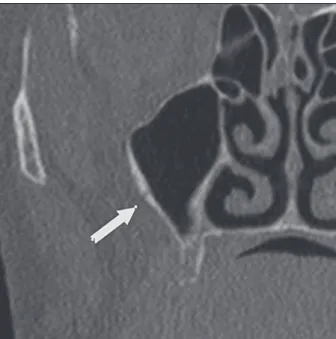
On coronal scans, evaluation was made between where the artery was irst and last seen. The dis-tance between the lower border of the artery and the alveolar crest (A), bone height from the sinus loor to the ridge crest (B), distance from the artery to the medial sinus wall (C), and the diameter and location of the artery were determined (Figure 1); occurrence of sinus septa and membrane thickening were also recorded from CBCT scans. Locations of the artery were:
a. intraosseous (Figure 2),
b. below the membrane (Figure 3), and
c. on the outer cortex of the lateral sinus wall (Fig-ure 4).
A and B were calculated for only the edentulous alveolar crest.
A dentomaxillofacial radiology specialist evalu-ated the images in a darkened quiet room with dual monitors (HP LP2475W, resolution 1920 × 1200; Hewlett-Packard, Houston, USA). Each viewing ses-Figure 1- Distance between the lower border of the artery
Figure 2 - Coronal view of the maxillary sinus reveals the
intraosseous artery (arrow). Figure 3 - Coronal view of the maxillary sinus reveals the artery (arrow), which is below the membrane.
Figure 4 - Coronal view of the maxillary sinus reveals the artery (arrow), which is on the outer cortex of the lateral sinus wall.
sion lasted 30 minutes. Care was taken to ensure that 24 hours elapsed between all sessions. For in-tra-examiner calibration and determination of reli-ability and reproducibility of the measurements, the images were evaluated a second time by the same observer 2 weeks later.
SPSS 15.0 (Statistical Package for Social Scienc-es, IBM, New York, USA) for Windows 2007 (Mi-crosoft, New Mexico, USA) was used for statistical analysis of the results. Prior to the study, all param-eters were evaluated with the Kolmogorov-Smirnov test, and the data were found to be normally dis-tributed. Simple Random Sampling Sample Size Es-timation was done. While evaluating the data using descriptive statistical methods, parameters with normal distribution for the comparison of quantita-tive data were evaluated using one-way analysis of variance, the Tukey test, and Student’s t-test. Quali-tative data were evaluated using the Chi-square test. The Pearson correlation was used to assess poten-tial correlations between parameters. Intra-observer agreement was calculated using the intraclass cor-relation coeficient. Signiicance was accepted at p < 0.05.
Results
scores of measurements A, B, and C were 0.95, 0.98, and 0.97, respectively. Kappa values for the diam-eter and location of the artery were 81.5% and 98%, respectively, and for the occurrence of septa and pa-thology they were 86.5% and 93%, respectively.
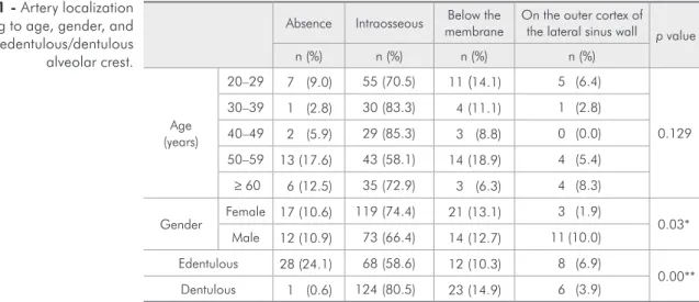
The PSAA was observed in 89.3% of all sinuses; 71.1% of arteries were intraosseous, 13% were be-low the membrane, and only 5.2% were on the outer cortex of the lateral sinus wall. The artery diameters ranged from 0.4 to 1.7 mm with a mean value of 0.94 ± 0.26 mm. The artery diameters were most-ly ≤ 1 mm (68.9%). The percentages of artery diam-eters ≤ 1 mm were 73.5% and 64.5% for the right and left sinuses, respectively, a difference that was not statistically signiicant (p > 0.05). More males (39.8%) than females (25.2%; p < 0.05) had an ar-tery diameter of 1 to 2 mm. No signiicant correla-tion was found between the presence of the artery and age/gender (p > 0.05). A statistically signiicant difference was found between locations of the artery and gender (p < 0.05). Although the prevalence of an intraosseous PSAA was signiicantly higher in fe-males (74.4%) than males (66.4%), the PSAA was lo-cated on the outer cortex of the lateral sinus wall in males (10%) signiicantly more often than in females (1.9%; Table 1; p < 0.05).
Regarding the prevalence of the artery, there was a statistically signiicant difference between edentu-lous and dentuedentu-lous alveolar crest where the artery was identiied. The artery was observed less often
in the edentulous crest (75.9%) than dentulous crest (99.4%; p < 0.01). No statistically signiicant dif-ference was observed between gender and distance from the artery to the alveolar crest (p > 0.05; Table 2). The distance from the artery to the medial si-nus wall in males was signiicantly higher than in females (p < 0.05). With regard to the edentulous crest, the mean distance from the artery to the al-veolar crest was 16.88 ± 3.46 mm (A), and the bone height from the sinus loor to the ridge crest (B) was 7.84 ± 3.20 mm. The mean distance from the artery to the medial sinus wall (C) in dentu-lous crests (13.92 ± 2.84 mm) was signiicantly higher than that measured for the edentulous crest (13.00 ± 2.32 mm; p < 0.05). There also was a sta-tistically signiicant difference among age groups re-garding the distance from the artery to the medial sinus wall (p < 0.01; Table 3). According to the Pear-son correlation test, a negative (−20.1%) correlation Absence Intraosseous membraneBelow the On the outer cortex of the lateral sinus wall
p value
n (%) n (%) n (%) n (%)
Age (years)
20–29 7 (9.0) 55 (70.5) 11 (14.1) 5 (6.4)
0.129
30–39 1 (2.8) 30 (83.3) 4 (11.1) 1 (2.8)
40–49 2 (5.9) 29 (85.3) 3 (8.8) 0 (0.0)
50–59 13(17.6) 43 (58.1) 14 (18.9) 4 (5.4)
≥ 60 6(12.5) 35 (72.9) 3 (6.3) 4 (8.3)
Gender Female 17(10.6) 119 (74.4) 21 (13.1) 3 (1.9) 0.03*
Male 12(10.9) 73 (66.4) 14 (12.7) 11 (10.0)
Edentulous 28(24.1) 68 (58.6) 12 (10.3) 8 (6.9)
0.00**
Dentulous 1 (0.6) 124 (80.5) 23 (14.9) 6 (3.9)
*p < 0.05; **p < 0.01 (chi-square test).
Table 1 - Artery localization according to age, gender, and edentulous/dentulous alveolar crest.
Table 2 - Measurements according to gender.
Female Male
p value Mean ± SD Mean ± SD
A** 16.79 ± 3.79 17.00 ± 2.94 0.785
B*** 7.50 ± 3.03 8.34 ± 3.43 0.233
C**** 13.27 ± 2.82 14.03 ± 2.44 0.031*
was found between these two parameters (p < 0.01). Increased age was negatively associated with the dis-tance between the artery and the medial sinus wall. In other words, older participants tended to have a shorter distance between their artery and the medial sinus wall. The prevalence of sinus septa was 55.2% and that of sinus membrane thickening was 57.4%. The presence of pathology in men (68.2%) was sig-niicantly higher than in females (48.8%; p < 0.01).
Discussion
Sinus augmentation is a method with high pre-dictability for placing successful dental implants into atrophic posterior maxillae.3-6 Knowledge of
the anatomic structure of the area is important for this procedure. In the present study, we were able to observe the presence and location of the PSAA with CBCT scans. The artery was observed in 89.3% of the sinuses and was mostly intraosseous (71.1%). The success rate for identifying the artery was higher than that reported by Güncü et al.13 (64.5%), Elian
et al.14 (52.9%), Mardinger et al.15 (55%), and Kim et
al.16 (52%). This may be related to the methods the
other groups used to detect and describe the artery. Collectively, these results suggest that an undetected intrabony canal in a CT scan does not exclude its existence but that it may not be visible owing to its small diameter.15 CBCT provides accurate and
reli-able linear measurements for reconstruction and im-aging of dental and maxillofacial structures.7-9
According to Kim et al.,16 the prevalence of the
PSAA differs signiicantly between males (64%) and females (40%). In the present study, no signiicant correlation was found between gender and presence of the artery. The reason may be that the number of males/females differs between the two studies.
Table 2 shows that the mean distance of the
ar-tery from the alveolar ridge was 16.79 ± 3.79 mm for females and 17.00 ± 2.94 mm for males. In anatomic studies, this distance was reported to be 18.9–19.6 mm.1,2 We observed a shorter distance
than in these anatomic studies. The differences may be due to the small number of cases evaluated with CT in previous studies as reported by Güncü et al.13
(18 mm), Elian et al.14 (16.4 mm), Mardinger et al.15
(16.9 mm), and Kim et al.16 (18.9 mm). Our current
results are close to those of these studies.
In the present study, the mean distance from the artery to the crest was 7.50 ± 3.03 mm in females and 8.34 ± 3.43 mm in males. The mean distance from the artery to the antral loor was 9.29 mm in females and 8.66 mm in males. This distance re-ported by Güncü et al.13 was 7.8 ± 0.3 mm and by
Mardinger et al.15 was 7–8 mm. These differences
may be explained by the anatomic variation in the positions of arteries.
Damage to the bony vessel can cause bleeding, may obscure the physician’s line of sight, and may lead to perforation of the Schneiderian membrane, all of which prolong the operation and assessment of the sinus membrane relection.17 According to
our current results, the mean artery diameter was 0.94 ± 0.26 mm. However, Güncü et al.,13 Ella et
al.,7 and Kim et al.16 reported larger diameters of
1.3 mm, 1.2 mm, and 1.52 mm, respectively. Simi-larly, in anatomic studies, the diameter was reported to be 1.6 mm at the exit from the maxillary artery.1,2
In our study group, artery diameter was general-ly ≤ 1 mm (68.9%). Güncü et al.13 reported that only
36.1% of arteries were ≤ 1 mm, Mardinger et al.15
reported that 26% of sinuses have vessels ≤ 1 mm, and Kim et al.16 detected 13.9% of vessels with
diam-eters < 1 mm. We found that the artery was ≥ 1 mm in 31.1% of patients, which may suggest that the
in-20–29 30–39 40–49 50–59 ≥ 60 years
p value Mean ± SD Mean ± SD Mean ± SD Mean ± SD Mean ± SD
A** 17.01 ± 3.01 17.52 ± 4.18 17.05 ± 2.93 15.97 ± 3.42 17.40 ± 3.54 0.553 B*** 8.29 ± 3.20 9.62 ± 3.28 7.74 ± 1.70 7.65 ± 3.76 7.50 ± 2.92 0.546 C**** 14.18 ± 2.48 14.89 ± 2.40 13.00 ± 2.27 12.60 ± 3.05 13.34 ± 2.45 0.001*
*p < 0.01 (one-way analysis of variance). **Distance between the lower border of the artery and the alveolar crest. ***Bone height from the sinus floor to the ridge crest. ****Distance from the artery to the medial sinus wall.
cidence of intense bleeding during a sinus augmenta-tion is low.
In our study group, the average diameter of ar-teries was higher in males (39.8%) than females (25.2%). Although Güncü et al.13 and Kim et al.16
reported similar results, Mardinger et al.15 found no
difference between men and women regarding the diameter of the artery. Anatomic variations in the maxillary sinus may be important for dental im-plant planning. In some instances, dental imim-plant planning may require modiications related to si-nus morphology. Results from our present study re-vealed that sinus septa were observed in 55.2% of the 270 sinuses. The prevalence of sinus septa found by Güncü et al.13 was 16.1%, by Krennmair et al.18
was 16%, and by Kim et al.19 was 26.5% when CT
was used to assess the sinuses. These results are not consistent with those of the present study. On the other hand, much higher percentages have been re-ported with CBCT, and they are close to the results obtained in our present study. Orhan et al.20
report-ed that the prevalence of sinus septa was 58%, and Neugebauer et al.21 reported that it was 47%. Lana
et al.22 stated that the prevalence of antral septa was
44.4%. The data in these reports may be due to the use of CBCT to identify the septa. Lana et al.22 also
found that the prevalence of mucosal thickening was 54.8% (≤ 3 mm) and 62.6% (≥ 3 mm) with CBCT. The prevalence of sinus membrane thickening was 57.4% in our present study. However, we evaluated only the existence of sinus mucosa thickening, and the thickness was not measured. The prevalence of thickening reported by Lana et al.22 was consistent
with those of the present study.
Conclusion
We conclude that preoperative imaging with CBCT seems to be helpful for assessing the location of the PSAA and the maxillary sinus morphology, which may be used to adjust the surgical treatment plan to yield more successful dental implant treat-ments.
References
1. Solar P, Geyerhofer U, Traxler H, Windisch A, Ulm C, Watzek G. Blood supply to the maxillary sinus relevant to sinus floor elevation procedures. Clin Oral Implants Res. 1999 Feb;10(1):34-44.
2. Traxler H, Windisch A, Geyerhofer U, Surd R, Solar P, Firbas W. Arterial blood supply of the maxillary sinus. Clin Anat. 1999;12(6):417-21.
3. Wallace SS, Froum SJ. Effect of maxillary sinus augmentation on the survival of endosseous dental implants A systematic review. Ann Periodontol. 2003 Dec;8(1):328-43.
4. Aghaloo TL, Moy PK. Which hard tissue augmentation tech-niques are the most successful in furnishing bony support for implant placement?. Int J Oral Maxillofac Implants. 2007;22 Suppl:49-70.
5. Del Fabbro M, Rosano G, Taschieri S. Implant surviv-al rates after maxillary sinus augmentation. Eur J Orsurviv-al Sci. 2008 Dec;116(6):497-506. DOI: 10.1111/j.1600-0722.2008.00571.x.
6. Pjetursson BE, Tan WC, Zwahlen M, Lang NP. A systematic review of the success of sinus floor elevation and survival of implants inserted in combination with sinus floor eleva-tion. J Clin Periodontol. 2008 Sep;35(8 Suppl):216-40. DOI: 10.1111/j.1600-051X.2008.01272.x.
7. Ella B, Sédarat C, Noble RC, Normand E, Lauverjat Y, Si-berchicot F, et al. Vascular connections of the lateral wall of
the sinus: surgical effect in sinus augmentation. Int J Oral Maxillofac Implants. 2008 Nov-Dec;23(6):1047-52. 8. van den Bergh JP, ten Bruggenkate CM, Disch FJ, Tuinzing
DB. Anatomical aspects of sinus floor elevations. Clin Oral Implants Res. 2000 Jun;11(3):256-65.
9. Garg AK. Augmentation grafting of the maxillary sinus for placement of implants: anatomy, physiology, and procedures. Implant Dent. 1999;8(1):36-46.
10. Lorenzoni DC, Bolognese AM, Garib DG, Guedes FR, Sant’anna EF. Cone-beam computed tomography and radio-graphs in dentistry: aspects related to radiation dose. Int J Dent. 2012;2012:813768. DOI: 10.1155/2012/813768. Epub 2012 Apr 4.
11. Ludlow JB, Ivanovic M. Comparative dosimetry of dental CBCT devices and 64-slice CT for oral and maxillofacial ra-diology. Oral Surg Oral Med Oral Pathol Oral Radiol Endod. 2008 Jul;106(1):106-14.
12. Valentin J. The 2007 Recommendations of the International Commission on Radiological Protection. Publication 103. Ann ICRP. 2007;37:1-34.
14. Elian N, Wallace S, Cho SC, Jalbout ZN, Froum S. Dis-tribution of the maxillary artery as it relates to sinus floor augmentation. Int J Oral Maxillofac Implants. 2005 Sep-Oct;20(5):784-7.
15. Mardinger O, Abba M, Hirshberg A, Schwartz-Arad D. Prev-alence, diameter and course of the maxillary intraosseous vascular canal with relation to sinus augmentation proce-dure: a radiographic study. Int J Oral Maxillofac Surg. 2007 Aug;36(8):735-8. Epub 2007 Jul 12.
16. Kim JH, Ryu JS, Kim KD, Hwang SH, Moon HS. A radio-graphic study of the posterior superior alveolar artery. Implant Dent. 2011 Aug;20(4):306-10.
17. Chanavaz M. Sinus grafting related to implantology. Statisti-cal analysis of 15 years of surgiStatisti-cal experience (1979–1994). J Oral Implantol. 1996;22(2):119-30.
18. Krennmair G, Ulm C, Lugmayr H. Maxillary sinus septa: incidence, morphology and clinical implications. J Cranio-maxillofac Surg. 1997 Oct;25(5):261-5.
19. Kim MJ, Jung UW, Kim CS, Kim KD, Choi SH, Kim CK, et al. Maxillary sinus septa: prevalence, height, location, and mor-phology. A reformatted computed tomography scans analysis. J Periodontol. 2006 May;77(5):903-8.
20. Orhan K, Kusakci Seker B, Aksoy S, Bayindir H, Berberoğlu A, Seker E. Cone beam CT evaluation of maxillary sinus septa prevalence, height, location and morphology in children and an adult population. Med Princ Pract. 2013;22(1):47-53. DOI: 10.1159/000339849. Epub 2012 Jul 24.
21. Neugebauer J, Ritter L, Mischkowski RA, Dreiseidler T, Scherer P, Ketterle M, et al. Evaluation of maxillary sinus anatomy by cone-beam CT prior to sinus floor elevation. Int J Oral Maxillofac Implants. 2010 Mar-Apr;25(2):258-65. 22. Lana JP, Carneiro PM, Machado VC, Souza PE, Manzi FR,