ORIGINAL ARTICLE
31
Stent thrombosis in aortic aneurysm: evaluation by multidetector CT
ROBERTODE MORAES BASTOS1, ALVARO RAZUK FILHO2, ROBERTO BLASBALG3, ROBERTO AUGUSTO CAFFARO4, WALTER KHEGAN KARAKHANIAN5,
FERNANDO PINHO ESTEVES6, ANDRE PACIELLO ROMUALDO6, ANTONIO JOSÉDA ROCHA7 1 Professor/Instructor; Radiologist
2 M.D.; Assistant Professor of Faculdade de Ciências Médicas da Santa Casa de Misericórdia de São Paulo, São Paulo, SP, Brazil 3 Physician of Hospital das Clínicas da Faculdade de Medicina da Universidade de São Paulo – USP, São Paulo, SP, Brazil 4 M.D.; Professor of Faculdade de Ciências Médicas da Santa Casa Misericórdia de São Paulo, São Paulo, SP, Brazil 5 M.D.; Professor of Faculdade de Ciências Médicas da Santa Casa de São Paulo, São Paulo, SP, Brazil 6 M.D.; Irmandade da Santa Casa de Misericórdia de São Paulo, SP, Brazil
7 M.D.; Professor of Faculdade de Ciências Médicas da Santa Casa de Misericórdia de São Paulo, São Paulo, SP, Brazil
Received from Irmandade da Santa Casa de Misericórdia de São Paulo and Fleury Medicina e Saúde, São Paulo, SP
Submitted on: 07/07/2010
Approved on: 11/07/2010
Corresponding author:
Roberto de Moraes Bastos Rua Bergamota, 470 - Apt. 81
Bloco C CEP: 05468 000 São Paulo, SP, Brazil Phone: (11) 3021-1658 /
99047102 Fax: (11) 2176-7333 [email protected]
Conlict of interest: None.
ABSTRACT
Objective: To evaluate the demographic characteristics and imaging indings of throm-bosis in a series of patients submitted to endovascular stent-grat repair of abdominal
aortic aneurysm (ESGAAA). Methods: We evaluated the imaging features, which
al-lowed the diagnosis of endoluminal thrombosis in a series of 30 patients undergoing ESGAAA and followed from 5 to 29 months, using 64-channel multidetector computed
tomography (MDCT). Results: hrombosis was diagnosed in 10 patients (33.3%), and
in 3 of those patients, total thrombosis was observed in an iliac branch. Conclusion:
MDCT allowed the diagnosis of diferent types of endoluminal thrombosis in patients submitted to ESGAAA. he use of this minimally invasive diagnostic technique should be encouraged in clinical practice.
ROBERTO DE MORAES BASTOS ETAL.
32 Rev Assoc Med Bras 2011; 57(1):31-34 INTRODUCTION
Until the early 90’s, open surgery was the only treatment for abdominal aortic aneurysm (AAA), with periopera-tive mortality rate for patients undergoing elecperiopera-tive sur-gery ranging from 4% to 8.4% in specialized centers1,2,3.
he continuous search for surgical techniques with lower mortality and morbidity rates led to the development of endovascular prosthesis and techniques to treat abdomi-nal aortic aneurysm. hey have been used in diferent re-gions of the world, being approved by the United States Food and Drug Administration (FDA) since 19994,5.
Endovascular surgery for AAA repair is not devoid of complications, which can develop during or ater the procedure. However, the choice of this treatment modal-ity has been justiied when compared to conventional surgery1. Parietal thrombosis may develop as a small
circumferential thrombus on grat’s wall, or as a luminal occlusion of the stent, demonstrated in digital angiogra-phy (DA) or computed tomograangiogra-phy (CT), with an esti-mated incidence of 3-19% of the cases of endovascular stent-grat repair of abdominal aortic aneurysm (ES-GAAA)6,7,8,9,10,11.
Using a 64-channel multidetector CT (MDCT), we investigated a series of consecutive patients who under-went ESGAAA in order to assess the demographic char-acteristics and images of endoluminal thrombosis.
METHODS
PATIENTS
his transversal study was approved by the Research Eth-ics Committee of the Institution, and all patients signed an informed consent. From February to September 2007, 30 consecutive patients followed-up ater ESGAAA (26 males and 4 females), ages 55 to 83 years (mean 70.9 years, median 72 years), were included in this study. All patients agreed to participate in the study. he follow-up period ranged from 5 to 29 months ater surgery (mean 14.4 months, median 13.5 months). he proposed followed-up protocol included MDCT at 1, 3, 6, and 12 months in the irst year ater the surgery, followed by an-nual MDCT in cases with aneurism regression or stabil-ity. In those cases in which expansion on the aneurysm was observed, patients were followed-up with MDCT every three months. All patients were treated with aorto-iliac abdominal stent grats of the following brands:
Tal-ent®, Meditronic Vascular, Sunrise, Fla (n = 18); Excluder
endoprothesis®, W.L. Gore & Associates, Sunnyvale, Calif
(n = 1); Zenith®, Cook Inc, Bloomington, Ind (n = 8); and
Apolo®, Nano Endoluminal, Florianópolis, Brasil (n = 3).
To analyze the development of endoluminal thrombosis, grats were grouped according to their material, and they were separated in Polytetraluorethylene (PTFE) (Apolo®
and Excluder endoprosthesis®) and Dacron/Polyester (
Tal-ent® and Zenith®).
Patients who refused to participate, and those who presented contraindications to the injection of iodinated contrast media or exams with ionizing radiation, as well as those with renal failure were excluded from the study.
EXAMS
MDCT 64-channel Brilliance equipment (Phillips, Eindhoven, Holland) was used for the exams. The exam covered the area between the diaphragm and common femoral arteries in three phases: pre-contrast, with collimation of 2.5 mm, 120 Kv, and 322 mAs; and post-contrast, arterial and venous phases, both with acquired collimation of 0.625 mm and reconstructed with 1.0 mm thickness, increment of 1.0 mm, Pitch of 0.703, rotation velocity of 0.75 per second, 120 Kv, and 350 mAs.
A dose of 1.5 mL/kg of contrast medium (mean of 100 ml per patient), with concentration of 300 mg of iodine per ml, was injected through a venous puncture with a 20-Gauge catheter in the antecubital vein, with an infusion pump at a rate of 5 ml per second, followed by 30 ml of normal saline. Post-contrast administra-tion phases were performed using an attenuaadministra-tion de-tector mechanism placed in the area of the aorta, at the level of the celiac branch (bolus tracker) that, when reached 180 Hounsfield units (HU) determined by the arrival of the contrast medium, began scanning the ar-terial phase, followed by the venous phase attained 60 seconds after the first one. In all phases, apnea time ranged from 12 to 18 seconds.
IMAGEANALYSIS
All images were independently analyzed at a worksta-tion (GE Medical Systems, Milwaukee, WI) by two ra-diologists (RMB and RB), with 11 and 15 years experi-ence, respectively, and final results were obtained by consensus.
Data related to the integrity of the stents, presence or absence of luminal thrombus, characteristics and lo-cation of thrombus, as well as information related to the type of stent and its constituents were analyzed. All arterial phase images were used for tridimensional angiographic reconstitutions using appropriate volume rendering (VR) and maximum intensity projection (MIP) reconstruction algorithms.
STATISTICALANALYSIS
To determine the site were thrombi are formed, the in-cidence and percentage were calculated. Fisher’s exact test was used to compare the occurrence of thrombo-sis with stent’s brand, as well as the group of stents ac-cording to the constituent material, formed by PTFE (Apolo® and Excluder endoprosthesis®) and Dacron/
STENT THROMBOSIS IN AORTIC ANEURYSM: EVALUATION BY MULTIDETECTOR CT
33
Rev Assoc Med Bras 2011; 57(1):31-34
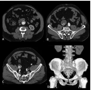
Figure 1 – Axial MDCT images showing a
semi-circumferential thrombus on the anterior internal wall of aortic endoprosthesis (A); right common iliac artery aneurysm associated with occlusion of the endoprosthesis and presence of an occluder in the right internal iliac artery (B and C); and angiographic reconstruction with volume rendering algorithm (D). Note the absence of contrast column in the right common iliac artery and the presence of a patent crossed femorofemoral graft (D).
RESULTS
All prosthesis used were intact and without signs of frac-ture on MDCT. Ten cases (33.3%) of endoluminal throm-bosis were identiied. Five of those (16.7%) showed partial aortic thrombosis; one (3.3%) had partial thrombosis of the aorta and right iliac branch; one (3.3%) had partial thrombosis of the right iliac branch; and three (10%) had partial thrombosis of the aorta and total thrombosis of the right iliac branch (Figure 1). Data are presented in Table 1.
All patients with complete right iliac branch thrombo-sis were smokers and had been treated for aneurysms of external iliac arteries by placing stent extensions and oc-cluders of the internal iliac arteries. In two of them, throm-bosis was diagnosed between 30 and 60 days ater surgery, requiring surgical limb revascularization with crossed
femorofemoral grat. he other patient presented throm-bosis, diagnosed on control exam at six months, evolving with retroperitoneal and abdominal collateral circulation, without the need of new surgery.
Out of the 10 patients with endoluminal thrombosis, 8 were smokers, 2 had DM, and 1 had hypertension. No statistically signiicant relationship between the presence of thrombosis and these clinical conditions was identiied using Fisher’s exact test.
Among the brands of prosthesis associated with thrombosis, it was identiied 1 case with Apolo®, 7 with
Talent®, and 2 with Zenith®. A statistically signiicant
dif-ference in the presence of thrombosis among the brands was not observed.
Among the group of materials, one case of thrombosis in PTFE prosthesis (Apolo® and Excluder endoprosthesis®)
and 9 cases in Dacron/Polyester prosthesis (Talent® and
Zenith®) were identiied. A signiicant diference in the
presence of thrombosis among the material of the pros-thesis was not observed.
DISCUSSION
Endoluminal vascular prosthesis thrombosis represents one of the main complications of ESGAAA diagnosed by CT scan. Specialized literature highlights the following key factors determining the characteristic of stents throm-bogenic material12, smoking, the need of adjuvant
proce-dures, and the experience of the surgical team13. According
to hurnher et al.14, formation of semicircular thrombi can
occur in up to 19% of the cases, usually not determinant of low changes or clinical repercussion. Our results is in agreement with this observation, since all seven patients with partial thrombosis (23.3%) remained asymptomatic in clinical evaluations and subsequent MDCTs.
Occlusion of the blood vessel by thrombosis, which has an incidence of 1.5% to 10%, in general extending to the iliac branch, has as main triggering factor the marked tortuosity of the prosthesis or even prosthesis bending by aneurysmal remodeling14. It has been reported that
the evolution of luminal thrombosis may be spontaneous resolution of the thrombus, embolization of distal limb branches, or even total occlusion of blood vessel10.
Accord-ing to Schunn et al.12, bifurcated prostheses have a greater
Prosthesis
Intraluminal thrombosis
No 20 (66.7%)
Yes 10 (33.3%)
Partial aortic circumference + total R iliac branch 3 (10%) Partial aortic circumference 5 (16,7%) Partial R iliac branch circumference 1 (3.3%) Parietal aortic and partial R iliac branch D 1 (3.3%)
Total 30
ROBERTO DE MORAES BASTOS ETAL.
34 Rev Assoc Med Bras 2011; 57(1):31-34
chance of thrombosis- and embolization-related compli-cations than tubular prostheses. he author reported an incidence of 10.5% of luminal thrombosis, 60% in bifur-cated prosthesis and 13.3% in tubular prosthesis12.
In the present study, we identiied an elevated rate of thrombosis (33%), regardless of the type of material or prosthesis used, which is above those reported in international literature.14 Despite the absence of
statis-tical signiicance in this case series, the literature has emphasized that superposition of some clinical condi-tions, especially smoking, may contribute to endolumi-nal thrombi formation13.
We believe that the high incidence of endoluminal thrombosis in this series might be due to the greater diag-nostic accuracy of the high-resolution of the 64-channel MDCT images used in this study. High-resolution ine cuts (1.0 mm) allowed the detection of small intralumi-nal thrombi, in addition to tridimensiointralumi-nal angiographic reformatting and reconstruction using volume rendering (VR) and maximum intensity projection (MIP) recon-struction algorithms. In a recent study using the same speciication equipment and similar protocol, homaz et al.10 observed an elevated rate of luminal thrombosis in
ESGAAA follow-up.
Speciic studies on inherent particularities of the di-agnosis and late treatment of AAA in the Brazilian popu-lation are required. Tortuosity with greater longitudinal and transversal dilation of aortic aneurysm has been con-sidered a risk factor for the development of endoluminal thrombosis14. Nevertheless, our results do not allow close
evaluation of this aspect.
Despite the limitations inherent in this type of study, we believe that our results indicate relevant fac-tors regarding the diagnosis of endoluminal thrombosis by MDCT. In the opinion of the authors, studies with larger series of patients, with longer follow-up, using the high resolution MDCT protocol, may contribute for better understanding of endoluminal thrombosis, be-sides providing precise diagnostic parameters and more adequate therapy.
CONCLUSION
Multidetector CT allowed the diagnosis of diferent types of endoluminal thrombosis in this series of patients who underwent ESGAAA. he use of MDCT in clinical prac-tice should be encouraged in order to obtain the best use of this minimally invasive diagnostic tool in the follow-up and diagnosis of individuals undergoing ESGAAA.
REFERENCES
1. Kaufman JA, Geller SC, Brewster DC, Fan CM, Cambria RP, La-Muraglia GM, et al. Endovascular repair of abdominal aortic aneu-Endovascular repair of abdominal aortic aneu-rysms: current status and future directions. AJR Am J Roentgenol 2000; 175:289-302.
2. Ernst CB. Abdominal aortic aneurysm. N Engl J Med 1993; 328:1167-72.
3. Lawrence PF, Gazak C, Bhirangi L, Jones B, Bhirangi K, Oderich
G, et al. he epidemiology of surgically repaired aneurysms in the
United States. J Vasc Surg 1999; 30:632-40.
4. Lederle FA. Abdominal aortic aneurysm - open versus endovascular repair. N Engl J Med 2004; 351:1677-9.
5. Parodi JC, Marin ML, Veith FJ. Transfemoral, endovascular stent-ed grat repair of an abdominal aortic aneurysm. Arch Surg 1995; 130:549-52.
6. Dorfner R, hurnher S, Polterauer P, Kretschmer G, Lammer J. Treatment of abdominal aortic aneurysms with transfemoral place-ment of stent-grats: complications and secondary radiologic inter-vention. Radiology 1997; 204:79-86.
7. Dofner R, hurnher S, Youssefzadeh S, Winkelbauer F, HÖlzenbein T, Polterauer P, et al. Spiral CT angiography in the assessment of ab-dominal aortic aneurysms ater stent grating: value of maximum intensity projections. J Comput Assist Tomogr 1997; 21:472-7. 8. Sakai T, Dake MD, Semba CP, Yamada T, Arakawa A, Kee ST, et al.
Descending thoracic aortic aneurysm: thoracic CT indings ater en-dovascular stent-grat placement. Radiology 1999; 212:169-74. 9. Silberzweig JE, Marin ML, Hollier LH, Mitty HA, Parsons RE,
Cooper JM, et al. Aortoiliac aneurysms: endoluminal repair-clinical evidence for a fully supported stent-grat. Radiology 1998; 209:111-6.
10. homaz FB, Lopez GE, Marchiori E, Magalhães IF, Kuroki IR, et al. Avaliação pós-operatória do tratamento endovascular de aneuris-mas da aorta abdominal por angiotomograia com multidetectores. Radiol Bras 2008; 41:213-7.
11. Mita T, Arita T, Matsunaga N, Furukawa M, Zempo N, Esato K, et al. Complications of endovascular repair for thoracic and abdomi-Complications of endovascular repair for thoracic and abdomi-nal aortic aneurysm: an imaging spectrum. Radiographics 2000; 20:1263-78.
12. Schunn CD, Krauss M, Heilberger P, Ritter W, Raithel D. Aortic aneurysm size and grat behavior ater endovascular stent-grating: clinical experiences and observations over 3 years. J Endovasc her 2000; 7:167-76.
13. Buth J, Laheij RJF. Early complications and endoleaks ater endo-vascular abdominal aortic aneurysm repair: report of a multicenter study. J Vasc Surg 2000; 31:134-46.