IMAGINGFEATURESOFIDIOPATHICGRANULOMATOUSMASTITIS– CASEREPORT
REV ASSOC MED BRAS 2016; 62(4):303-306 303
IMAGE IN MEDICINE
Imaging features of idiopathic granulomatous mastitis – Case report
LUCIANA GRAZIANO1, ALMIR GALVÃO VIEIRA BITENCOURT2*, CAROLINE BAPTISTADA SILVA3, CAMILA SOUZA GUATELLI1, JULIANA ALVES SOUZA1,MIRIAM ROSALINA BRITES POLI1, ELVIRA FERREIRA MARQUES1
1MD – Physician, Member of the Department of Imaging, A.C. Camargo Cancer Center, São Paulo, SP, Brazil 2PhD – Physician, Member of the Department of Imaging, A.C. Camargo Cancer Center, São Paulo, SP, Brazil 3MD – Resident Physician of the Department of Imaging, A.C. Camargo Cancer Center, São Paulo, SP, Brazil
S
UMMARYStudy conducted at A.C. Camargo Cancer Center, São Paulo, SP, Brazil
Article received: 7/4/2015
Accepted for publication: 7/6/2015
*Correspondence:
Address: Rua Prof. Antonio Prudente,
211, Liberdade São Paulo, SP – Brazil
Postal code: 09015-010 [email protected]
http://dx.doi.org/10.1590/1806-9282.62.04.303
Idiopathic granulomatous mastitis is a rare disorder of unknown etiology. This disease occurs mostly in young women and often after the lactation period. Wom-en usually presWom-ent with a fixed, painful mass, sparing the retroareolar region, as-sociated with skin thickening and possible ulceration that mimics carcinoma. Nipple discharge can be present and bilateral involvement may occur in up to 25% of cases. In this case report, we present a typical case of histologically con-firmed idiopathic granulomatous mastitis, highlighting the imaging findings, including magnetic resonance imaging (MRI), which may favor this diagnosis and enable better clinical management of these patients.
Keywords: breast, mastitis, granulomatous mastitis, magnetic resonance imaging.
I
NTRODUCTIONIdiopathic granulomatous mastitis, also known as gran-ulomatous lobular mastitis, is a rare disease of unknown etiology. This disease was first described by Kessler and Wollocb in 1972, and its diagnosis is made by ruling out other known causes of granulomatous disease. It occurs mostly in young women and often after the lactation period.1-3
The primary cause of the disease is speculated to be an injury to the ductal epithelium, leading to extravasa-tion of glandular secreextravasa-tions into the lobe tissue, creating inflammatory lesions. An autoimmune process has been suggested in patients after pregnancy, with the disease generally occurring within 6 years after pregnancy.1,3
Clinically, these women present with a hardened, fixed and painful mass, sparing the retroareolar region, with 0.5 cm to 15 cm in size, associated with skin thick-ening and possible ulceration that mimics carcinoma. Nipple discharge can be present and bilateral involve-ment may occur in up to 25% of cases. Secondary axil-lary lymphadenopathy may occur in up to 40-60% of cases.4,5
The aim of this study is to report a case of idiopath-ic granulomatous mastitis, highlighting the image as-pects, especially magnetic resonance imaging (MRI), and conduct a literature review.
C
ASE REPORTFemale patient, 38 years old, G2P2A0, last delivery two years earlier, admitted to an oncological reference center with complaints of pain and volume increase of the left breast for 30 days associated with fever. She reported use of hot compresses, anti-inflammatory drugs and oral an-tibiotics without clinical improvement. She reported hav-ing thalassemia minor. She denies any family history of breast or ovary cancer. On physical examination, the breasts showed areas of asymmetric bulging with areas that were hardened, mobile and painful on palpation, and discreet erythema on the skin in the upper-lateral quad-rant of the left breast.
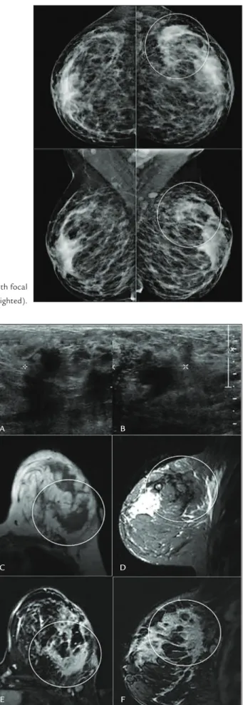
Mammogram revealed heterogeneously dense breasts and focal asymmetry in upper-outer quadrant of the left breast (Figure 1). Ultrasonography revealed heteroge-neous, predominantly hypoechoic areas, with indistinct margins and increased echogenicity of the adjacent tis-sues in the upper-outer quadrant of the left breast, with internal vascularization on color Doppler (Figure 2). Ax-illary and intramammary lymph nodes were found, with unusual appearance, rounded and hypoechoic with pre-served dimensions and reactive appearance.
inten-GRAZIANO L ETAL.
304 REV ASSOC MED BRAS 2016; 62(4):303-306
FIGURE 2 Ultrasonography showing a heterogeneous,
predominan-tly hypoechoic area, with indistinct margins, in the upper-outer quadrant of the left breast (A and B). MRI showing an area of non-mass enhancement with regional distribution occupying the lateral quadrants of the left breast (highlighted), with low signal intensity on axial T1 (C) and sagittal T2 (D) scans, presenting an heterogeneous internal enhancement pattern (E – axial; F – sagittal).
FIGURE 1 Mammogram revealing heterogeneously dense breasts, with focal
asymmetry in the upper-lateral quadrant of the left breast (highlighted).
A B
C D
IMAGINGFEATURESOFIDIOPATHICGRANULOMATOUSMASTITIS– CASEREPORT
REV ASSOC MED BRAS 2016; 62(4):303-306 305
FIGURE 3 MRI (A) showing a mass enhancement at the upper quadrants of the right breast (arrow). Targeted ultrasound (B) revealing a
heterogeneous isoechoic mass in the same topographic of the MRI inding.
sity on T1 and T2 (Figure 2). MRI also revealed a mass enhancement at the upper quadrants of the right breast, which was subsequently identified in targeted ultrasound (Figure 3).
The patient underwent ultrasound-guided percuta-neous core needle biopsy on both breasts, which showed marked chronic inflammation associated with granulo-mas and microabscesses in breast lobules, more evident to the left. No evidence of malignancy was found. Search for etiological agents using Grocott, PAS and Ziehl-Neelsen special staining yielded negative results. Thus, the final pathological diagnosis was idiopathic granulomatous mastitis.
D
ISCUSSIONThe case described above is typical of idiopathic granu-lomatous mastitis in a patient with recent pregnancy his-tory, who showed prolonged symptoms of a palpable mass in the breast associated with inflammatory signs, and no improvement after treatment with antibiotics. This diag-nostic possibility should always be remembered in such cases because the clinical and imaging findings can mim-ic malignancy. Bilateral involvement is unusual and may be associated with increased risk of recurrence and resis-tance to medical treatment.6
The most common mammographic finding is focal asymmetry, not associated with distortions or microcal-cifications; nodules with defined margins are less com-mon. Sonographic findings include one or more irregular hypoechoic masses associated with increased echogenici-ty of the parenchyma without posterior acoustic shadow-ing. Doppler study showed an increase of vascularization. These imaging findings are nonspecific and mimic breast carcinoma.1,2,4
These findings on MRI are little known because there are few reports in the literature. The most common is a non-mass enhancement with the following distribution:
segmental, ductal and regional. The most common mass findings are irregular margins with intense post-con-trast peripheral enhancement. The kinetic curve is usu-ally suspicious. However, the morphological findings of lesions often do not distinguish them from malignant lesions.7 In the case above, MRI showed involvement of
the contralateral breast that had not initially been iden-tified on conventional tests.
As described above, diagnostic confirmation can be done by percutaneous core needle biopsy, which is more accurate than fine-needle aspiration.2,5 Microscopic
anal-ysis showed mixed inflammatory cell infiltrate (mononu-clear and polymorphonu(mononu-clear), abundant with histiocytes within and outside of the breast lobules, non-caseating granulomas and microabscesses (neutrophil cluster), and absence of infectious etiologic agents.2,3
Prognosis and treatment depend on the disease pre-sentation. Steroid therapy is indicated for extensive cases, but not for localized forms. Surgical removal is indicated in refractory cases, but may be associated with the occur-rence of fistulas and scars. Recuroccur-rence decreases when the surgical margins are not compromised. The course of the disease is usually long with a significant impact on qual-ity of life. Prognosis is good; however, sometimes there is delay in the improvement of the disease, and possible re-currences and interventions can leave sequelae.1,2,8
There-fore, knowing this pathological entity, its clinical course and imaging findings is important for proper medical management, since this is a benign entity.
R
ESUMOAspectos de imagem na mastite granulomatosa idiopática – Relato de caso
A mastite granulomatosa idiopática é uma afecção rara e de etiologia desconhecida. Essa doença ocorre
GRAZIANO L ETAL.
306 REV ASSOC MED BRAS 2016; 62(4):303-306
mente em mulheres jovens e frequentemente após o pe-ríodo de lactação. As mulheres apresentam clinicamente massa endurecida, fixa, dolorosa, poupando a região re-troareolar, associada a espessamento cutâneo, podendo ulcerar, simulando carcinoma. Descarga papilar pode es-tar presente e o envolvimento bilateral pode ocorrer em até 25% dos casos. Neste relato, apresentamos um caso típico de mastite granulomatosa idiopática, com confir-mação histológica, destacando os aspectos de imagem, incluindo a ressonância magnética (RM), que possam fa-vorecer o diagnóstico e possibilitar um melhor manejo clínico dessas pacientes.
Palavras-chave: mama, mastite, mastite granulomatosa, imagem por ressonância magnética.
R
EFERENCES1. Gautier N, Lalonde L, Tran-Thanh D, El Khoury M, David J, Labelle M, et al. Chronic granulomatous mastitis: imaging, pathology and management. Eur J Radiol. 2013; 82(4):e165-75.
2. Larsen LJH, Peyvandi B, Klipfel N, Grant E, Iyengar G. Granulomatous lobular mastitis: imaging, diagnosis, and treatment. Am J Roentgenol. 2009; 193(2):574-81.
3. Ocal K, Dag A, Turkmenoglu O, Kara T, Seyit H, Konca K. Granulomatous mastitis: clinical, pathological features, and management. Breast J. 2010; 16(2):176-82.
4. Cheng L, Reddy V, Solmos G, Watkins L, Cimbaluk D, Bitterman P, et al. Mastitis, a radiographic, clinical, and histopathologic review. Breast J. 2015; 21(4):403-9. 5. Aghajanzadeh M, Hassanzadeh R, Alizadeh Sefat S, Alavi A, Hemmati H, Esmaeili
Delshad MS, et al. Granulomatous mastitis: presentations, diagnosis, treatment and outcome in 206 patients from the north of Iran. Breast. 2015; 24(4):456-60. 6. Velidedeoglu M, Kilic F, Mete B, Yemisen M, Celik V, Gazioglu E, et al. Bilateral
idiopathic granulomatous mastitis. Asian J Surg. 2016; 39(1):12-20. 7. Yildiz S, Aralasmak A, Kadioglu H, Toprak H, Yetis H, Gucin Z, et al. Radiologic