1109 Arq Neuropsiquiatr 2009;67(4):1109-1110
Letter
SpontaneouS carotid diSSection with
hypogloSSal nerve palSy aS reSidual deficit
The importance of magnetic resonance evaluation
Marcio Luiz Escorio Bezerra, José Luiz Pedroso, Alexandre Pieri
diSSecÇÃo eSpontÂnea de carÓtida coM paraliSia reSidual do nervo hipogloSSo: a iMportÂncia da avaliaÇÃo por reS-SonÂncia MagnÉtica
Department of Neurology, Federal University of São Paulo, São Paulo SP, Brazil. Received 16 February 2009, received in inal form 3 July 2009. Accepted 23 July 2009.
Dr. Marcio Luiz Escorio Bezerra – Rua Dr. Diogo de Faria 671 / 106 - 04037-002 São Paulo SP - Brasil. E-mail: marciobzrra@gmail.com
Spontaneous dissection of the carotid artery was once considered uncommon, but nowadays neuroimaging prog-ress has lead to an increased recognition of the pathology. It is an important cause of stroke in young patients, rep-resenting 10 to 25% of the cases1,2. Most patients
affect-ed are in the ifth decade of life2,3. It is not deinitely
es-tablished whether the cardiovascular risk factors are im-portant in spontaneous carotid dissection; however, to-bacco use, migraine and respiratory tract infections have been shown to be relevant in different studies1-4. In spite
of the term spontaneous dissection, minor precipitating events, such as rotation or hyperextension of the neck, are frequently found1,5. The most frequent initial
symp-tom is headache, which is reported in up to 90% of the cases, usually preceding the neurological deicit. Few pa-tients present the classical triad of headache, ischemic stroke and Horner syndrome. Cranial nerve palsy is found
in about 12% of the cases, but unusually it is the sole man-ifestation1-3.
We report a case of carotid artery dissection present-ing as lower cranial nerve palsy and headache. The diagno-sis was made through magnetic resonance imaging (MRI), using fat suppression technique, highlighting its impor-tance in such cases.
caSe
A 45 year-old right handed man with previous histo-ry of systemic hypertension experienced severe right cer-vical pain followed by pain in the ipsilateral ear. He re-ported no traumatic or strenuous event in the previous days. The pain remitted with regular analgesics, but two days later it returned with the same qualities, what made the patient seek medical counseling. The systemic blood pressure was high, and analgesics and antihypertensive
Arq Neuropsiquiatr 2009;67(4)
1110
Spontaneous carotid dissection Bezerra et al.
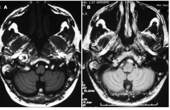
medications were administered. On this same day, he felt that his tong was numb on the right posterior area and he noticed dificulty in manipulating food in the mouth. The next morning he woke up with tong paresthesia, dys-phagia and dysarthria. He was then taken to the hospital. Upon admission, general examination was unremarkable. Neurological examination disclosed right hypoglossal pal-sy, in addition to ipsilateral palsy of the glossopharynge-al and vagus nerves. Craniglossopharynge-al CT showed no abnormglossopharynge-alities and brain MRI was highly suggestive of right carotid ar-tery dissection (Fig 1A). Carotid and vertebral Doppler ul-trasonography was also performed, but showed no abnor-malities. Antiplatelet therapy was started. Four months later a second MRI and MR angiography showed no signs of dissection (Fig 1B). Follow-up clinical examination de-tected a mild hypoglossal palsy (Fig 2), but there was no longer dysarthria or dysphagia.
diScuSSion
Lower cranial palsy has a large range of differential diagnoses, such as malignant tumors in the base of the skull, neurinomas, leptomeningeal carcinomatosis, infec-tious diseases or may even be idiopathic6,7, but carotid or
vertebral dissection should always be considered. The ab-sence of ischemic stroke and the fact that clinically it pre-sented as lower nerve palsy, may have delayed the diag-nosis in this case.
Carotid dissection usually arises from an intimal tear of the artery. The intramural hematoma may grow towards the intima, which cause stenosis of the arterial lumen or towards the adventicia, resulting in aneurismal dilatation1.
Considering that there were only local symptoms
(crani-al nerve p(crani-alsies) and that brain MRI showed only intra-mural hematoma and no vascular lumen abnormalities, it was probably an adventicia dissection. In contrast, with the intimal tear, the vascular lumen is affected and tends to cause distal ischemia due to embolization.
The gold standard diagnostic method for carotid dis-section is angiography, which is limited by its invasive-ness. This case highlights the importance of MRI with MR angiography as a diagnostic method. Its resolution approaches the conventional angiography and is able to show intramural hematoma, especially when fat suppres-sion techniques are used8. The complementary exam also
showed that the dissection extended to the petrous por-tion of the carotid artery, which is an uncommon event because usually the temporal bone represents a barrier to the pathologic process1. Also, it became evident that
even thought ultrasonographic techniques are useful, as the dissection is a dynamic process, the abnormalities may appear only in an initial moment9.
The follow-up MRI with no abnormalities emphasizes the dissection as a transitory process with a good prog-nosis3. Treatment involves antithrombotic or antiplatelet
therapy, as was the case in our patient. Systemic antico-agulation may bring some beneits by reducing the risk of embolization and aiding intimal healing, but there is no conclusive evidence in current literature for one choice over the other1-3,10.
referenceS
1. Schievink WI. Spontaneous dissection of the carotid and vertebral ar-teries. N Engl J Med 2001;344:898-906.
2. Campos CR, Evaristo EF, Yamamoto FL, Pugli Jr P, Lucato LT, Scaff M. Dissecção espontânea cervical carotídea e vertebral. Arq Neuropsiquiatr 2004;62:492-498.
3. Pieri A, Spitz M, Valiente RA, Avelar WM, Silva GS, Massaro AR. Spon-taneous carotid and vertebral arteries dissection in a multiethnic pop-ulation. Arq Neuropsiquiatr 2007;65:1050-1055.
4. Grau AJ, Brandt T, Buggle F, et al. Association of cervical artery dissec-tion with recent infecdissec-tion. Arch Neurol 1999;56:851-856.
5. Norris JW, Beletsky V, Nadareishvili ZG, Canadian Stroke Consor-tium. Sudden neck movement and cervical artery dissection. CMAJ 2000;163:38-40.
6. Combarros O, Alvarez de Arcaya A, Berciano J. Isolated unilateral hy-poglossal nerve palsy: nine cases. J Neurol 1998;245:98-100. 7. Schoenen J, Sándor PS. Headache with focal neurological signs or
symp-toms: a complicated diferential diagnosis. Lancet Neurol 2004;3:237-245. 8. Rizzo L, Crasto GS, Savio D, et al. Dissection of cervicocephalic arter-ies: early diagnosis and follow-up with magnetic resonance imaging. Emerg Radiol 2006;12: 254–265.
9. Benninger DH, Georgiadis D, Gandjour J, Baumgartner RW. Accuracy of color Duplex ultrasound diagnosis of spontaneous carotid dissection causing ischemia. Stroke 2006;37:377-381.
10. Redekop GJ. Extracranial carotid and vertebral artery dissection: a re-view. Can J Neurol Sci 2008;35:146-152.