13 artigo 039
OrigiNAl ArticlE
cOmparative anatOmical study betWeen the right and left
sides Of the axillary nerve in relatiOn tO deltOpectOral
apprOach and acrOmiOn
Antonio Carlos Tenor Junior1, Fabiano Rebouças Ribeiro1, Rômulo Brasil Filho1, Cantídio Salvador Filardi Filho2, Hilton Vargas Lutfi3, Eduardo Angoti Magri1
The authors declare that there was no conflict of interest in conducting this work
1 – Attending Physicians in the Shoulder and Elbow Group, Orthopedics and Traumatology Service of the State of São Paulo Public Servants’ Hospital (HSPE), São Paulo, Brazil. 2 – Head of the Shoulder and Elbow Group, Orthopedics and Traumatology Service of the State of São Paulo Public Servants’ Hospital (HSPE), São Paulo, Brazil. 3 – Trainee Physician in the Shoulder and Elbow Group, Orthopedics and Traumatology Service of the State of São Paulo Public Servants’ Hospital (HSPE), São Paulo, Brazil.
Work performed in the Orthopedics and Traumatology Service of the State of São Paulo Public Servants’ Hospital (HSPE). Correspondence: Rua Francisco Leitão, 474, apto. 92 – Pinheiros – 05414-020 – São Paulo, SP. E-mail: [email protected]
Work received for publication: March 01, 2009; accepted for publication: April 21, 2009. AbStrAct
Objective: To establish anatomical parameters for the axillary nerve by measuring the distances to the acromion and the deltopectoral access, and to ascertain whether there are any differences in comparative measurements between the left and right sides. Method: An anatomical study on the path of the axillary nerve was conducted by dissecting 30 shoulders of 20 fresh adult cadavers. For comparative study, bilateral dissection was performed on 10 cadavers. Digital caliper gauges, accurate to the nearest 0.05 cm, were used to measure the distances between the lateral extremity of the acromion and the anterior and posterior branches of the axillary nerve, and between the deltopecto-ral space and the anterior branch of the axillary nerve. Re-sults: The shortest distance between the acromion and the axillary nerve was 5.47 cm, and the greatest distance was
7.06 cm. The shortest distance between the deltopectoral groove and the axillary nerve was 3.94 cm. A statistically significant difference was found using Wilcoxon’s test in comparative measurements between the left and right si-des for the distances between the posterior branch of the axillary nerve and the midpoint of the lateral border of the acromion (A-E), and between the anterior branch of the axillary nerve and the anterior extremity of the acromion (B-C), both of which were larger on the right side. Con-clusions: The axillary nerve was situated between 5.47 and 7.06 cm distally to the acromion, and 3.94 cm laterally to the deltopectoral space. There was a statistically significant difference in the comparison between the left and right sides, and both measurements were larger on the right side.
Keywords – Shoulder; Brachial Plexus; Measures; Cadaver
Rev Bras Ortop. 2011;46(3):309-14
iNtrOdUctiON
The axillary nerve originates from the fibers of the fifth and sixth cervical roots, which form the posterior fascicle of the brachial plexus. Its proximal portion is located laterally to the radial nerve, posteriorly to the axillary artery and anteriorly to the subscapular mus-cle. It emerges through the quadrangular space formed medially and laterally by the brachial triceps muscle, superiorly by the teres minor muscle and inferiorly by the teres major muscle, accompanied by the posterior humeral circumflex artery. It comes into contact with
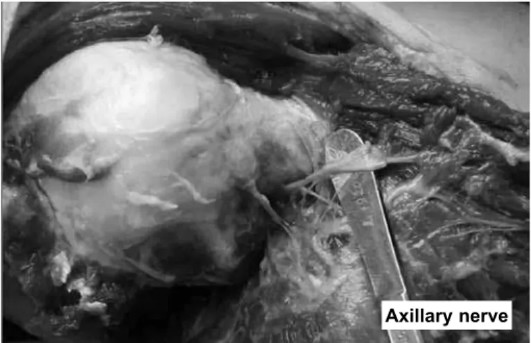
and the clavicle, and was pulled back laterally Figure 2). The axillary nerve and its branches were dissected from their emergence through the quadran-gular space to the point at which its fibers penetrate the belly of the deltoid muscle (Figure 2).
Figure 1 – horizontal incision, parallel to the later margin of the
acromion.
Deltoid muscle
Figure 2 – Deltoid muscle deinserted from the acromion and cla
-vicle, and pulled back laterally; axillary nerve dissected starting from its emergence through the quadrangular space.
Axillary nerve
Rev Bras Ortop. 2011;46(3):309-14
According to Steimann et al, the anterior bran-ch of the axillary nerve is located between 3 and 7 cm inferiorly to the lateral margin of the acromion(3). Bono et al(4) measured the distance between the most cranial point of the head of the humerus and the point where the axillary nerve divides into its anterior and posterior branches and found that this distance was 6.01 ± 0.7 cm. According to Burkhead et al(2), the axillary nerve is located 5 cm from the lateral margin of the acromion.
Certain procedures may cause damage to the axillary nerve, and these include: proximal intrame-dullary locking nails for the humerus; fixation with percutaneous wires and screws; fixation with plates; arthroscopic portals; intramuscular injections; and even poor positioning of the spacers during surgical approaches to the proximal humerus(5,6).
The aims of the present study were:
1) To establish a “safety zone” for the axillary ner-ve, by measuring its distance from the acromion and from the deltoid-pectoral access route;
2) To investigate whether there is any difference in comparative measurements between the right and left sides.
MAtEriAl ANd MEthOdS
Material – Between March 2004 and August 2005, 30 shoulders without previous scars from 20 fresh adult cadavers were dissected at the São lo Autopsy Service, at the University of São Pau-lo, with approval for the work from the Research Ethics Committee. Fifteen cadavers were male and five were female. Their weights ranged from 48 to 80 kg, with a mean of 63.05 kg, and their heights ranged from 1.60 to 1.90 m, with a mean of 1.74 m. Their ages ranged from 44 to 61 years, with a mean of 53 years (Tables 1 and 2).
Method – The cadavers were positioned in dorso-lateral decubitus, with the shoulder in neutral rotation,
the elbow flexed at 90˚ and the forearm in mid-range
pronosupination.
A horizontal access was used, parallel to the lateral margin of the acromion. The skin and subcutaneous tissue were pulled back and the deltoid muscle was exposed (Figure 1). The deltoid muscle was totally deinserted from the lateral margin of the acromion
The following points were marked out using 12 x 7 mm needles:
1) On the axillary nerve:
A – at the humerus, 2 cm laterally to its emergence through the quadrangular space, using a Codman® pen (Figure 3).
B – at the belly of the deltoid muscle, in the most proximal portion of its anterior branch in relation to the acromion (Figure 4).
311
DELTOPECTORAL APPROACH AND ACROMION
Rev Bras Ortop. 2011;46(3):309-14
3) On the deltopectoral groove:
F – at the point most proximal to point B (Figure 4). Using a Mistainless® digital caliper gauge, the following distances were measured:
A – C (Figure 6).
A – D (Figure 6).
A – E (Figure 6).
B – C (Figure 6).
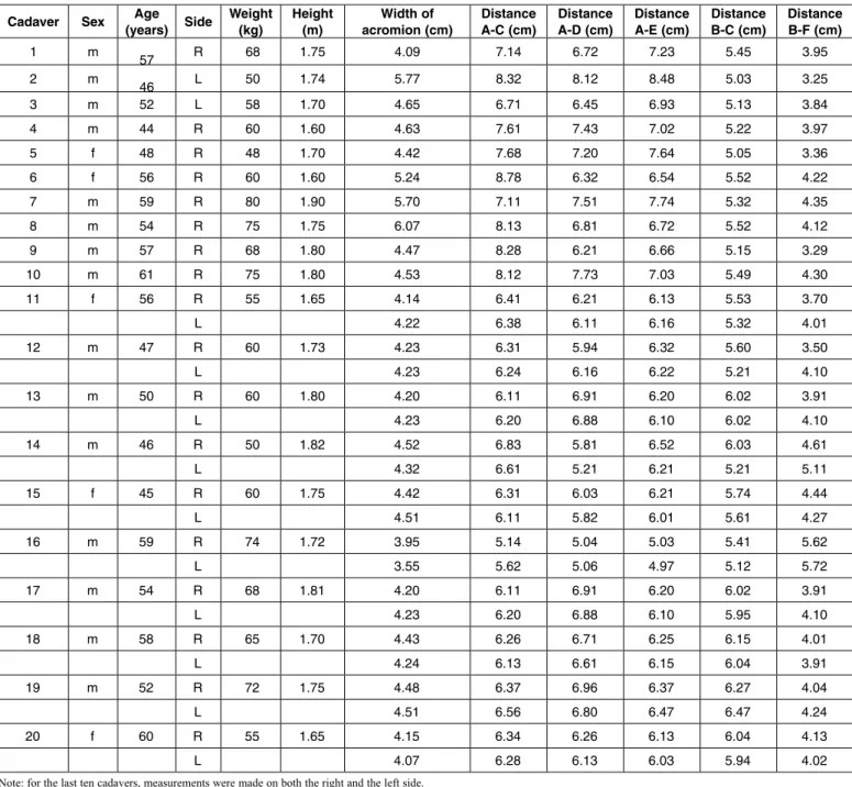
B – F (Figure 7). table 1 – Data and results obtained.
Note: for the last ten cadavers, measurements were made on both the right and the left side.
table 2 – Biometrics on the cadavers.
*95% CI: 95% confidence interval for the study population; LL: lower limit of the confi-dence interval; UL: upper limit of the conficonfi-dence interval.
cadaver Sex (years) SideAge Weight (kg) height (m) acromion (cm)Width of distance A-c (cm) distance A-d (cm) distance A-E (cm) distance b-c (cm) distance b-F (cm)
1 m
57 R 68 1.75 4.09 7.14 6.72 7.23 5.45 3.95 2 m
46 L 50 1.74 5.77 8.32 8.12 8.48 5.03 3.25
3 m 52 L 58 1.70 4.65 6.71 6.45 6.93 5.13 3.84
4 m 44 R 60 1.60 4.63 7.61 7.43 7.02 5.22 3.97
5 f 48 R 48 1.70 4.42 7.68 7.20 7.64 5.05 3.36
6 f 56 R 60 1.60 5.24 8.78 6.32 6.54 5.52 4.22
7 m 59 R 80 1.90 5.70 7.11 7.51 7.74 5.32 4.35
8 m 54 R 75 1.75 6.07 8.13 6.81 6.72 5.52 4.12
9 m 57 R 68 1.80 4.47 8.28 6.21 6.66 5.15 3.29
10 m 61 R 75 1.80 4.53 8.12 7.73 7.03 5.49 4.30
11 f 56 R 55 1.65 4.14 6.41 6.21 6.13 5.53 3.70
L 4.22 6.38 6.11 6.16 5.32 4.01
12 m 47 R 60 1.73 4.23 6.31 5.94 6.32 5.60 3.50
L 4.23 6.24 6.16 6.22 5.21 4.10
13 m 50 R 60 1.80 4.20 6.11 6.91 6.20 6.02 3.91
L 4.23 6.20 6.88 6.10 6.02 4.10
14 m 46 R 50 1.82 4.52 6.83 5.81 6.52 6.03 4.61
L 4.32 6.61 5.21 6.21 5.21 5.11
15 f 45 R 60 1.75 4.42 6.31 6.03 6.21 5.74 4.44
L 4.51 6.11 5.82 6.01 5.61 4.27
16 m 59 R 74 1.72 3.95 5.14 5.04 5.03 5.41 5.62
L 3.55 5.62 5.06 4.97 5.12 5.72
17 m 54 R 68 1.81 4.20 6.11 6.91 6.20 6.02 3.91
L 4.23 6.20 6.88 6.10 5.95 4.10
18 m 58 R 65 1.70 4.43 6.26 6.71 6.25 6.15 4.01
L 4.24 6.13 6.61 6.15 6.04 3.91
19 m 52 R 72 1.75 4.48 6.37 6.96 6.37 6.27 4.04
L 4.51 6.56 6.80 6.47 6.47 4.24
20 f 60 R 55 1.65 4.15 6.34 6.26 6.13 6.04 4.13
L 4.07 6.28 6.13 6.03 5.94 4.02
Weight
(kg) height (m) (kg/mbMi2 )
Mean 63.05 1.74 20.92 Standard deviation 9.23 0.08 2.73
Median 60.00 1.75 20.87 Minimum 48.00 1.60 15.09 Maximum 80.00 1.90 25.01
Figure 3 – Mark-up of the axillary nerve on the humerus, 2 cm laterally to its emergence through the quadrangular space.
Deltoid muscle
Figure 4 – Mark-up of the anterior branch of the axillary nerve, at the end that is most proximal to the acromion (b), and on the del-topectoral groove (f) at the point that is most proximal to point b.
F
Figure 5 – Mark-up of the lateral margin of the acromion: anterior extremity (c) and posterior extremity (d), and the
mi-dpoint between the extremities (E).
Axillary nerve
b
C
E
D
Figure 7 – Measurement of the distance b-f, using a digital
caliper gauge.
F
b
Figure 6 – Measurement of the distances A-c, A-d, A-E and b-c, using a digital caliper gauge.
A
b C
E
D
rESUltS
The data and the results obtained are in Table 1 and the statistical analysis is in Tables 2, 3 and 4.
Anterior branch – The distance between the acro-mion and the anterior branch of the axillary nerve ranged from 5.03 to 6.47 cm, with a mean of 5.23 cm. The standard deviation was 4.09 cm. Comparative analysis between the right and left sides showed that the difference between the means of the measure-ments was 0.19 cm (p = 0.027).
Posterior branch – The distance between the an-terior extremity of the acromion and the posan-terior branch of the axillary nerve ranged from 5.14 to 8.78 cm, with a mean of 6.5 cm. The standard deviation was 8.79 cm. Comparative analysis between the ri-ght and left sides showed that the difference between
313
the means of the measurements was 0.14 cm (p = 0.922). The distance between the posterior extremi-ty of the acromion and the posterior branch of the axillary nerve ranged from 5.04 to 8.12 cm, with a mean of 6.50 cm. The standard deviation was 7.34 cm. Comparative analysis between the right and left sides showed that the difference between the means of the measurements was 0.65 cm (p = 0.084). The distance between the midpoint of the anterior and posterior extremities of the acromion and the posterior branch of the axillary nerve ranged from 4.97 to 8.48
cm, with a mean of 6.46 cm. The standard deviation was 7.05 cm. Comparative analysis between the right and left sides showed that the difference between the means of the measurements was 0.1 cm (p = 0.014).
Deltopectoral space – The distance between the anterior branch of the axillary nerve and the delto-pectoral space ranged from 3.25 to 5.72 cm, with a mean of 4.14 cm. The standard deviation was 5.60 cm. Comparative analysis between the right and left sides showed that the difference between the means of the measurements was 0.56 cm (p < 0.065).
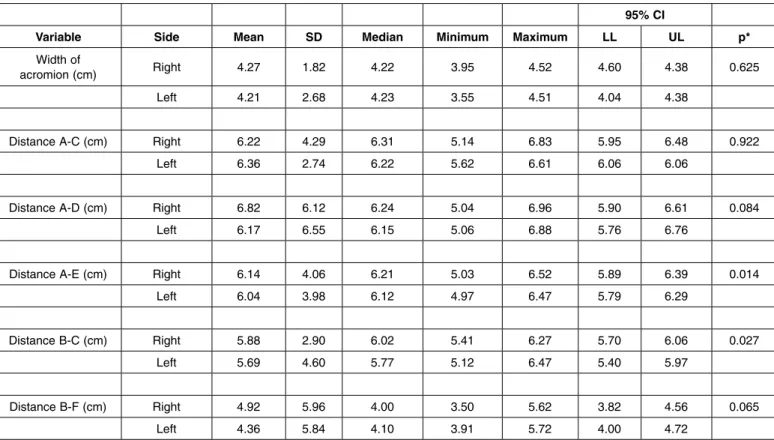
table 3 – Statistical analysis on the results obtained.
*95% CI: 95% confidence interval for the study population; LL: lower limit of the confidence interval; UL: upper limit of the confidence interval.
table 4 – Comparative analysis between the right and left sides (last ten cadavers).
* Descriptive probability level of the Wilcoxon nonparametric test7.
Rev Bras Ortop. 2011;46(3):309-14
Width of
acromion (cm) distance A-c (cm) distance A-d (cm) distance A-E (cm) distance b-c (cm) distance b-F (cm)
Mean 4.48 6.50 6.50 6.46 5.23 4.14
Standard deviation 5.44 8.79 7.34 7.05 4.09 5.60
Median 4.37 6.37 6.53 6.23 5.52 4.0.7
Minimum 3.55 5.14 5.04 4.97 5.03 3.25
Maximum 6.07 8.78 8.12 8.48 6.47 5.72
*95% cI LL 4.28 6.43 6.24 6.21 5.47 3.94
UL 4.67 7.06 6.76 6.71 5.76 4.34
95% ci
variable Side Mean Sd Median Minimum Maximum LL UL p*
Width of
acromion (cm) Right 4.27 1.82 4.22 3.95 4.52 4.60 4.38 0.625
Left 4.21 2.68 4.23 3.55 4.51 4.04 4.38
distance A-c (cm) Right 6.22 4.29 6.31 5.14 6.83 5.95 6.48 0.922
Left 6.36 2.74 6.22 5.62 6.61 6.06 6.06
distance A-d (cm) Right 6.82 6.12 6.24 5.04 6.96 5.90 6.61 0.084
Left 6.17 6.55 6.15 5.06 6.88 5.76 6.76
distance A-E (cm) Right 6.14 4.06 6.21 5.03 6.52 5.89 6.39 0.014
Left 6.04 3.98 6.12 4.97 6.47 5.79 6.29
distance b-c (cm) Right 5.88 2.90 6.02 5.41 6.27 5.70 6.06 0.027
Left 5.69 4.60 5.77 5.12 6.47 5.40 5.97
distance b-f (cm) Right 4.92 5.96 4.00 3.50 5.62 3.82 4.56 0.065
Left 4.36 5.84 4.10 3.91 5.72 4.00 4.72
diScUSSiON
Studying the path of the axillary nerve and esta-blishing its location in relation to the anatomical pa-rameters used here may help surgeons to avoid inju-ring the nerve duinju-ring open surgical procedures. Such injuries could occur if the access route lies within the safety zone, or because of inadequate or prolonged positioning of spacers on the deltoid muscle.
In arthroscopy, the axillary nerve may also be injured, in positioning an accessory distal posterior arthroscopic portal, at the point where it comes out from the quadrangular space and turns anteriorly, approximately 5 cm distally to the acromion, through using heat-generating instruments on the lower capsule. This may also occur while removing lower humeral spurs.
According to Albritton et al(6), the mean distance between the axillary nerve and proximal locking screw of the intramedullary nail used is 0.26 cm, and nerve injuries may occur during the proximal locking, in treating fractures of the humerus. According to these authors, the risk of injury is greater for smaller humerus bones. For greater safety, they recommended that the deltoid muscle should be dissected and the lateral cortical bone of the humerus should be brought into direct view, with use of soft-tissue protection.
Meireles et al(5) reported a case of axillary nerve injury while applying an injection to the deltoid mus-cle. In our study, the anterior branch of the axillary nerve was located at a distance of between 5.47 cm and 7.06 cm from the lateral margin of the acromion.
This is precisely the location recommended in the past for application of intramuscular injections, which makes this now-proscribed technique dangerous(5,7).
We agree with Ferreira Filho et al(1), who demons-trated that the axillary nerve can be injured in cases of lateral access through the deltoid muscle, in proce-dures that require greater surgical exposure. Surgeons need to know how far it is possible to expand the operative incision caudally without compromising the integrity of the axillary nerve(8).
In orthopedic surgery, the deltopectoral access rou-te is used for a variety of pathological conditions of the shoulder, such as osteosynthesis of fractures, ar-throplasty and repairs to labral lesions(8). The axillary nerve needs to be protected, thereby avoiding paraly-sis of the deltoid muscle and functional impairment of the shoulder(1,9-12). Knowledge of its path and anato-mical relationships is important in this respect(1,12-14). In a study carried out on 67 cadavers at the Univer-sity of Crete, in Greece, Kontakis et al(15) found three cases (two women) in which there was a difference of 1-2 cm in the position of the axillary nerve between the right and left sides.
cONclUSiONS
The axillary nerve was located between 5.47 and 7.06 cm distally to the acromion and 3.94 cm laterally to the deltopectoral space. There was a statistically significant difference in comparing between the right and left sides, such that both distances were greater on the right side.
rEFErENcES
1. Ferreira Filho AA, Suzuki JM, Ferreira M. O nervo axilar na abordagem cirúrgica do ombro. Rev Bras Ortop 1989; 24(5):176-8.
2. Burkhead W, Scheinberg R, Box G. Surgical anatomy of the axillary nerve. J Shoulder Elbow Surg. 1992;1(1):31-6.
3. Steinmann SP, Moran EA. Axillary nerve injury: diagnosis and treatment. J Am Acad Orthop Surg. 2001;9(5):328-35.
4. Bono M, Grossmam MG, Hochwald N, Tornetta P. Radial and axillary ner-ves: anatomic considerations for humeral fixation. Clin Orhtop Relat Res. 2000;(300):259-64.
5. Meirelles H, Motta Filho GR. Lesão do nervo axilar causada pela injeção in-tramuscular no deltóide: relato de caso. Rev Bras Ortop. 2004;39(10):615-9.
6. Albritton MJ, Barnes CJ, Karas SG. Relationship of the axillary nerve to the proximal screws of a flexible humeral nail system: an anatomic study. J Orthop Trauma. 2003;17(6):411-4
7. Pery AG, Potter PA. Clinical nursing skills and technique. St. Louis: Mosby; 1998. p. 631.
8. Rocha RP, Lenza MV, Lenza M, Mongon ML, Ribeiro FB, Braga EV, et al.
Relações do nervo axilar com o músculo axilar: sua importância no acesso delto-peitoral. Rev Bras Ortop 2002;37(3):79-82.
9. Bryan WJ, Schauder R, Tullos HS. The axillary nerve and its relationship to com-mom sport medicine shoulder procedures. Am J Sports Med. 1986;14(2):113-6.
10. Flatow EL, Bigliani LU. Tips of the trade. Locating and protecting the axillary nerve in shoulder surgery: the tug test. Orthop Rev. 1992;21(4):503-5.
11. Loomer R, Graham B. Anatomy of axillary nerve and its relation to inferior caspsular shift. Clin Orthop Relat Res. 1989; (243):100-5.
12. Durval MJ, Parker AW, Drez D Jr, Hinton MA. The anterior humeral circumflex vessel and the axillary nerve: an anatomic study. Orthop Rev. 1993;22(9):1023-6.
13. Rowles DJ. McGrory JE. Percutaneous pinning of the proximal part of the humerus. An anatomic study. J Bone Joint Surg Am. 2001;83(11):1695-9.
14. Moran MC. Distal interlocking during intramedullary nailing of the humerus. Clin Orthop Relat Res. 1995;(317):215-8.
15. Kontakis, GM, Steriopoulos K , Damilakis J, Michalodimitrakis E. The position of the axillary nerve in the deltoid muscle: A cadaveric study. Acta Orthop. 1999;70(1):9-11.