artigo 291
CASE REPORT
The authors declare that they did not have any conflict of interests in producing this article
1 – Neurology Resident at the Hospital Santa Casa de Misericórdia de Belo Horizonte – Minas Gerais. 2 – Nephrology Resident at the Hospital Vera Cruz de Belo Horizonte – Minas Gerais.
3 – Student in the Fifth Year of Medicine of the Faculdade de Ciências Médicas de Minas Gerais.
4 – General Surgeon of the Instituto Mário Penna – Hospital Luxemburgo de Belo Horizonte – Minas Gerais. 5 – Surgery Resident at the Hospital LifeCenter and Hospital Vera Cruz de Belo Horizonte – Minas Gerais.
Work carried out at the Hospital Santa Casa de Belo Horizonte.
Correspondence:Rua Santo Antônio do Monte, 139/001 – Bairro Santo Antônio – 30330-220 – Belo Horizonte – MG. E-mail: [email protected].
Work received for publication: February 26, 2010; accepted for publication: April 12, 2010.
gianT cell TuMor in The proXiMal phalanX WiTh pulMonary
MeTasTasis: case reporT and liTeraTure reVieW
Frederico Carvalho de Medeiros1, Fernando Carvalho de Medeiros2, Izabella de Campos Carvalho Lopes3,
Guilherme Carvalho de Medeiros4, Eduardo Carvalho de Medeiros5
InTRODUCTIOn
Giant cell tumor (GCT) is a benign neoplasia in the majority of cases, and accounts for around 5% of primary bone tumors. It is generally located in the epiphysis and metaphysis of the long bones. In a large number of cases, it extends close to the arti-cular cortex, and its occurrence in the bones of the hand is rare. GCT occurs more frequently in the third and fourth decades of life, with a slight prevalence in women(1).
Clinically, its behavior is locally aggressive(1) and
the main complaint is pain, followed by increased limb size and functional impairment(2).In rare cases,
pulmonary metastasis occurs(1).
The diagnosis is established through clinical
correla-ABSTRACT
This is a case report on a giant cell tumor (GCT) in the proximal phalanx of the third finger of the left hand, with pulmonary metastasis. The patient presented pain in the finger without any previous history of trauma. Clinical examination, radiographic imaging and magnetic resonance imaging were carried out. A histological evaluation was carried out from an incisional biopsy, taking the hypothesis of GCT. The patient underwent amputation of the finger and the diagnosis was confirmed by means of microscopy on
the specimen. The patient was followed up because of the risk of lung metastasis, which was shown by radiographic examination and computed tomography on the chest, and thoracotomy was performed. Since then, there has been an improvement in the symptoms that had been reported preoperatively, and no local recurrence or new metastasis has been found.
Keywords – Giant cell tumors/surgery; Finger Phalanges; Amputation; Neoplasm Metastasis
tion, imaging exams and histopathological data to rule out other lesions with a similar histological pattern(1).
The treatment is surgical with a recurrence rate of around 5% to 10%(1).
The objective of this work is to report on a case of GCT in the proximal phalanx of the third finger of the left hand, which evolved to pulmonary metastasis.
CASE REPORT
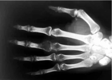
Figure 1 – Hypertransparency in the proximal phalanx of the third finger.
Figure 2 – Evolution of the lesion in one month.
Figure 3 – Amputation of the third finger, transposing the index finger to the resection site.
not taking any regular medication. On examination, there was pain on palpation in the proximal phalanx and metacarpophalangeal joint, and limited flexion and extension of the third finger.
The x-ray performed in May 2007 showed hyper-transparency with increased bone volume (Figure 1). After one month, the lesion showed significant growth (Figure 2).
imaging (MRI) showed a large expansive lesion in the two proximal thirds of the phalanx of the third finger of the left hand, with destructive appearance and clear margins, measuring at its widest points: 27.0 x 24.0 x 24.0 mm. Characterized by hyposignal in T1 and hypersignal in T2, containing areas with cystic appearance in the center. The adjacent extensor tendons and other bone and joint structures were preserved. At the end of July 2007, the patient was submitted to amputation of the third finger, transposing the index finger to the resection site (Figure 3).
The piece was sent for anatomapathological study, which confirmed the diagnosis of GCT. Macroscopic examination of the surgical piece revealed a whitish, elastic tumoration with extensive areas of hemorrha-ge, measuring 2.0 cm at its widest diameter to 5.0 cm from the proximal margin of the sample. The lesion presented impairment of the proximal phalanx, with destruction of the cortex and bone marrow, and inva-sion to the surrounding soft tissues. Microscopically, characteristics of incisional biopsy were apparent, with the presence of angiolymphatic tumor invasion, but without evidence of malignant sarcomatous trans-formation. Surgical margins free (Figure 4).
In August 2007, the patient underwent full-body bone scintigraphy revealing hyperemia and inflam-mation of the soft tissues of the left hand, compatible with non-specific post-surgical alterations, secondary to exeresis of the GCT. The scintigraphic pattern was not compatible with additional lesions of the under-In July 2007, incisional biopsy was performed,
207
Figure 4 – Microscopy of the giant cell tumor affecting the proximal phalanx.
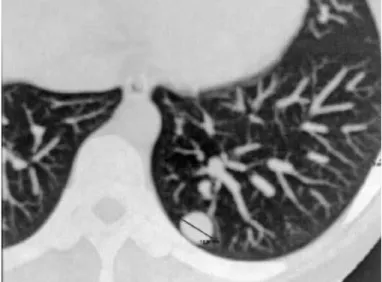
Figure 5 – Multislice computed tomography of the thorax with metastasis in the left lung.
Figure 6 – Pulmonary metastasis of intraparenchymal giant cell tumor of the bone.
AND LITERATURE REVIEW
lying pathology in the other skeletal segments (absen-ce of multi(absen-centric GCT).
In the postoperative evolution, the patient conti-nued to have free movements in the left hand, without complaints, and showed improvement with physio-therapy. During postoperative follow-up, a semestral radiological study was carried out which, 11 months after excision of the lesion, showed nodular forma-tions in both lungs, one in the right middle/upper third (around 5.0 mm) and the other in the left lower third (around 14.0 mm). Multislice computed tomography of the thorax was performed, showing three nodular images in pulmonary parenchyma with density of the soft tissues, one located in the anterior segment of the right upper lobe (6.0 mm) and two located in the posterior basal segment of the left lower lobe (15.0 and 4.0 mm) (Figure 5).
In July 2008, the patient was submitted to explo-ratory thoracotomy of the left lung, and in the follo-wing month, the same procedure was carried out in the right lung. Exeresis was performed of the four nodules on the left and six nodules on the right, all benign pulmonary metastasic GCT, without high de-gree transformation, as confirmed in the anatomopa-thological tests (Figure 6). The patient evolved well, without complaints, and is in tomographic control without evidence of local recurrence or metastasis in the lungs. She has resumed her normal activities and continues in follow-up: every three months in the first year, every six months up to five years after the treatment, and then annually after this period.
DISCUSSIOn
GCT of the bone is a benign neoplasia, locally aggressive, with uncertain biological behavior(1,3,4).
Approximately 5% to 10% of patients present malig-nant evolution(1).
GCT represents 5% to 8% of all primary bone tu-mors, and 20% of all benign bone tumors(1,3-7). Some
studies show a slight prevalence in women compared to men (ratio of 1.3:1)(1,4,5), and a prevalence in the
third and fourth decades of life(3,7,8). The lesion is rarely
found before fusion of the epiphyseal plate(3,7).
location
cases. Other locations include: the distal portion of the radius (10-12%), sacrum (4-9%), proximal humerus (4-8%), proximal femur (4%) and less frequently in the vertebral bodies (2.5%) and bones of the hands and feet(1,7).
The condition is rare in the bones of the hand (2-4%)(2,3,7,9,10).Out of 1547 cases of GCT of the bone
collected in various studies, only 70 were located in the hands or feet(11). It affects patients 10 years
young-er than the avyoung-erage age (32 years) for GCT occurring in other locations(11), with more aggressive behavior(9)
and earlier recurrence(2).The most common locations
are the metaphyseal region of the metacarpals and phalanges. The tumor grows in the direction of the diaphysis, which complicates the treatment(10).
Clinical aspects
The majority of patients with GCT are oligosymp-tomatic with localized pain, with insidious onset and progressive deterioration(1,4). The disease evolves with
increased volume and functional incapacity of the limb, particularly if the lesion is near the joint(2,3). However,
there may be acute pain as a result of pathological frac-ture(3,7,9). In some cases, the tumor grows quickly, with
thinning and rupture of the bone cortex and invasion of the adjacent soft tissues, but without invading and ulcerating the skin and subcutaneous cell tissue(10,11).
In the hand, the clinical aspect has peculiar characte-ristics. Pain and swelling occur sooner than in GCT in other locations, due to the fact that the tumor is close to the surface, and the high mobility of the fingers(11).
image
The classical radiographic aspects of GCT of the bone define it as an eccentric, swollen, lytic epiphy-seal-metaphyseal lesion, not always with clearly-de-fined margin(3,4,7,9). Its appearance is radiotransparent,
with thinning of the cortical bone and absence of in-ternal calcifications, marginal sclerosis, or periosteal reaction. However, there is no pathognomonic cha-racteristic(9). Although computed tomography (CT) is
of little use in the differential diagnosis, it confirms the absence of calcified matrix, shows the margins of the lesion, and reveals the extent of subcortical bone loss prior to surgical reconstruction(1). MRI is also
used in the preoperative evaluation(1). This is
consi-dered the best method for delineating intraosseous, extraosseous and intra-articular tumors(1,7). Its greatest
utility is for demonstrating recurrence of the tumor(1).
In relation to skeletal screening by bone scintigra-phy, GCT presents local hypercaptation due to the increased reactive osteoblastic activity, with broad concentration of the radioindicator in the lesion. In
up to 57% of the cases, the “donut sign” of central
photopenia is present. The exam should be carried out to search for other bone lesions(12,13).
The radiological classification of GCT of the bone was described by Enneking, and subsequently by Cam-panacci. There are three stages that correlate the local tumor according to its aggressiveness and risk of local recurrence(7,8). Stage 1 is the less common. The tumor
is small and the cortical layer is preserved. There is no extension to the joint cartilage. Symptoms are absent or minimal. Stage 2 corresponds to 75% of the GCT in its initial presentation. The tumor has extended to the cortex, which is deformed by the expansion of the tumor. There is no extension to the adjacent soft tissues. Stage 3 is characterized by extreme aggressiveness; the tumor is large in volume, destroying the bone and invading the surrounding tissues(7,8); as presented in
the case reported here. The highest rate of recurrence is observed in tumors that are in this phase(7,8).
Histomorphological aspects
In its morphological aspect, GCT of the bone is solid, large, brown in color, with yellow areas of necrosis and dark red areas of hemorrhage. In cases with slower evolution, there are areas of whitish, star-shaped fibrosis, with firm consistency at the core(14).
Histologically, the neoplasic tissue is highly vascula-rized, consisting of stroma with oval or fusiform cells, and numerous multinucleated giant cells, the nuclei of which are identical to those of the stroma cell(14).
The-re is little or no intercellular substrate, except for some collagen fibers. Mitotic figures may be numerous, but without abnormalities(7).
differential diagnosis
Differential diagnosis should be performed with tumors that contain giant cells, like aneurismal bone cyst, giant cell reparative granuloma and brown tumor of hyperparathyroidism. These pathologies present a similar radiological pattern. Through the histologi-cal findings, differentiation is possible, except with brown tumor of hypoparathyroidism, which requires biochemical studies for its diagnostic definition(7,8,11).
209
TREATMEnT
The treatment of GCT is surgical(1,4,5,10). The
con-duct should be well-planned and individualized, due to the unpredictable nature of the neoplasia in terms of its local prognosis(4). The greatest challenge is due to the
aggressive local behavior of the tumor and its tendency to recur(6,8). Compared with GCT in proximal locations,
tumors of the hand are presented in advanced stages with greater bone destruction and extension for diaphy-sis, which complicates the treatment. The objective of the treatment in the hands is to achieve local control of the tumor and avoid recurrences, preserve the functions of the hand, and maintain a good esthetic standard(3,10).
The types of treatment described in the literature are: curettage, curettage with bone graft, radiotherapy, am-putation and resection with reconstruction(5).
Curetta-ge, whether in isolation or associated with bone graft, is the most common form of treatment, but it presents a rate of recurrence of around 90%(2,8). Curettage is
not, therefore, the ideal intervention. Radiotherapy should also not be used as the primary procedure as it is associated with malignant transformation. Am-putation, although it reduces the recurrence rates, is esthetically disfiguring and causes loss of functionality of the limb(2,5). In the present case, this procedure was
carried out in order to prevent recurrence, due to the clinical condition presented. Resection with recons-truction of the articulation of the proximal phalanx with the metacarpus can be carried out with a bone graft, prosthesis or with polymethylacrilate, which re-establishes the structural and functional integrity of the metacarpophalangeal joint(2,5). These last two
forms of treatment are considered the treatments of choice in stage 3 lesions of the hand, where there is invasion of the surrounding soft tissues(8).
Prognosis
The rate of recurrence with the appropriate
surgical technique is approximately 5% to 10%(1,7)
and the majority occur around 12 to 18 months after the therapy(7). There is a study that reports a peak of
recurrence three years after surgery(1). In the hands,
the recurrence rate is higher(8,11), approximately
70%(11). A literature review indicates that patients with
recurrence should be carefully monitored due to the higher malignant potential of the recurrent disease, and the higher propensity to metastasis(3). Thus, due
to the small, but definite risk of pulmonary metastasis,
clinical follow-up and imaging exams are necessary for the rest of the patient’s life(15).
Pulmonary metastasis
The development of pulmonary metastasis is rare, its incidence varying between studies from 2% to 6%(7,16). In the series of Bertoni et al(15) involving 327
cases of GCT of the bone, only six presented pulmo-nary metastasis. They generally occur in the first years after discovery of the primary bone tumor, at around 3.5 years, on average(16).In relation to the surgical
procedure, the appearance of pulmonary metastasis presented an interval of 17 to 40 months, according to a study by Athanasian et al(17), and 16.4 months in
the series of Bertoni et al(15). The progression is
gene-rally slow(7). The increased incidence of pulmonary
metastasis is associated with the location of the pri-mary tumor in the distal radius, sacrum and hands, stage 3 lesions, aggressive local behavior, and multi-ple recurrences, characteristics of the tumor(3,7,10). The
clinical presentation (age, sex and tumor location) of GCT with pulmonary metastasis is similar to that of GCT without metastasis(15). The majority of pulmonary
lesions are asymptomatic, and are incidentally detected during follow-up of the tumor. However, some patients present pulmonary symptoms before the discovery of the primary tumor(16). Regular chest x-ray and CT are
therefore recommended(16).The histomorphological
findings of the pulmonary metastatic nodules present the same benign morphology as the primary tumor in terms of cellularity, abundance of osteoid, anaplasia and mitotic aspects(7,16,18).
In the search for metastasis, the x-ray shows no-dules of homogenous opacity, occasionally with peri-pheral calcification. Most of the nodules occur in the basal and peripheral regions of the lungs. CT is the most sensitive method and can help determine more accurately the number of lesions and whether there is mediastinal involvement. It is extremely important in the surgical planning for removal of the metastases(16).
are inaccessible to surgical treatment, rapidly recurring lesions, lesions with incomplete resection, or multiple lesions may respond to chemotherapy, including Interferon. High doses of Cisplatin, Cyclophosphamide, Methotrexate and Doxorubicin can prolong the disease-free time. The response is variable. Many patients die from the side effects of chemotherapy, such as secondary sepsis due to agranulocytosis(7,15,16). Interferon alpha-2
is a therapeutic option due to its antiangiogenic action, which results in a major reduction in tumor size(4).
Radiotherapy may also be used in these situations, despite the risks of malignant sarcomatous transformation(15,16).
This possibility of sending metastases is still a source of confusion for many authors, who start to consider GCT and treat it like a high malignity neoplasm. Commonly, descriptions of chemotherapy and even radiotherapy treatment are seen, in cases considered as malignization of a GCT. Benign histological analysis of the pulmonary nodule corroborates the affirmation that there is no malignant transformation of the GCT(19), therefore,
these two last therapies are little used, as a series of studies describes that the absence of treatment of the pulmonary metastasis from the GCT of bone does not reduce long-term survival(15,16,18).
Follow-up
Due to the high incidence of local recurrence and the risk of pulmonary metastasis, it is extremely
important to closely monitor these patients .
Follow-up after treatment should initially be done every three to four months and should include phy-sical and x-ray tests on the affected bone. This will enable any recurrence to be detected at an early stage. CT or MRI are useful for clarifying a suspected recur-rence. After two to three years, the follow-up should be every six months, and then annually after five years of treatment. Chest x-ray should be requested in each follow-up visit due to the risk of pulmonary metastasis. Chest CT can be performed on diagnosis to check for early signs of pulmonary metastasis since CT is more sensitive than x-ray. Subsequently, in the absence of lesions, the patient is followed-up with x-ray at the above-mentioned intervals(7).
COnCLUSIOn
GCT in the hand is a rare, benign, but locally aggressive entity. It evolves earlier than GCT in other locations. It is oligosymptomatic, with prevalence of pain and increased volume. It mainly affects young women, who are more subject to pulmonary metastasis. The diagnosis is based on the clinical, radiological and histopathological aspects. The treatment is surgical, but there is no consensus in the literature. Cases should be assessed individually to ensure safe intervention, seeking to prevent recurrences and functional limitations.
REFEREnCES
1. Gruenwald N, Demos TC, Lomasney LM, Rapp T. The case. Giant-cell tumor. Orthopedics. 2006;29(2):167-71.
2. Gadegone WM, Salphale YS, Sonwalker HA, Nagtode PP, Navghare SM. Giant cell tumor of proximal phalanx of ring finger in immature skeleton – Resection and reconstruction by iliac crest graft – A case report. J Orthopaedics [Internet Journal]. 2007 [quoted on 05/25/2009]; 4(2): [about 3p.] Available at: http://www. jortho.org/2007/4/2/e5.
3. Slesarenko YA, Sampson SP, Gould ES. Giant cell tumour of the distal phalanx of the hand. Hand Surg. 2005;10(2-3): 289-91.
4. Camargo OL, Croci AT, Oliveira CRGCM, Baptista AM, Caiero MT, Giannotti MA. Tumor de células gigantes – evolução histórica do seu diagnóstico e tratamento junto ao Instituto de Ortopedia e Traumatologia da FMUSP. Acta Ortop Bras. 2001;9(4):46-52.
5. Mendenhall WM, Zlotecki RA, Scarborough MT, Gibbs CP, Mendenhall NP. Giant cell tumor of bone. Am J Clin Oncol. 2006;29(1):96-9.
6. Boons HW, Keijser LCM, Schreuder HWB, Pruszezynski M, Lemmens JAM, Veth RPH. Oncologic and functional results after treatment of giant cell tumors of bone. Arch Orthop Trauma Surg. 2002;122(1):17-23.
7. Turcotte ER. Giant cell tumor of bone. Orthop Clin North Am. 2006;37(1):35-51.
8. Ropars M, Kaila R, Cannon SR, Briggs TWR. Primary giant cell tumours of the digital bones of the hand. J Hand Surg Eur. 2007;32 (2):160-4.
9. Biscaglia R, Bacchini P, Bertoni F. Giant cell tumor of the bones of the hand and foot. Cancer. 2000;88(9):2022-32.
10. Wittig JC, Simpson BM, Bickles J, Kellar-Graney KL, Malawer MM. Giant cell
tumor of the hand: superior results with curettage, cryosurgery, and cementation. J Hand Surg. 2001;26(3):546-55.
11. López-Barea F, Rodriguez-Peralto JL, García-Girón J, Guemes-Gordo F. Benign metastasizing giant-cell tumor of the hand: report of a case and review of the literature. Clin Orthop Relat Res. 1992;(274):270-74.
12. Catalan J, Fonte AC, Lusa JR, Oliveira AD, Melo ES, Júnior RO et al. Tumor de células gigantes ósseo: aspectos clínicos e radiográficos de 115 casos. Radiol Bras. 2006;39(2):119-22.
13. Cordeiro SZ, Cordeiro PB, Sousa AM, Lannes DC, Pierro GS. Tumor de células gigantes costal ocupando todo o hemitórax. J Bras Pneumol. 2008;34(3): 185-8. 14. Andrade-Filho JS, Barbosa CSP, Aymoré IL, Lemos C. Sistema ósteo-articular. In:
Filho GB. Bogliolo patologia. 7a. ed. Rio de Janeiro: Guanabara; 2006. p. 1014-6.
15. Bertoni F, Present D, Sudanese A, Baldini N, Bacchini P, Campanacci M. Giant-cell tumor of bone with pulmonary metastases: six case reports and a review of the literature. Clin Orthop Relat Res. 1988;(237):275-85.
16. Cheng JC, Johnston JO. Giant cell tumor of bone: prognosis and treatment of pulmonary metastases. Clin Orthop Relat Res. 1997;(338):205-14.
17. Athanasian EA, Wold LE, Amadio PC. Giant cell tumors of the bones of the hand. J Hand Surg Am. 1997;22:91-8.
18. Seethalakshmi V, Jambhekar NA. Metastatic giant cell tumor of bone: are there associated factors and best treatment modalities? Clin Orthop Relat Res. [Inter-net Journal]. 2009 [quoted on 07/14/2009]: [about 7p.] Available at: http://www. springerlink.com/content/q263g3096648m181/.