Inflammation and poor response to treatment with
erythropoietin in chronic kidney disease
Authors
Wander Valadares de Oliveira Júnior1
Adriano de Paula Sabino2
Roberta Carvalho Figueiredo1
Danyelle Romana Alves Rios1
1 Universidade Federal de
São João Del-Rei.
2 Universidade Federal de
Minas Gerais.
Submitted on: 08/11/2014. Approved on: 02/06/2015.
Correspondence to: Danyelle Romana Alves Rios. Campus Centro Oeste Dona Lindu - Universidade Federal de São João Del-Rei.
Rua Sebastião Gonçalves Coelho, 400 Chanadour, Divinópolis, MG. CEP: 35501-296.
E-mail: danyelleromana@gmail. com
FAPEMIG.
I
NTRODUCTIONChronic kidney disease (CKD) is a serious public health problem and its incidence has increased in recent years. It is progressive and is associated with high morbidity and mortality rates.1
There are several conditions associated with CKD onset and progression. We stress the following: obesity, hypertension and diabetes mellitus.
In addition to these conditions, there are signs of inflammation in the pathophysiology of CKD.1 This disease
can cause numerous complications: cardiovascular disease (CVD) and anemia are the most prevalent and
DOI: 10.5935/0101-2800.20150039
The prevalence of kidney chronic disease (CKD) has increased in recent years and several risk factors have been associated with the onset and progression of CDK, such as obesity, hypertension and
diabetes mellitus. In addition, anemia is one of the complications of CRD, mainly by iron and erythropoietin (EPO) deficiency and the management of this situation is with exogenous erythropoietin, but patients undergoing dialysis present chronic inflammatory process followed by EPO resistance and anemia, malnutrition, worse of atherosclerosis and increased mortality ratio. The aim of this study was to review the association of erythropoietin resistance and chronic inflammatory process in patients with chronic renal disease.
A
BSTRACTKeywords: anemia; cytokines; erythro-poietin; inflammation; polymorphism, ge-netic; renal insufficiency, chronic.
severe, because they are associated with worse outcome and even death.2
Chronic disease anemia is charac-terized by the development of anemia in patients with chronic inflamma-tory diseases such as cancer, autoim-mune diseases, chronic infection and chronic kidney disease.3 This type of
anemia progresses with low serum iron concentrations, despite abundant amounts of iron present inside macro-phages.4 In the pathogenesis of chronic
disease anemia there are at least three prevalent mechanisms: changes in erythropoiesis, decreased survival of red blood cells and inadequate bone marrow response to hemolysis.3,5
Iron deficiency prevalence in CKD is around 50%, and together with the relative EPO deficiency, they represent the two key causes of anemia in CKD. This anemia is one of the complications of CKD, since there is evidence of inflammation in the pathophysiology of CKD and various cytokines and chemokines have been identified in the plasma and urine of patients in early stages of CKD - which are also related to disease complications.2
Thus, measurement of circulating levels of immune inflammatory mediators as well as the investigation of polymorphisms of the genes encoding these immune inflammatory mediators show that patients with CKD are in a pro-inflammatory state, according to the phenotype, which is more evident wherein the renal damage progresses to end-stages.7
Single nucleotide polymorphisms (SNPs) or microsatellites may alter transcription factor links to their internal sites of promoter genes, thus affecting the amount of cytokines produced. SNPs are single base variations in a sequence of one chromosome allele; whereas microsatellites are repeated sequences from one to four nucleotides scattered throughout the genome (GA, TC, GT, CA).8 These polymorphisms are responsible for
phenotypic variations associated with various diseases, and may stimulate cytokine production in a greater or lesser manner, which can lead to poor response to the use of EPO in patients with chronic disease anemia.8,9
This study aims to review the scientific evidence regarding the association of poor clinical response to EPO use and the presence of inflammation in CKD.
C
HRONIC KIDNEYDISEASEKidneys are vital organs for maintaining human body homeostasis and the progressive decline in renal function peaks with impairment in all other organs.10 CKD is defined as the presence
of structural or functional changes in the kidney, for a period longer than three months, with implications on the individual’s health. This disease is increasing epidemically in the world, especially due to the global increase in the prevalence of the main causes of CKD, such as hypertension, diabetes mellitus and obesity.11
Kidney function is measured by glomerular filtration rate (GFR) and its decrease is evident in CKD, coupled with the loss of kidney regulatory, excretory and endocrine functions. When the GFR reaches very low values, less than 15 ml/min/1.73 m2, it means the patient
developed functional renal failure, or end-stage
CKD and dialysis is recommended at this stage.12 According to the Brazilian Society of
Nephrology, today there are approximately 100,397 patients on renal replacement therapy in Brazil.13
CKD progresses with complications and comorbidities, which can be causes or consequences of disease, such as malnutrition, metabolic acidosis, peripheral vascular disease, inflammation, infection and increased cardiovascular risk, which may lead to death.14
Another common complication in patients with terminal CKD is anemia, which leads to increased mortality and morbidity in these patients. Correction of anemia by recombi-nant human erythropoietin improves quality of life, exercise capacity and leads to reduced left ventricular hypertrophy.9
A
NEMIA INCHRONIC KIDNEYDISEASEAnemia in CKD is clinically recognized as a hypoproliferative process, accompanied by sideropenia and hyperferritinemia in the presence of adequate reserves of iron in the bone marrow.15 This type of anemia is seen
in patients with chronic infections, inflam-matory conditions, cancer and autoimmune diseases.16
Chronic disease anemia has the following laboratory parameters: it shows normochro-mic/normocytic or possibly slightly microcytic/ hypochromic cells and low or normal reticulo-cyte count. The iron binding capacity to trans-ferrin and serum iron are reduced, and serum ferritin levels are normal or elevated.17 This
anemia is the second most prevalent type after iron-deficiency anemia.18
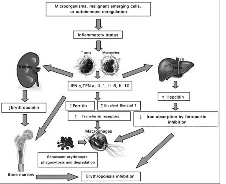
Figure 1. Pathophysiological mechanism in Chronic Disease Anemia. INF-γ: interferon-γ; TNF-α: tumor necrosis factor-α; IL: interleukin.
IL-6 stimulates hepatic expression of an acute phase protein, hepcidin, inhibiting duodenal absorption of iron. Hepcidin, when released by the liver inhibits ferroportin. This is a transmembrane protein found in enterocytes, macrophages and hepatocytes, which is responsible for the transfer of Fe2+ absorbed into
the circulation. As a result, there is a low level of serum iron, leading to decreased release of iron to the bone marrow and thus favoring anemia, even in the presence of total body iron stores, i.e., the so-called functional iron deficiency. IFN-g also decreases the expression of ferroportin.20-22
IFN-gamma enhances the expression of the divalent metal transporter-1 (DMT-1) protein in macrophages by stimulating the uptake of iron in the ferrous state. IL-10 anti-inflammatory
cytokine regulates transferrin receptor expres-sion, enhancing the uptake thereof connected to the iron, mediated by monocyte transferrin receptor. Furthermore, activated macrophages phagocyte and degrade senescent erythrocytes for iron recycling, a process that is mainly in-duced by TNF-α.19
differentiation of erythrocyte progenitor cells. Then, together with the limited availability of iron and decreased biologically active EPO, they will inhibit erythropoiesis and hence foster the development of anemia.19,22
Chronic disease anemia is a phenomenon that plays an important role in CKD. Several pathophysiological mechanisms underlie this condition, including those described previously, as well as reducing EPO receptor expression and possibly a deficiency in EPO signal transduction.23
However, the main cause is inadequate EPO synthesis, with disproportionately low serum levels of this hormone vis-à-vis the degree of anemia.18 Anemic patients with normal renal
function have EPO levels 10-100 fold higher compared with anemic CKD patients.24
Other causes of anemia in patients with CKD are infections and absolute blood loss leading to iron deficiency. This blood loss includes gastrointestinal bleeding with loss of occult blood in the stool, blood retained in the extracorporeal circulation during dialysis, blood taken for laboratory tests, hemolysis, B12 vitamin and folic acid deficiency, vitamin D deficiency, hyperparathyroidism, hemoglobin diseases and neoplasia.18
Secondary hyperparathyroidism is a common complication in CKD, and contributes to the de-velopment of anemia, and it can contribute to greater resistance to the action of erythropoietin. Vitamin D analogues administration has been associated with an improvement in anemia and/ or a reduction in EPO needs. The positive effects of vitamin D, both in anemia as in the required doses of EPO during CKD may be related to their action on the suppressive effect of PTH. Another possible explanation is that active vitamin D di-rectly stimulates erythrocyte progenitor cells.18,25
Hemolysis, although mild, can contribute to anemia. In CKD patients, intra and extracellular factors decrease red blood cells survival in 30% to 50%. This is probably due to the red blood cell membrane inability to pump sodium to the extracellular medium.14
Clinicians should consider that anemia causes are not a result of EPO deficiency when: 1) the
severity of anemia is disproportionate to the def-icit of renal function, 2) there is evidence of iron deficiency, or 3) there is evidence of leukopenia or thrombocytopenia. Assessing the cause of ane-mia should precede the initiation of therapy with EPO and as per the latest Brazilian guidelines the recommendation is to not raise Hb levels above 11.5 g/dL11,26 (Table 1).
Advances in understanding the pathophysiol-ogy of chronic disease anemia led to the devel-opment of new therapeutic strategies. These in-clude treatment of the underlying disease, the use of erythropoietic drugs, iron supplementation or blood transfusions.27
Although the positive effects of short-term therapy with erythropoiesis-stimulating agents in correcting anemia and to avoid blood transfusions are well documented, little data is available about possible effects on the course of the disease, particularly if EPO may exert additional biological effects, including interference with signal transduction and in the cytokine cascade.28
E
RYTHROPOIETIN RESISTANCE IN CHRONIC KIDNEY DISEASEEPO is mainly expressed by hepatocytes during the fetal stage. After birth, peritubular fibroblasts in the renal cortex become its main production site.29 The stimulus for the EPO gene to start
producing erythropoietin is related to the kidney oxygen pressure (renal pO2). When renal pO2 decreases, as in anemia, the EPO gene is stimulated to synthesize this hormone. On the other hand, when renal pO2 normalizes, the synthesis of this hormone is reduced.23
The biological effect of EPO on hematopoi-etic cells is mediated by its binding to their spe-cific receptors on the cell surface.30 The binding
TABLE 1 DIFFERENTIALDIAGNOSISOFANEMIAS
Microcytic and hypochromic Normocytic and normochromic Macrocytic
MCV < 80 FL MCV 80-95 FL MCV > 95 FL
MCH < 27 pg. MCH > 26 pg. Megaloblastic
Iron deficiency Hemolytic anemia Non-megaloblastic
Beta thalassemia Chronic disease anemia (some cases)
Chronic disease anemia (some cases) Acute hemorrhage
Lead poisoning Nephropathy
Sideroblastic anemia Medullar insufficiency, chemotherapy,
infiltration
protein kinase and phosphatidylinositol-3 ki-nase. The main effect of EPO is the reduction of physiological apoptosis associated with cell transformation into erythroid progenitors. Nevertheless, in conjunction with other growth factors, EPO stimulates cell proliferation, sur-vival and differentiation.30,31
The introduction of EPO treatment in clinical practice in 1986 completely changed the monitoring of patients with CKD. The successful correction of anemia in CKD resulted in reduced morbidity associated with improved cardiovascular function, exercise tolerance and cognitive function.9,32
Anemia, even after the administration of exogenous EPO in CKD, points to the fact that peripheral resistance or decreased response to EPO can be the true reason for its development.33
EPO resistance is sometimes found from causes such as functional iron deficiency, secondary hyperparathyroidism, blood loss or interactions with other drugs.34
It is known that CKD involves a chronic inflammatory state with increased levels of inflammatory markers such as C reactive protein (CRP), IL-1, IL-6, IFN-g and TNF-α.35 Cytokines
have a direct effect on cell differentiation of the erythroid pathway and mediate apoptosis induction, suggesting that the cytokine-mediated pro-inflammatory signaling also affects EPO activity. They interfere with the EPO-mediated signaling pathway, inhibiting the expression and regulation of specific transcription factors involved in erythrocyte differentiation control.36
Cytokines can affect different erythropoiesis stages. Immune activation involves accessory
cells of the hematopoietic microenvironment and T cells produce TNF-α and IFN-g, and monocytes produce TNF-α and IL-6. These pro-inflammatory cytokines inhibit proliferation of erythrocyte progenitor cells and antagonize the antiapoptotic actions of EPO. Moreover, this direct negative effect on erythrocyte progenitor cells may be primarily due to changes in sensitivity to the action of EPO.25
The responsiveness of erythrocytic progenitor cells to EPO appears to be inversely related to the chronic disease severity and the amount of circulating cytokines. High concentrations of IFN-gamma or TNF-α causes higher amounts of EPO to be required to restore the formation of erythrocyte colony forming units.37
Inflammation is also associated with increased serum ferritin, which leads to a reluctance of doctors to administer iron to these patients, and then the best therapeutic approach is to administer EPO and have cytokine production. This inflammatory condition can lead to a poor response to treatment with EPO;38 and the end
result of EPO resistance is cachexia, increasing in the number of patients with CVD and reduced quality of life.35
I
NFLAMMATORY CYTOKINES,
GENETIC POLYMOR-PHISMS ANDEPO RESISTANCE
even the use of catheters, buildup of advanced glycation end-products. In addition, progres-sive GFR decrease may contribute to the deve-lopment of inflammation in CKD, with conse-quent production of cytokines in response to this inflamation.1,6,9,12
Cytokines are glycoproteins or low molecular weight regulatory polypeptides secreted by lymphocytes and various other body cells in response to many stimuli.39 They initiate their
action by binding to specific receptors on the target cell membrane, triggering a signal transduction pathway, leading to changes in gene expression in the target cell. They act as positive or negative regulators of the immune, inflammatory and reparative host responses to lesions.40 Thus, cytokines may generally
be characterized as having stimulating (pro-inflammatory) or inhibitory (anti-(pro-inflammatory) effects, depending on the clone subtype of the activated T helper cells (Th).41
Proinflammatory IL-1, IL-2, IL-6, IL-8, IL-12 cytokines, TNF-α and IFN-g promote activation of the inflammatory process, assisting in the elimination of pathogens and in the resolution of inflammation. Elevation in the levels of proinflammatory cytokines lead to activation of macrophages, natural killer (NK) cells, T and B lymphocytes, T and B cells proliferation and secretion of immunoglobulins. In systemic levels, cytokines have been shown to induce fever and enhance the synthesis of acute phase proteins. Locally, they promote the recruitment of inflammatory cells to inflammation sites. Anti-inflammatory cytokines (4, 10, IL-13, TGF-β) reduce the inflammatory response by decreasing pro-inflammatory cytokines and suppression of monocyte activation.42
IL-17 is a pro-inflammatory cytokine with ability to promote the activation and maturation of neutrophils. It is produced by T-helper cells 17, which are a lineage of T CD4+, different
from the ones already studied and the known 1 and 2 T helper. IL-17 may play a role in chronic disease anemia in which the prolonged immune stimulation and increase in IL-17 production
result in decreased hematopoiesis, playing a critical role in the regulation of hematopoietic inflammation, and it may also inhibit the Burst Erythrocyte colony forming units - (BFU-E).43,44
There is evidence of immune system activation in early and late stages of CKD and the presence of inflammation is an independent predictor of mortality.45 Patients on dialysis have an increased
production of TNF-α, IL-6, IL-10 and IL-12 due to the presence of inflammatory processes.46
Furthermore, it has been shown that some polymorphisms of genes encoding inflammatory mediators are associated with worse outcomes in CKD patients.7
Functional Polymorphisms in cytokine genes, which can confirm interindividual differences in synthesis and secretion of these cytokines have been associated with an inflammatory disease pathogenesis. It is believed, therefore, that genetic, cultural and environmental factors influence the inflammatory status of CKD patients.7
Cytokine polymorphisms arise from the coding region and can lead to an amino acid substitution and changes in protein function or activity.47 Other polymorphisms are in the
promoter region and can disrupt or abolish transcriptional regulation, either by regulatory elements or by other genes involved in translation signaling pathways.8
TNF-α recruits neutrophils and mast cells to the infected environment and acts on the vascular endothelial cells and leukocytes, promoting inflammation and apoptosis.40 The position -308
of the promoter region has guanine (G) as the normal allele which, when replaced by adenine (A), results in the A/G heterozygous mutant or the A/A homozygote. Mutation at position -308 increases plasma levels of this cytokine, speculated to be responsible for the development of more aggressive diseases such as rheumatoid arthritis and hepatitis B and C,48 and high levels
of this cytokine may be involved with a poor response to the use of EPO.49
cells and T CD8+ cells. Among the biological ac-tion is the activaac-tion of macrophages, facilitating the action of cytotoxic T cells and NK cells in the elimination of phagocyted micro-organisms.50
Polymorphism in the IFN-g gene is located in the region +874 of intron 1. The wild allele in this region is thymine (T), which can mutate to A, resulting in lower serum levels of IFN-g, which would make the hosts more susceptible to in-fections.51 In addition; increased levels of
IFN-gamma are associated with a worse response to the use of EPO to treat chronic disease anemia.49
The type beta transforming growth factor (TGF-β) is an anti-inflammatory cytokine of adaptive immunity, produced mainly by T cells and activated phagocytes. They inhibit T-lymphocyte proliferation, differentiation, and macrophage activation, and stimulate the production of IgA and extracellular matrix synthesis, such as collagen, metalloproteinases, cell receptors for the matrix and integrins. With this, it performs its main function, which is to promote tissue healing after inflammatory or immune reactions have been controlled.40,52
The most studied polymorphisms of TGF-β are located in the coding region of the gene at positions +869 [T to cytosine (C)] and +915 (G to C). Combinations of these polymorphisms form the TG haplotypes associated with an activity/ production of this citokine.53
Cytotoxic T-lymphocyte-activated mac-rophages and other non-lymphoid cell types secrete IL-10. It has Th1 immune response in-hibitory action seen in macrophage suppres-sion, returning to resting states as the infection is controlled, in IL-12 production suppression by activated macrophages and dendritic cells and inhibition of the expression of co-stimulators, and molecules of the greater histocompatibility complex (MHC) II in these cells.40 Among these
IL-10 polymorphisms, the most investigated are located in the promoter region at positions -1082 (G to A), -819 (C to T), and -592 (C to A), from the transcription starting site influencing IL-10 expression. These single nucleotide polymor-phisms produce three major haplotypes: GCC
associated with the increased production of cy-tokine, this polymorphism being associated with EPO increases required for chronic disease ane-mia treatment,54 ACC, associated with
interme-diate production, and ATA, to low production.55
IL-6 is synthesized by mononuclear cells, vascular endothelial cells, fibroblasts and other cells.56,57 It is involved in various
physiologi-cal and pathologiphysiologi-cal processes such as infec-tion, inflammainfec-tion, trauma, bone metabolism, C-reactive protein synthesis and carcinogenesis.57
In the promoter region of this gene, a substitu-tion from C to G at posisubstitu-tion -174 is associated with different levels of production of this cito-kine58 and the GG genotype is associated with
the need for higher doses of EPO in an attempt to correct Hb levels in chronic disease anemia.54
The common CKD outcome is shown by the progressive glomerular and/or tubulointerstitial fibrosis, peritubular capillary damage by hypoxia and loss of nephron function by glomerular sclerosis and tubular atrophy, regardless of the primary mechanism that triggered the renal injury.6
Inflammatory mechanisms have increasingly being discovered in these pathophysiological processes of renal progression, which imply development of chronic kidney disease anemia, and subsequent poor response to the use of EPO in the treatment of anemia as was explained earlier,3 as well as electrolyte disturbances,
oxidative stress, overt and hidden infections.14
In this context, the role of inflammation as well as that of cytokines in the progression of CKD is highlighted in glomerulopathies - disorders in which inflammation is classically recognized in congenital malformations of the urinary tract and kidneys, diseases whose main mechanism of injury was traditionally related to mechanical obstruction.12 The potential of certain cytokines
and chemokines function as CKD progression biomarkers, such as IL-6, IL-17, TNF-α and TGF-β should be considered together with the use of these parameters, whenever possible in clinical practice.36,37,39,40
have been considered. Different stimuli such as infection, physical-chemical and antigenic changes or traumatic damage is evidenced in response to a physiological process of inflammation. And this response has to be regulated accurately, since shortage or excess of this response are related to mortality and morbidity.59
C
ONCLUSIONThe discovery of new diagnostic and therapeutic approaches for the treatment of anemia in CKD becomes increasingly necessary. Anemia is a common complication in CKD patients and treatment with EPO brings about great benefits for the patient. By clinical and experimental evidence, the role of inflammation in CKD anemia and the presence of genetic polymorphisms of cytokines may be involved with a worse prognosis in therapy with EPO, leading to a poor response to its use and increased comorbidities such as CVD. Given the role of inflammation in CKD anemia, agents with anti-inflammatory properties and vitamin supplements, such as vitamin D, may be beneficial in treating patients who are bad responders to the use of EPO as well as the modulation of immune-inflammatory response, which could be a therapeutic target for CKD treatment, or also drugs that could antagonize the effects of hepcidin, which could be targets for prospective studies. However, despite major advances in understanding the mechanism of EPO, anemia in CKD and inflammation through the study of cytokines and their gene polymorphisms, many aspects remain to be clarified.
R
EFERENCES1. Vianna HR, Soares CMBM, Tavares MS, Teixeira MM, Silva ACS. Inflamação na doença renal crônica: papel de citocinas. J Bras Nefrol 2011;33:351-64. DOI: http://dx.doi.org/10.1590/ S0101-28002011000300012
2. Abensur H. Deficiência de ferro na doença renal crônica. Rev Bras Hematol Hemoter 2010;32:95-8. DOI: http://dx.doi. org/10.1590/S1516-84842010005000047
3. Means RT, Krantz SB. Progress in understanding the pathoge-nesis of the anemia of chronic disease. Blood 1992;80:1639-47 PMID: 1391934
4. Lee JR. Deficiência de ferro e anemia ferropriva. In: Lee GR, Bithell TC, Foerster J, Athens JW, Lukens JN. Wintrobe - He-matologia Clínica. São Paulo: Manole; 1998. p.884-919. 5. Alvin RC, Azevedo WM, Silva CM. Manifestações
hematológi-cas das doenças infecciosas na infância. In: Tonelli E. Doenças infecciosas na infância. Rio de Janeiro: Medsi; 1987. p.1274-88.
6. Bruchfeld A, Carrero JJ, Qureshi AR, Lindholm B, Barany P, Heimburger O, et al. Elevated serum macrophage migration inhibitory factor (MIF) concentrations in chronic kidney disea-se (CKD) are associated with markers of oxidative stress and endothelial activation. Mol Med 2009;15:70-5. DOI: http:// dx.doi.org/10.2119/molmed.2008.00109
7. Rao M, Wong C, Kanetsky P, Girndt M, Stenvinkel P, Reilly M, et al. Cytokine gene polymorphism and progression of renal and cardiovascular diseases. Kidney Int 2007;72:549-56. PMID: 17579660 DOI: http://dx.doi.org/10.1038/sj.ki.5002391 8. Pravica V, Brogan IJ, Hutchinson IV. Rare polymorphisms in the
promoter regions of the human interleukin-12 p35 and inter-leukin-12 p40 subunit genes. Eur J Immunogenet 2000;27:35-6. DOI: http://dx.doi.org/10.1046/j.1365-2370.2000.00190.x 9. Sharples EJ, Varagunam M, Sinnott PJ, McCloskey DJ, Raftery
MJ, Yaqoob MM. The effect of proinflammatory cytokine gene and angiotensin-converting enzyme polymorphisms on erythro-poietin requirements in patients on continuous ambulatory pe-ritoneal dialysis. Perit Dial Int 2006;26:64-8. PMID: 16538877 10. Sesso R, Lopes AA, Thomé FS, Bevilaqua JL, Romão Jr JE,
Lugon J. Relatório do censo Brasileiro de diálise, 2008. J Bras Nefrol 2008;30:233-8.
11. Coyne DW. The KDOQI US commentary on KDIGO anemia guideline and quality of life. Am J Kidney Dis 2014;63:540. DOI: http://dx.doi.org/10.1053/j.ajkd.2013.08.037
12. Meguid El Nahas A, Bello AK. Chronic kidney disease: the global challenge. Lancet 2005;365:331-40. PMID: 15664230 DOI: http://dx.doi.org/10.1016/S0140-6736(05)17789-7 13. Sociedade Brasileira de Nefrologia. Censo da Sociedade
Bra-sileira de Nefrologia 2013 [Acesso 21 Mar 2015]. Disponível em: http://http://www.sbn.org.br/pdf/censo_2013-14-05.pdf 14. Bandeira MFS. Consequências hematológicas da uremia. In:
Riella MC. Princípios de nefrologia e distúrbios eletrolíticos. 4a ed. Rio de Janeiro: Guanabara Koogan; 2003. p.691-701. 15. Sears DA. Anemia of chronic disease. Med Clin North Am
1992;76:567-79. PMID: 1578957
16. Zarychanski R, Houston DS. Anemia of chronic disease: a harmful disorder or an adaptive, beneficial response? CMAJ 2008;179:333-7. PMID: 18695181
17. Bron D, Meuleman N, Mascaux C. Biological basis of ane-mia. Semin Oncol 2001;28:1-6. PMID: 11395845 DOI: http:// dx.doi.org/10.1016/S0093-7754(01)90205-2
18. Weiss G. Pathogenesis and treatment of anaemia of chro-nic disease. Blood Rev 2002;16:87-96. DOI: http://dx.doi. org/10.1054/blre.2002.0193
19. Weiss G, Goodnough LT. Anemia of chronic disease. N Engl J Med 2005;352:1011-23. DOI: http://dx.doi.org/10.1056/NE-JMra041809
20. Grotto HZ. Anaemia of cancer: an overview of mechanisms involved in its pathogenesis. Med Oncol 2008;25:12-21. DOI: http://dx.doi.org/10.1007/s12032-007-9000-8
21. Jairam A, Das R, Aggarwal PK, Kohli HS, Gupta KL, Sakhu-ja V, et al. Iron status, inflammation and hepcidin in ESRD patients: The confounding role of intravenous iron thera-py. Indian J Nephrol 2010;20:125-31. DOI: http://dx.doi. org/10.4103/0971-4065.70840
22. Silverberg DS, Wexler D, Iaina A, Schwartz D. Correction of iron deficiency in the cardiorenal syndrome. Int J Nephrol 2011;2011:365301. PMID: 21603160 DOI: http://dx.doi. org/10.4061/2011/365301
23. Sulikowska B, Odrowaz-Sypniewska G, Manitius J. Interpreta-tion of erythropoietin levels in patients with various degrees of renal anemia. Kidney Int 2005;67:1635-6. DOI: http://dx.doi. org/10.1111/j.1523-1755.2005.247_3.x
24. Cotes PM. Erythropoietin: the developing story. Br Med J (Clin Res Ed) 1988;296:805-6. DOI: http://dx.doi.org/10.1136/ bmj.296.6625.805
26. Kirsztajn GM, Filho NS, Draibe SA, Netto MV, Thomé FS, Souza E, et al. Fast reading of the KDIGO 2012: guidelines for evaluation and management of chronic kidney disease in clini-cal practice. J Bras Nefrol. 2014;36:63-73. DOI: http://dx.doi. org/10.5935/0101-2800.20140012
27. Salvarani C, Baricchi R, Lasagni D, Boiardi L, Piccinini R, Brunati C, et al. Effects of desferrioxamine therapy on chronic disease anemia associated with rheumatoid arthritis. Rheu-matol Int 1996;16:45-8. DOI: http://dx.doi.org/10.1007/ BF01816434
28. Aguilera A, Selgas R, Ruiz-Caravaca ML, Bajo MA, Cuesta MV, Plaza MA, et al. Effects of recombinant human erythro-poietin on functional and injury endothelial markers in perito-neal dialysis patients. Perit Dial Int 1999;19:S161-6.
29. Noguchi CT, Asavaritikrai P, Teng R, Jia Y. Role of erythro-poietin in the brain. Crit Rev Oncol Hematol 2007;64:159-71. PMID: 17482474 DOI: http://dx.doi.org/10.1016/j.critre-vonc.2007.03.001
30. Hardee ME, Arcasoy MO, Blackwell KL, Kirkpatrick JP, Dewhirst MW. Erythropoietin biology in cancer. Clin Cancer Res 2006;12:332-9. DOI: http://dx.doi.org/10.1158/1078-0432.CCR-05-1771
31. Westenbrink BD, Visser FW, Voors AA, Smilde TD, Lipsic E, Navis G, et al. Anaemia in chronic heart failure is not only rela-ted to impaired renal perfusion and blunrela-ted erythropoietin pro-duction, but to fluid retention as well. Eur Heart J 2007;28:166-71. PMID: 17158825 DOI: http://dx.doi.org/10.1093/eurhear-tj/ehl419
32. Jeong KH, Lee TW, Ihm CG, Lee SH, Moon JY. Polymorphis-ms in two genes, IL-1B and ACE, are associated with erythro-poietin resistance in Korean patients on maintenance hemo-dialysis. Exp Mol Med 2008;40:161-6. PMID: 18446054 DOI: http://dx.doi.org/10.3858/emm.2008.40.2.161
33. Lacombe C, Mayeux P. The molecular biology of erythropoie-tin. Nephrol Dial Transplant 1999;14:22-8. PMID: 10334664 DOI: http://dx.doi.org/10.1093/ndt/14.suppl_2.22
34. Jones C, Roderick P, Harris S, Rogerson M. An evaluation of a shared primary and secondary care nephrology service for managing patients with moderate to advanced CKD. Am J Kid-ney Dis 2006;47:103-14. PMID: 16377391 DOI: http://dx.doi. org/10.1053/j.ajkd.2005.09.020
35. Agarwal N, Prchal JT. Anemia of chronic disease (anemia of inflammation). Acta Haematol 2009;122:103-8. PMID: 19907147 DOI: http://dx.doi.org/10.1159/000243794
36. Macdougall IC, Cooper AC. Erythropoietin resistance: the role of inflammation and pro-inflammatory cytokines. Nephrol Dial Transplant 2002;17:39-43. DOI: http://dx.doi.org/10.1093/ ndt/17.suppl_11.39
37. Minoo P, Zadeh MM, Rottapel R, Lebrun JJ, Ali S. A novel SHP-1/Grb2-dependent mechanism of negative regulation of cytokine-receptor signaling: contribution of SHP-1 C-terminal tyrosines in cytokine signaling. Blood 2004;103:1398-407. DOI: http://dx.doi.org/10.1182/blood-2003-07-2617
38. Kalantar-Zadeh K, McAllister CJ, Lehn RS, Lee GH, Nis-senson AR, Kopple JD. Effect of malnutrition-inflammation complex syndrome on EPO hyporesponsiveness in mainte-nance hemodialysis patients. Am J Kidney Dis 2003;42:761-73. PMID: 14520627 DOI: http://dx.doi.org/10.1016/S0272-6386(03)00915-6
39. Opal SM, DePalo VA. Anti-inflammatory cytokines. Chest 2000;117:1162-72. PMID: 10767254 DOI: http://dx.doi. org/10.1378/chest.117.4.1162
40. Abbas AK, Lichtman AH, Pillai S. Imunologia celular e molecu-lar. 6a ed. Rio de Janeiro: Elsevier; 2008.
41. Hauser SL. Tumor necrosis factor: immunogenetics and disea-se. Ann Neurol 1995;38:702-4. PMID: 7486860 DOI: http:// dx.doi.org/10.1002/ana.410380503
42. Remick DG. Cytokine therapeutics for the treatment of sepsis: why has nothing worked? Curr Pharm Des. 2003;9:75-82.
43. Broxmeyer HE, Starnes T, Ramsey H, Cooper S, Dahl R, Williamson E, et al. The IL-17 cytokine family members are inhibitors of human hematopoietic progenitor proliferation. Blood 2006;108:770. PMID: 16822904 DOI: http://dx.doi. org/10.1182/blood-2006-01-0292
44. Miossec P. IL-17 and Th17 cells in human inflammatory di-seases. Microbes Infect 2009;11:625-30. DOI: http://dx.doi. org/10.1016/j.micinf.2009.04.003
45. Tashiro T, Nakamura K, Morishige N, Iwakuma A, Tachi-kawa Y, Shibano R, et al. Off-pump coronary artery bypass grafting in patients with end-stage renal disease on hemo-dialysis. J Card Surg 2002;17:377-82. DOI: http://dx.doi. org/10.1111/j.1540-8191.2001.tb01162.x
46. Stites DP, Terr AI, Parslow TG. Imunologia médica, 9ª ed. Rio de Janeiro: Guanabara Koogan; 2000.
47. Hollegaard MV, Bidwell JL. Cytokine gene polymorphism in human disease: on-line databases, Supplement 3. Genes Immun 2006;7:269-76. DOI: http://dx.doi.org/10.1038/sj.gene.6364301 48. Migita K, Miyazoe S, Maeda Y, Daikoku M, Abiru S, Ueki T,
et al. Cytokine gene polymorphisms in Japanese patients with hepatitis B virus infection--association between TGF-beta1 polymorphisms and hepatocellular carcinoma. J Hepa-tol. 2005;42:505-10. PMID: 15763337 DOI: http://dx.doi. org/10.1016/j.jhep.2004.11.026
49. Cooper AC, Mikhail A, Lethbridge MW, Kemeny DM, Ma-cdougall IC. Increased expression of erythropoiesis inhibiting cytokines (IFN-gamma, TNF-alpha, IL-10, and IL-13) by T ce-lls in patients exhibiting a poor response to erythropoietin the-rapy. J Am Soc Nephrol 2003;14:1776-84. DOI: http://dx.doi. org/10.1097/01.ASN.0000071514.36428.61
50. Schoenborn JR, Wilson CB. Regulation of interferon-γ during in-nate and adaptive immune responses. In: Frederick WA, ed. Ad-vances in immunology. Oxford: Academic Press; 2007. p.41-101. 51. Natividad A, Wilson J, Koch O, Holland MJ, Rockett K, Faal
N, et al. Risk of trachomatous scarring and trichiasis in Gam-bians varies with SNP haplotypes at the interferon-gamma and interleukin-10 loci. Genes Immun 2005;6:332-40. DOI: http:// dx.doi.org/10.1038/sj.gene.6364182
52. Wells RG. Fibrogenesis. V. TGF-beta signaling pathways. Am J Physiol Gastrointest Liver Physiol 2000;279:G845-50. PMID: 11052979
53. Guan X, Zhao H, Niu J, Tan D, Ajani JA, Wei Q. Polymorphis-ms of TGFB1 and VEGF genes and survival of patients with gastric cancer. J Exp Clin Cancer Res 2009;28:94. DOI: http:// dx.doi.org/10.1186/1756-9966-28-94
54. Girndt M, Stenvinkel P, Ulrich C, Axelsson J, Nordfors L, Barany P, et al. Influence of cytokine gene polymorphisms on erythropoetin dose requirements in chronic haemodialysis pa-tients. Nephrol Dial Transplant 2007;22:3586-92. DOI: http:// dx.doi.org/10.1093/ndt/gfm244
55. Liu J, Song B, Wang JL, Li ZJ, Li WH, Wang ZH. Polymor-phisms of interleukin-10 promoter are not associated with prognosis of advanced gastric cancer. World J Gastroente-rol 2011;17:1362-7. PMID: 21455338 DOI: http://dx.doi. org/10.3748/wjg.v17.i10.1362
56. Sugimoto M, Yamaoka Y, Furuta T. Influence of interleukin polymorphisms on development of gastric cancer and peptic ulcer. World J Gastroenterol 2010;16:1188-200. DOI: http:// dx.doi.org/10.3748/wjg.v16.i10.1188
57. Vishnoi M, Pandey SN, Choudhury G, Kumar A, Modi DR, Mittal B. Do TNFA -308 G/A and IL6 -174 G/C gene polymor-phisms modulate risk of gallbladder cancer in the north Indian population? Asian Pac J Cancer Prev 2007;8:567-72.
58. Gu F, Qureshi AA, Niu T, Kraft P, Guo Q, Hunter DJ. Inter-leukin and interInter-leukin receptor gene polymorphisms and sus-ceptibility to melanoma. Melanoma Res 2008;18:330-5. DOI: http://dx.doi.org/10.1097/CMR.0b013e32830658b2