Gene therapy: what it is,
what it is not and what it
will be
R
afael
l
inden
Introduction
F
rom its foundation by the monk Johann (Gregor) mendel in thenineteenth century to date, genetics has evolved dramatically and earned an important place among the sciences. the sequencing of the human genome (Lander et al., 2001, Venter et al., 2001), a great achievement that promises to accelerate the progress of biology and medicine in the twenty-first century, was completed ten years ago.
Every day modern medicine makes important discoveries in research areas aimed at developing new paradigms for treating diseases that are still incurable. Among them, the expectation of curing genetic diseases rests on the identification of genes responsible for its pathogenesis and on the advancement of recombi-nant DNA technology or “genetic engineering”, which enable manipulating the genome in an increasingly efficient and safer way (Watson et al. 2006). in paral-lel, the determination of genetic susceptibility to certain diseases, the course and clinical manifestations thereof (NCBi, 2009), as well as the tremendous advance in understanding the cellular and molecular biology of fundamental pathological events such as inflammatory processes, proliferative disorders and programmed cell death (tsongalis & Coleman, 2009), all increase the expectation that genome manipulation may someday be applied to a wide range of diseases.
this is still an incipient area of medicine, practiced especially in funda-mental research laboratories, and its application is still strictly experifunda-mental. Al-though there area in this area commercial products approved for medical use (Pearson et al., 2004), the expectation of scientists, as well as of the pharma-ceutical and biotechnology industry, is that the release of genome manipulation protocols for medical practice and the respective market of biological products should advance cautiously over the next 5-10 years, but still limited to a re-stricted number of applications.
context called “therapeutic genes”) to replace, manipulate or supplement inac-tive or dysfunctional genes (Linden, 2008).
The beginning of gene therapy
From the 1940s genetics gained great momentum, and discoveries about the nature, chemical composition and properties of the genetic material, as well as the first manipulations of the DNA of bacteria began to generate expectations of novel therapeutic advances.
the mid-1960s were marked by speculation about the possibility of using viruses to transfer genes to human patients and cure genetic diseases (Fried-mann, 1997). Back then, scientists already believed that the genes of certain vi-ruses could be effective and that it was possible to insert healthy human genes in viruses which, in turn, would transfer them to the patient. However, it was only in the beginning of the following decade that Paul Berg succeeded in actually manipulating a DNA molecule (Jackson et al., 1972), creating the recombinant DNA technology.
two initial attempts to apply the gene therapy concept to clinical practice failed, one for relying on an assumption about the properties of a virus, which later proved to be false (rogers, 1952; rous & rogers, 1951; Andrewes, 1966; Friedman, 2001; scaglia & Lee, 2006); the other, although technically justifi-able and already using recombinant DNA methodologies, was marred by a seri-ous ethical lapse (mercola & Cline, 1980). But in 1989 a new test conducted in accordance with the rules in force at the time restored positive expectations in this research area.
the patient treated in 1989 was a four-year-old girl who had been deprived of a normal life because of a genetic disorder caused by a deficiency of the ad-enosine deaminase enzyme (ADA), which is essential for the development of the immune system. several mutations in the gene encoding the enzyme cause ADA deficiency, which results in degeneration of the t cells of the immune system (Buckley, 2004) and is one of the main causes of severe combined immunode-ficiency syndrome (sCiD). in this case, the disease is known by the acronym ADA-sCiD. Children affected by various forms of sCiD (ibid.) have very low resistance to infection and, if untreated, usually die before six months of age. they are known as “bubble children” because they need to be isolated, often in a plastic bubble. treatment usually entails replacing the enzyme with weekly injections. in the case in question, after a period of one year of relative success, in the second year of treatment the child was again plagued by frequent infections and devel-oped an allergy to the enzyme preparation used for injections, indicating that the enzyme replacement therapy was failing. Dr. William French Anderson, from the University of southern California, was then authorized by the ethics committees to initiate a clinical gene therapy trial (Anderson et al., 1990).
inserted the ADA gene, induced the proliferation of these cells in the laboratory and then re-infused the treated cells into the patient’s blood stream (Culver et al. 1,991). After seven infusions there was a six-month break, and then the infusions were resumed up to two years of treatment. For safety purposes, the girl continued to receive weekly injections of the enzyme. Gene therapy in this patient, as well as that started in 1991 in a second nine-year old patient, yielded positive results. there was clinical improvement with a reduction in the amount of enzyme that needed to be replaced. it was observed that enzyme levels in the patients’ blood increased progressively as a result of the gene therapy and re-mained stable during the six-month break period (Blaese et al. 1995; mullen et al. 1996). Finally, twelve years after the end of the infusions, when the two cases were reevaluated, large numbers of t cells continued to express the therapeutic gene in the first patient’s blood, whose treatment was more successful than that of the second girl (muul et al. 2003).
it should be noted that there are still technical issues related to this study that prevent us from considering it a complete clinical success. As the children continued to receive enzyme replacement, although in smaller doses, there is cause for doubt about how much the gene therapy has actually contributed, for example, for the first patient, now 24 years old, to be leading a healthy and ac-tive life. However, since the treatment of these first two patients, gene therapy for ADA-sCiD has evolved and today is considered a clinical success (Aiuti et al., 2009; Candotti & Kohn, 2009). Although in its infancy, the study started in 1989 and which has produced at least some positive results in compliance with ethical requirements, is a milestone in the history of gene therapy and has inspired the subsequent growth of this area of scientific research.
Forms of gene therapy
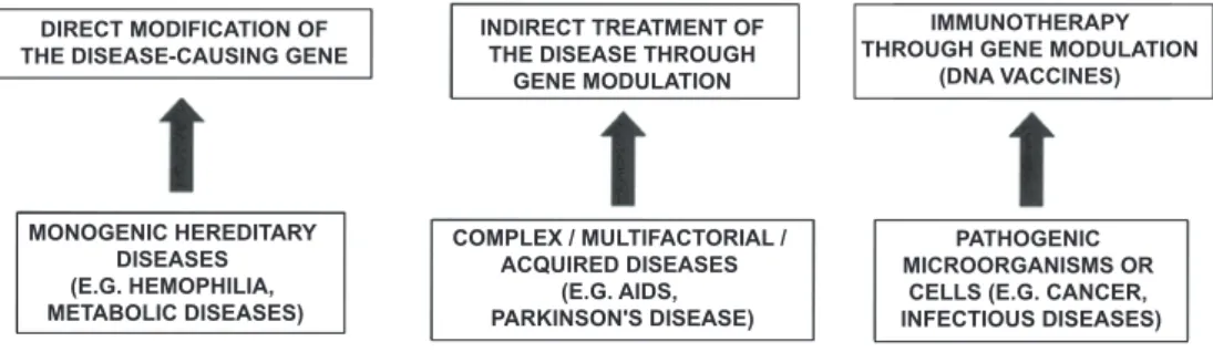
the idea of using recombinant DNA techniques to repair the genome was inspired by diseases caused by mutation in a single gene (the so-called mono-genic diseases). in this case, the idea is to replace or supplement dysfunctional gene expression by inserting one or more copies of the therapeutic gene (Por-teus et al. 2,006; o’Connor & Crystal, 2006; Brinkman et al. 2006). the treat-ment of ADA-sCiD is a successful application of this idea.
by modulating genes not necessarily related to the cause of the disease (Bagley et al., 2008; Lundberg et al. 2008). in the case of tumors, the main objective is the selective induction of cell death in proliferating cell populations (Bauzon & Hermiston, 2008; Cattaneo et al., 2008; ribacka et al., 2008).
Finally, there is a peculiar form of gene therapy called DNA vaccine, in which instead of using a protein or a whole inactivated virus, as is the case in conventional vaccines, the patient receives the gene encoding a protein typical of the aggressor agent. the patient’s body will then begin to permanently produce the exogenous protein, stimulating its own immune system. these vaccines can have a preventive purpose, similarly to classical vaccines, or a curative purpose, leading the immune system to attack the aggressor agents already established in the body (Atkins et al., 2008 sykes, 2008; silva et al., 2009).
Figure 1 – main forms of gene therapy
Cell therapy, stem cells and gene therapy
stem cells are currently the most-commonly referenced medical topic in the media. At the same time, there is some confusion as to the actual meaning of the terms stem cells, cell therapy and gene therapy. in the so-called cell therapy, whole cells are used to treat an illness, based on the regenerative properties of stem cells or on other effects - most of which have not yet been explained - of the transplanted cells. the classic example, whose foundation is well known, is leukemia, but there is expectation that many classes of diseases will be treated with the use of cell therapy in the coming years (torrente & Polli, 2008; Grib-ben, 2008; Einstein & Ben-Hur, 2008; reffelmann et al., 2008).
in this context, it is important to note that cell therapy does not necessar-ily involve genetic modification. Gene therapy, in turn, is based on the introduc-tion or modificaintroduc-tion of genes. this can be done directly in vivo, without the aid of whole cells from the patient or from a donor.
that is, gene therapy and cell therapy are two different concepts. However, there are methods that combine the two techniques. An example of combined gene therapy and cell therapy was again the aforementioned ex vivo procedure that started gene therapy. Novel gene therapy technologies for ADA-sCiD are
DIRECT MODIFICATION OF THE DISEASE-CAUSING GENE
INDIRECT TREATMENT OF THE DISEASE THROUGH
GENE MODULATION
IMMUNOTHERAPY THROUGH GENE MODULATION
(DNA VACCINES)
PATHOGENIC MICROORGANISMS OR
CELLS (E.G. CANCER, INFECTIOUS DISEASES) MONOGENIC HEREDITARY
DISEASES (E.G. HEMOPHILIA, METABOLIC DISEASES)
COMPLEX / MULTIFACTORIAL / ACQUIRED DISEASES
based on the genetic manipulation of bone marrow-derived stem cells, instead of the t cells used in the initial studies (Aiuti et al. 2009). therefore, under certain circumstances cells can be used as a vehicle to introduce the therapeutic gene. However, it is the introduction of genes and the use of recombinant DNA technologies that characterize a treatment as gene therapy.
Vectors for gene therapy
the basis of gene therapy lies in delivering genes to cells. However, the entry of pure DNA through the plasma membrane of eukaryotic cells is ex-tremely rare (Vellai & Vida, 1999). this difficulty, of course, is beneficial for the body, as it hinders spurious changes in cellular metabolism and even changes similar to those observed in the evolution of species.
therefore, in general, a carrier is needed to facilitate the delivery of DNA to living cells. this carrier is called “vector”. there are three main classes of vectors currently under development: plasmids, viral vectors and nanostructured vectors.
Plasmids
Plasmids are relatively simple DNA sequences, but effective for gene ex-pression, in which it is possible to deliver a therapeutic gene using recombinant DNA techniques (Voss, 2007; Clanchy & Williams, 2008; Gill et al., 2009). But breaking the resistance of cells to the introduction of plasmids requires weaken-ing the cell membrane, which can be done by various methods such as the use of electroshock or chemical substances that chemically weaken the cell membrane (Dass, 2004; & Cemazar & sersa, 2007; Favard et al., 2007; Wu & Lu, 2007). Another alternative is to apply a large number of plasmids in the vicinity of the cells so that, even with very low efficiency, a small fraction that succeeds in cross-ing the membrane will be effective, or, still, to rapidly inject a large amount of a solution containing plasmid (Herweijer & Wolff, 2007).
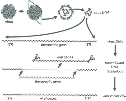
Figure 2 – Construction of a viral vector for gene therapy. the figure illustrates, as an example, the adeno-associated virus, whose genetic material is a single-stranded DNA. At the top is the scheme showing the outside and inside of a virus, in a section showing the location of viral DNA. this DNA contains several genes necessary for the life cycle of the virus, i.e., its mul-tiplication and rearrangement inside the cells. But to be used as a vector, the DNA of terminal regions, identified by the acronym itr, is enough. the process consists of replacing the viral genes with the therapeutic gene, using recombinant DNA technology. the essential component of the viral vector is therefore produced. However, as naked DNA does not enter the cells easily, it is necessary to reassemble a virus similar to the one shown in the upper left corner of this figure and in large quantities, as illustrated in Figure 3. reproduced from Linden (2008), with permission of the publishers.
Viral vectors
in contrast to the resistance of the cell membrane to the spontaneous entry of DNA into a cell, viruses are microorganisms specialized precisely in invading cells and introducing genetic material therein. they contain nucleic acid (DNA or rNA) surrounded by a layer of protein and, in some cases, by an additional envelope of proteins and lipids, and their life cycle involves releasing viral nucleic acid inside the host cell. this property is exploited for delivering therapeutic genes to cells using recombinant DNA technologies.
some vectors are derived from adenovirus. this family includes almost 50 different types of viruses that cause, for example, pharyngitis or conjunctivitis. infections by adenovirus are very common and, therefore, the majority of the population has antibodies against one or more types of this virus family. others
therapeutic gene therapeutic gene
viral genes
viral genes
virus DNA
recombinant DNA technology
are members of the retroviruses family, which includes the HtLV that causes a type of leukemia and the HiV that causes AiDs, a member of the lentivi-ruses subfamily, which have been widely studied as a source of vectors for gene therapy. there are also other vectors derived from viruses of the adenovirus-associated family, which are not pathogenic to humans.
the principle of production of viral vectors for gene therapy (Figs. 2 and 3) consists in removing the genes involved in pathogenic and viral prolifera-tion mechanisms and keeping only the necessary for the invasion of cells with-out multiplication, followed by the introduction of a therapeutic gene into the remaining viral DNA (machida, 2002). removing genes responsible for the pathogenic character and for multiplication enables, for example, a virus of the same subfamily of the dangerous HiV to give rise to a viral vector useful for gene therapy.
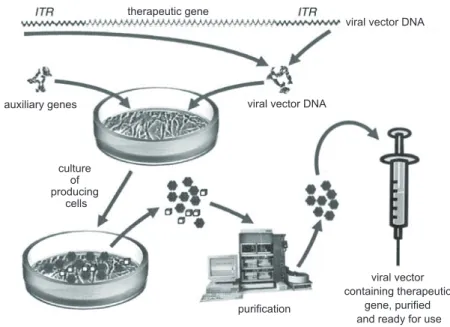
Figure 3 – mass production of viral vectors for gene therapy. the figure once again uses as an example a vector derived from adeno-associated virus. the viral vector DNA was constructed as shown in Figure 2. this DNA is introduced by precipitation or electroporation into producing cells to-gether with a plasmid containing auxiliary genes, which are required for packaging the vector DNA into the structure of viruses similar to the original adeno-associated viruses. Producing cells form large quantities of complete viral vectors together with contaminants, which are removed at a purification stage, after which trillions of viral particles containing the therapeutic gene free of impurities are obtained. the vector is therefore ready for use. reproduced from Linden (2008) with permission of the publishers.
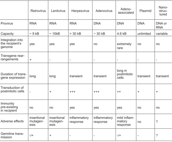
Viral vectors differ from one another (table 1). some are more efficient while others are more capable of carrying large genes. some are more likely to
auxiliary genes
purification culture
of producing
cells
viral vector containing therapeutic
gene, purified and ready for use viral vector DNA
cause inflammatory reactions than others. Finally, some vectors, such as those derived from retroviruses, have the property of integrating into the cell genome. this is positive when one wants a permanent expression of the therapeutic gene; however, it may cause serious adverse effects.
table 1 – Properties of various types of viral and non-viral vectors for gene therapy
Retrovirus Lentivirus Herpesvirus Adenovirus associatedAdeno- Plasmid Nano- struc-tured
Provirus RNA RNA RNA DNA DNA DNA DNA or
RNA
Capacity ~ 9 kB ~ 10kB > 30 kB ~ 30 kB 4.6 kB unlimited variable
Integration into the recipient’s
genome yes yes yes no extremely rare no no
Transgene
rear-rangements + - - -
-Duration of
trans-gene expression long long transient transient
long in postmitotic
cells transient transient
Transduction of
postmitotic cells - + +++ +++ ++ + +
Immunity pre-existing
in recipient no no yes yes yes no no
Adverse effects
insertional mutagen-esis
insertional mutagen-esis
inlammatory
response
inlammatory
response
mild inlam -matory
response no ?
Germline
trans-mission -/+ + - - -/+ - ?
Source: modified from Nathwani et al. (2005). Nanostructured vectors
Another way to introduce DNA into cells is being developed from preparations obtained using advanced nanotechnology techniques (sanvicens & marco, 2008). this includes polymers that form real networks that hold a gene and release their load when they penetrate the cells, as well as lipid vesicles containing DNA, which are ca-pable of fusing with the cell membrane, releasing their contents inside the latter.
Figure 4 – model of non-viral vector combined with a routing molecule. the figure uses as an example an immunoliposome for cerebral gene transduction (Pardridge, 2005). As the wall of cerebral blood vessels is highly resistant to drug penetration, including DNA, from the blood, the vector is composed of a vesicle formed by lipids containing DNA with the therapeutic gene. Antibodies against the transferrin receptor, which recognize this receptor on the surface of cells of the wall of cerebral blood vessels and of neurons are inserted on the surface of the vesicle. so, when the vector is injected into the blood stream, it attached strongly to the wall of cerebral blood vessels, thus facilitating its penetration into the brain tissue and, consequently, the introduction of the therapeutic gene into neural cells. this technique can be used to route vectors to the appropriate destination, based on the choice of the antibody inserted on the surface of the vector, which should be selec-tive for blood vessels of the organ to be treated. reproduced from Linden (2008) with permission of the publishers.
in other cases, cells modified by the insertion of a therapeutic gene can be encapsulated in compartments produced from inert polymers and then in-troduced into the body. the advantage of this technique is that the cells can produce and secrete therapeutic molecules while isolated from the patient’s im-mune system (Hauser et al. 2,004; Lindvall & Wahlberg, 2008). therefore, encapsulated cells do not need to be derived from the patient him or herself.
Gene therapy today
Gene therapies are novel procedures still in the experimental stage. Basic knowl-edge has been acquired in fundamental research laboratories through tests in experimental models and preclinical trials. these studies validate the potential efficacy of a therapeutic strategy, and enable detecting potential risks to humans, anticipating changes in vectors and other components of the therapeutic strategy that increase safety for human use.
Fundamental research in gene therapy is intense and growing in the world. Figure 5 illustrates the continued growth in the number of scientific
publica-facilitating penetration of the vector into the brain vector antibody recognizes and
attach to the wall of the cerebral blood vessel
vector is injected into the bloodstream
therapeutic gene and antibody are added to the lipid vesicle making up the vector
tions in this area. in the last three years, about 30 scientific papers, on average, have been published each day on topics related to gene therapy.
Figure 5 – Annual frequency of publication of scientific articles classified under the significant term “gene therapy”, registered in the database of the National Center for Biotechnology information (Pubmed) in the period 1980-2009.
As in other areas of research into novel therapeutic methods, the use of a gene therapy product or process depends on a series of clinical trials, which are classified by phases. it starts with the so-called phase i, which is aimed to test the safety of the procedure and identify any adverse effects attributed to the novel product or method. Next come trial phases ii, iii and iV which, progressively and always monitored for adverse effects, are intended to test the efficacy of the novel product or method in growing samples of patients, often distributed into multiple research centers.
prevent the inappropriate use of therapies and to control the production and im-port of raw materials from abroad. Presently, it is up to the health authorities to enforce standards established abroad to examine any license requests or to oversee clinical trials and inspect any gene therapy products in the country.
Worldwide, nearly 1,650 clinical gene therapy trials had been registered in the database of the Journal of Gene medicine (http://www.wiley.co.uk/ genmed/clinical/) until June 2010. Figures 6-11 illustrate the main aspects of the current state of clinical research in this area.
Figure 6 – Geographical distribution of countries hosting clinical gene therapy trials. reproduced from the website of the Journal of Gene medicine with per-mission of the publishers.
the distribution of countries hosting clinical trials (Figure 6) corresponds generally to the investment made in fundamental research in previous years. Among the countries that make up the “others” group, the JGm database in-cludes one trial hosted in mexico and none in south America. in fact, of the 38 clinical trials underway in south American countries identified at the end of 2009 on the database of the U.s. National institute of Health (www.clini-caltrials.gov), 37 are extensions of trials hosted in countries of the northern hemisphere and only one, started in 2009, is actually hosted in south America, specifically in Brazil (see below).
U.S 62.9% (n=1034) UK 11.9% (n=195) Germany 4.8% (n=79) Switzerland 2.9% (n=48) France 2.7% (n=44) Australia 1.7% (n=28) The Netherlands 1,6% (n=27) Belgium 1.5% (n=24) Canada 1.3% (n=22) China 1.2% (n=19)
Other countries 7.5% (n=124) Geographical distribution of clinical gene therapy trials (by country)
Phase I Phase I/II Phase II Phase II/III Phase III Phase IV Individual patient Phases of clinical gene therapy trials
The Journal of Gene Medicine, © 2010 John Wiley and Sons Ltd. www.wiley.co.uk/genmed/clinical
Figure 7 – Phases of clinical trials recorded in the database of the Journal of Gene medicine. reproduced with permission of the publishers.
the distribution into phases (Figure 7) clearly reflects the experimental character of gene therapy. For comparison purposes, data on the set of clinical trials registered on the webpage clinicaltrials.gov can be mentioned. Among these trials, which include mainly pharmaceuticals and conventional medical and surgical procedures, about 45 percent are phase ii trials and just over 30 percent are phase iii trials. in turn, as shown in the graph on Figure 7, the majority of clinical gene therapy trials are still in phase i and, to date, only about 4 percent have reached phases iii and iV. However, there are signs that the progression of experimental gene therapy towards medical practice is gaining speed (Figure 8).
safety is still the main barrier to the development of gene therapy into medical practice. the main obstacle is the fact that the safer non-viral vectors currently available are still little efficient or have very limited application, as is the case of the plasmids discussed above. the high efficiency of viral vec-tor transduction makes the latter the most promising for application. However, some types, especially of adenoviral and retroviral vectors, which are the most widely used to date, have produced adverse effects, some serious and even fatal, and contributed greatly to the interruption of many studies in Phase i.
PHASE
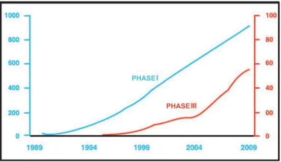
PHASE
Figure 8 – Cumulative curves of evolution of phase i and phase iii clinical trials in the area of gene therapy, developed from data registered in the database of the Journal of Gene medicine. While phase i trials show a linear growth, the red curve suggests an acceleration in the evolution of phase iii trials since 2004. Note that the vertical scales for the two phases are differ-ent.
Figure 9 – therapeutic indications of clinical trials registered in the database of the-Journal of Gene medicine. reproduced with permission of the publish-ers.
the distribution of clinical trials by therapeutic indication (Figure 9) confirms a point previously raised in this article. Although gene therapy was originally conceived with the aim of treating monogenic diseases, these today account for less than 10 percent of clinical trials. the prevalence of cancer may be explained in part by greater ease in the approval of clinical trials based on the
Cancer 64.5% (n=1060)
Cardiovascular diseases 8.7% (n=143) Monogenic diseases 8.2% (n=134) Infectious diseases 8% (n=131) Neurological diseases 1.8% (n=30) Eye diseases 1.1% (n=18) Other diseases 2.4% (n=40) Gene marking 3% (n=50) Healthy volunteers 2.3% (n=38) Clinical Indications of Clinical Gene Therapy Trials
compassionate use of experimental drugs or therapies in terminally ill patients, but also by the breakthrough in the design of oncolytic viruses (which destroy tumor cells) and suicide gene therapies (see below).
Figure 10 – Classification of genes used in clinical trials registered in the Journal of Gene medicine. reproduced with permission of the publishers.
Adenovirus 23.8% (n=400) Retrovirus 20.5% (n=344)
Plasmid / naked DNA 17.7% (n=304) Vaccinia virus 7.9% (n=133) Lipofection 6.5% (n=109) Poxvirus 5.5% (n=93)
Adeno-associated virus 4.5% (n=75) Herpes simplex virus 3.3% (n=56) Lentiviral 1,7% (n=29)
Other categories 4.9% (n=82) Unknown 3.3% (n=55)
Vectors Used in Clinical Gene Therapy Trials
The Journal of Gene Medicine, © 2010 John Wiley and Sons Ltd. www.wiley.co.uk/genmed/clinical
Figure 11 – Vectors used in clinical trials registered in the database of the Journal of Gene medicine. reproduced with permission of the publishers.
the variety of genes used in clinical trials (Figure 10) reflects the ad hoc nature of gene therapy. Progress in this area of medical research is probably strongly influenced by the trend towards the development of personalized medicine based on advances in genetics, pharmacogenomics and other fields of modern research. still, the prevalence of antigens, cytokines, tumor suppres-sor genes and suicidal genes corresponds to the prevalence of cancer as the most frequent indication in clinical research in this area. the topic of vectors is undoubtedly the most critical to the advancement of gene therapy as regards application to medical practice. the graph in Figure 11 compiles data obtained
Antigen 19.8% (n=325) Cytokine 18.4% (n=302) Tumor suppressor 10.5% (n=173) Growth factor 7.7% (n=173) Deficiency 7.2% (n=118) Suicidal 7.2% (n=118) Receptor 5.8% (n=95)
Replication inhibitor 4.1% (n=68) Marker 3% (n=58)
Other categories 12.9% (n=56) Unknown 3.4% (n=56)
Types of Genes Transferred in Clinical Gene Therapy Trials
over two decades, during which technological progress in the field of vectors was extraordinary. For example, in contrast to the type of adenovirus vector that led to the death of a patient in a clinical trial in 1999 (see below) and almost paralyzed gene therapy research, third-generation adenoviral vectors are avail-able today, which are radically modified in order to avoid adverse effects such as the one that killed the said patient. However, there is increased expectation in the use of viral vectors inherently safer, such as vectors derived from adeno-associated viruses.
Applications of gene therapy
to illustrate the potential applications of gene therapy as well as the un-derlying logic and the sequence of fundamental and pre-clinical research that led to clinical trials, some examples have been selected, which are described below.
Monogenic diseases
Haemophilia: since each type of hemophilia is a monogenic disease, the procedure is to introduce the respective healthy gene (Factor Viii or Factor iX, depending on the type of hemophilia) into the patient’s cells, so that these will start to produce the required protein. the therapy should not only get the body to produce the missing protein, but to produce it in sufficient quantity to restore the patient’s health and for a long time, ideally throughout the person’s lifetime.
After extensive preclinical trials in mice and dogs, which showed long-term recovery of factor iX infusion-mediated procoagulant activity using ex-perimental gene therapy trial, two Phase i / ii studies have been recently con-ducted by a group at the University of Pennsylvania, with the infusion of an adeno-associated virus vector (rAAV) containing the gene encoding factor iX of haemophilia B patients (manno et al., 2003, 2006, Hasbrouck & High, 2008). No serious adverse effects were observed in any of the patients tested.
the results indicate the potential efficacy of the treatment, since a patient who received a high dose of rAAV-F9 by liver infusion presented, between two and five weeks of treatment, therapeutic levels of circulating factor iX above 10 percent of normal activity, which is sufficient to sustain blood coagulation capacity. However, the therapeutic effect was transient, having disappeared six weeks after treatment, accompanied by a temporary and asymptomatic increase in transaminase levels (Figure 12). the results observed in this patient as well as in another patient in the same study indicated that the therapeutic effects disappeared as a result of degeneration of the liver cells in which the vector had been introduced, caused by an immune response to the viral vector proteins (mingozzi & High, 2007).
immune response observed in the patients. the result demonstrated the need for caution in the transition from preclinical to clinical trials, even in the absence of se-rious adverse effects, and provided critical data to advance the clinical application of gene therapy. New experimental studies are underway, with the aim to avoid this immune response with the use of vector variants and transient immunosup-pression, which will guide future clinical trials (Hasbrouck & High, 2008).
Figure 12 – Activity of circulating factor iX (red) and transaminase levels (blue and green) over time after gene therapy for hemophilia B in a clinical trial con-ducted with the use of adeno-associated virus vector containing the gene encoding factor iX. modified from Hasbrock & High (2008).
Leber’s congenital amaurosis: the first results of phase i/ii clinical tri-als for the treatment of Leber’s congenital amaurosis (LCA) began to be pub-lished in the end of April 2008. LCA is a disease that causes progressive blind-ness, starting with significant loss of vision in infants and progressing over time to total blindness. initially, photoreceptors, which are light-sensitive retinal cells essential for vision, are inactivated, but remain alive in the retina (den Hollander et al., 2008). over the years, inactive photoreceptors, predominantly rods that function in dim light, degenerate and disappear (spuy et al., 2005).
there are several forms of LCA, some of well-known genetic cause, such as rPE65 deficiency, an enzyme required to produce the vitamin derivative required for the functioning of photoreceptors (Poehner et al. 2000; Bereta et al. 2008). the photoreceptors of these patients gradually lose function, but de-generation usually only occurs around 30 years of age (Hollander et al., 2008). the course of the disease provides a therapeutic window for injecting healthy copies of the rPE65 gene into the retina of young adults with this form of LCA (Figure 13). the tests are still preliminary and, in principle, only three patients have been tested in each of the three Phase i clinical trials conducted in England and in the United states (Bainbridge et al., 2008; maguire et al., 2008; Cideci-yan et al., 2008; Hauswirth et al., 2008).
F.IX Infusion
F
.
IX
A
c
ti
v
ity
(%
)
Weeks
L
iv
e
r
e
n
zy
m
e
s
(U
l/
m
l)
Figure 13 – Diagram of gene therapy for Leber congenital amaurosis caused by rPE65 mutations. the figure illustrates clinical trials conducted since 2007 by teams from the University College in London, England, and the universi-ties of Pennsylvania and Florida, in the United states. this degenerative retinopathy initially produces inactivation of rod function, with progres-sive loss of scotopic vision and subsequent photoreceptor degeneration before 30 years of age, leading to blindness. the three research groups are testing the effects of the injection of healthy rPE65 gene into the retina of young adult patients with LCA. studies are underway and re-searchers are evaluating also the safety of the procedure, the patients’ vi-sual acuity compared to that presented before the gene therapy. in some cases there was an improvement in the results of ophthalmic exam (top right), pupillary reflexes and spatial orientation in dimly lit environments containing various obstacles (below right, in a frame extracted from the movie moorfields Hospital, courtesy of Prof. robin Ali, University Col-lege, London).
the first results showed that the infusion of adenovirus-associated virus vectors containing the healthy gene in the patients’ retina did not cause sig-nificant adverse effects. improvement was observed in ophthalmic exams and visual performance in some patients, who partially recovered sensitivity to light (Hauswirth et al., 2008) and the ability to orient themselves in dimly lit envi-ronments, something they were unable to do before the injection of the healthy gene (Bainbridge et al., 2008).
the results achieved so far, however, relate to few patients, and no signs of improvement have been observed in certain crucial ophthalmic exams (Haus-wirth et al., 2008), and rPE65 deficiency accounts for only 6 percent of LCA cases (Hollander et al., 2008). that is, the therapy that is being tested at the moment, if successful, can only be applied to a small fraction of patients.
treat-Mutations in the RPE65 gene cause loss of photoreceptor function and blindness
photoreceptors
retina
light light
ophthalmic exams
Patient in vision test in the dark Injection of viral vector containing the RPE65
ments for the other groups of patients need to be developed on a case by case basis. still, it is an important advance in the development of novel therapies for blindness-causing diseases, and a phase ii clinical trial is underway to confirm (or not) in a systematic way, the possible efficacy of the treatment.
Cancer
most clinical gene therapy trials have been conducted in cancer patients (Figure 9), usually in advanced stages of the disease. the desirable effect of any cancer treatment is to cause the selective death of tumor cells (Evan & Little-wood, 1998; Green & Evan, 2002). Cancer cells often multiply rapidly, which explains the growth of tumors. many drugs are used in treating cancer precisely because they selectively attack cells that multiply rapidly and, therefore, kill tu-mor cells (Wang et al. 2,008; Prochownik, 2008; Vazquez et al. 2008).
the physiological need for continuous renewal of blood cells from the prolif-eration of bone marrow precursors implies, however, severe adverse effects of che-motherapy. these effects are difficult to avoid because, among other factors, the drugs are injected into the bloodstream. For treating cancer it is desirable to reach, in some way, only the tumor cells. in the case of solid tumors, such as tumors in the central nervous system, this can be achieved through localized gene therapy (rainov & ren, 2003), and various strategies are being developed accordingly (table 2).
the procedure dubbed “suicide gene technique” consists in introducing into the tumor cells a gene that does not exist in the human genome and that encodes the thymidine kinase enzyme of the herpesvirus genome. the presence of this enzyme in a human cell kills the cell in the presence of a drug called ga ciclovir, as the thymidine kinase converts ganciclovir into a toxin. the toxin, in turn, affects only multiplying cells (Figure 14).
Figure 14 – Action mechanism of suicide genes. the concept, formulated in the late 1960s by the American researcher Frederick moolten from Boston University, is
after surgery, the therapeutic gene is injected in the remainder of the tumor
the therapeutic gene controls the production of an enzyme
the toxin kills tumor cells while preserving neurons the enzyme converts
shown schematically in the figure, following the arrows from the upper left corner. the example refers to glioblastomas, but also applies to other types of tumors. First, the neurosurgeon removes as much of the tumor as possible, leaving tumor cells scattered among normal brain neurons. the therapeutic gene (HsV-tK or thymidine kinase) is injected into the surgical area, petrating the cells and commanding the production of the enzyme. this en-zyme phosphorylates the ganciclovir injected, transforming it into a powerful toxin that is incorporated into the DNA of target cells, blocking DNA replica-tion and leading eventually to the death of the proliferating cell. reproduced from Linden (2008) with permission of the publishers.
Although the efficacy of suicide gene technology to treat tumors is still controversial, some studies have reported promising results. Among them is a phase i/ii clinical trial conducted in Finland, in which the resection of extreme-ly aggressive tumors of the central nervous system, known as glioblastomas, was followed by the injection into the surgical cavity of an adenoviral vector contain-ing the thymidine kinase gene of herpesvirus. the procedure continued with daily intravenous injections of ganciclovir for 14 days. Gene therapy resulted in a significant increase in the survival (Figure 15) of the group of 17 patients treated with gene therapy, as compared to a group of 19 patients treated with conventional therapy, or as compared to a control population of 36 patients pre-viously treated by conventional methods in the same neurosurgery unit, in the two years prior to the trial (immonen et al., 2004). the vector used in this study is being developed by the company Ark therapeutics, which recently reported significant positive results of a phase iii multicentric study with 250 patients, and in February 2009 was granted in France the first authorization for compas-sionate use of the product called Cerepro®.
table 2 – Genes and gene therapy strategies for tumors of the central nervous system
Strategy Examples Operation
Suicide genes - induction of programmed selective killing of tumor cells
HSV-TK (herpes virus thymidine kinase)
Blocking of DNA synthesis in the presence of a pro-drug
Oncolytic virus with conditional replication
HSV-1 Onyx-015
Replication only in dividing or tumor cells
Induction of apoptosis FasL, TRAIL Activation of apoptosis
High afinity ligands Transferrin receptor Drug speciically targeted to the tumor
Corrective strategy p53, Rb, p16, PTEN Correction of genes eliminated
from tumors
Immune gene therapy Interleukins, interferons, TNF-α Activation of antitumor immune
response
Suppression of angiogenesis Angiostatin, endostatin Blocking of blood vessel growth
Interference RNA VEGF, EGFR, IGFR Decreased expression of
oncogenes
Combination with cell therapy Neural stem cells or
mesenchymal as producers of viral vectors
Continuous and localized production of viral vectors
Figure 15 – increased survival of glioblastoma patients treated with gene therapy with the use of “suicide gene”. An adenoviral vector encoding the gene of thy-midine kinase enzyme was injected into the surgical cavity after resection of the tumors, followed by intravenous injections of ganciclovir for two weeks. the Kaplan-meier curve indicates the fraction of surviving patients over time for patients treated (red) and controls (black). the interpre-tation is that gene therapy was effective on part of the tumor cells that remained after surgery, which could not be attacked otherwise. modified from immonen et al. (2004).
Parkinson’s Disease
Neurodegenerative diseases are one of the most complex classes of diseases facing contemporary medicine. Despite the advances achieved since the 1990s, a period called “the brain decade” (Goldstein, 1994), and the extensive body of knowledge about various aspects of the pathogenesis, genetics, clinical course, complications and response to different treatments tested over years of research, there is a conspicuous lack of therapeutic options, particularly in the later stages of these diseases (radunovic et al., 2007; Cacabelos, 2007; Han & mcDonald, 2008; Jalbert et al., 2008; Gauthier & Poirier, 2008; olanow et al., 2008).
in turn, some neurodegenerative disorders illustrate the potential for the development of gene therapy for multifactorial and high complexity diseases. Parkinson’s disease (PD) is an example of this category.
PD is characterized by the progressive death of neurons in the substantia nigra pars compacta of the midbrain and functional changes in other brainstem nuclei (Figure 16), followed by the appearance of intracellular inclusions known as Lewy bodies. this results in loss of dopamine - the neurotransmitter used by neurons that degenerate - in the target of nigral extension neurons in the substantia nigra, which is called the striatum. As the disease progresses, other
Su
r
v
iv
a
l
Weeks Control
neurotransmitter systems become involved. motor disorders typical of the dis-ease, such as resting tremor, slowness of movement and muscle stiffness are fre-quently accompanied by postural instability, visceral dysfunction and cognitive disturbances (Guttman et al., 2003). the mechanisms that lead to the death of nigral neurons are still controversial (Dawson & Dawson, 2003; Dauer & Przedborski, 2003).
Pharmacological treatment with L-dopa, a precursor of dopamine synthe-sis, is effective in the short or medium term, but tends to become innocuous with the progressive loss of neurons, and can potentially cause additional motor disorders. the progression of the disease requires higher doses and combina-tions of drugs, which not always are effective (Poewe, 2009). Cellular therapies aimed at regenerating nigral dopaminergic neurons may eventually benefit PD patients, but so far clinical trials conducted with fetal nerve cell grafts have pro-duced discrete effects and suggested the possibility of disease transmission to the transplanted tissue (thajeb et al. 1,997, Li et al. 2,008; Kordower et al. 2,008; mendez et al. 2,008; Braak & del tredici, 2008).
Figure 16 – Diagram of main neurotransmitter connections and systems of the basal ganglia circuitry relevant for Parkinson’s disease. Degeneration (in red) of substantia nigra pars compacta dopaminergic neurons (sNc) reduces
CEREBRAL CORTEX - MOTOR AREAS
neurturin
T H A L A M U S
BRAINSTEM, SPINAL CORD
activation of dopamine receptors (D1 and D2) in the striatum (str). As a result, the activity of projection targets of the striatum becomes unbal-anced producing, among other effects, hyperactivity of the glutamatergic neurons of the subthalamic nucleus (stN), causing motor disturbanc-es. the strategies of gene therapy trials in PD patients are indicated by arrows and their respective targets are in blue. modified from Nakano (2000).
Gene therapy strategies for Parkinson’s disease include induction of lo-cal production of dopamine in the striatum, supply of neurotrophic factors to reduce the progressive loss of dopaminergic neurons, or even compensation of functional imbalance in the cell communication network of the basal ganglia (Chen et al. 2005).
the production of dopamine depends mainly on the activity of three en-zymes. the techniques aimed to produce dopamine in the depleted striatum in-volve, in general, viral-vector induction of one or more of these enzymes (Kang et al. 2001). Preclinical experimental models consist of chemical lesions of the substantia nigra in rats or primates. several types of viral vectors have been tested (Chen et al., 2005 for review). Based on the results of preclinical studies, a phase i clinical trial has been started to test the safety and, secondarily, the beneficial effects of gene therapy by expression of one of the producing enzyme (AADC), carried by an adenovirus-associated vector injected in the striatum of PD pa-tients who have had the diseases for 14 years on average (http://clinicaltrials. gov/show/nCt00229736). the results (Christine et al., 2009) showed clinical improvement without adverse effects of the gene therapy per se, although risks have been identified in the surgical procedure.
on some of these signs, justifying the use of the so-called deep brain stimula-tion in the treatment of advanced cases of PD (Jankovic & Diamond, 2005). Knowledge of the functional properties of neural circuits involved in the disease has led to a striking example of genetic intervention designed to modulate the physiology of the nervous system, regardless of the cause of the disease which, even today, remains controversial.
A gene therapy trial has been developed, which involves inducing the ex-pression of enzymes that produce an inhibitory neurotransmitter, with the aim of inhibiting excessive neural activity in the stN. the expression of these en-zymes in the stN produced beneficial functional effects in PD models in rats (Luo et al. 2002). Based on these results, a phase i clinical gene therapy trial was conducted in the period 2003-2005, using a recombinant adeno-associated virus vector containing the gene encoding one of these enzymes, injected into the stN (http://www. clinicaltrials.gov/ct/show/nCt00195143). results for 11 patients monitored for up to 12 months showed significant improvement in motor performance, accompanied by reduced metabolic activity in stN pro-jection targets, consistent with the results of preclinical studies. A significant improvement in daily activities was also reported, which reflects the views of pa-tients about their performance on tasks of everyday life. No adverse effects have been reported questioning the safety of the procedure (Kaplitt et al., 2007).
the results of the clinical trials described are still very preliminary, were obtained in small numbers of patients and require confirmation through broad-er trials with more stringent controls for placebo effects and othbroad-er variables. therefore, it is still early to conclude on the viability and particularly the efficacy of gene therapy for neurodegenerative diseases. However, these studies are in addition to other clinical trials suggesting that gene therapy may become an ef-fective alternative for treating currently incurable diseases.
Risk-beneit assessment of gene therapy
Among the hundreds of clinical gene therapy trials completed to date, most aimed to test the safety of the procedure. in certain cases, early detection of adverse effects during the study period was sufficient to immediately termi-nate the trial, thereby avoiding any risk of aggravation. But in many cases, the procedure used was considered safe, with only occasional, mild and tolerable adverse effects.
immune reactions, however, not only can cause adverse effects, but even in the absence of these can destroy the vectors or cells infected with viral vectors, despite the use of sophisticated recombinant DNA techniques in their production. this was the case of the clinical trial for hemophilia B previously described (mingozzi & High, 2007), but which did not bring significant consequences for patients. in other cases, however, the adverse effects can be quite severe or, in rare cases, even fatal.
in 1999 a patient died soon after the injection of a viral vector during a clini-cal gene therapy trial, victimized by a systemic inflammatory response syndrome caused by first-generation adenoviral vector (raper et al., 1998, 2003). in more recent clinical trials conducted in France and England (Hacein-Bey-Abina et al., 2002; Gaspar et al., 2004), of a total of 20 children under one year of age under-going gene therapy for X-linked severe combined immunodeficiency syndrome (XL-sCiD) (Buckley, 2004), five developed leukemia (Hacein-Bey-Abina et al., 2003; Howe et al., 2008). of these, one died and four went into complete remis-sion after chemotherapy. tests performed after the onset of leukemia revealed that the retroviral vectors used in both trials produced insertional mutagenesis, i.e., mutations produced by the injection of the DNA vector, breaking the continuity of the genetic sequence (Cavazzana-Calvo & Fischer, 2007; Howe et al., 2008).
the above mentioned cases are the most serious examples actually terized as direct adverse effects of gene therapy. Both are derived from charac-teristics of the viral vectors used. However, in both cases fundamental research coupled with careful observation of the events associated with the treatment and the clinical course of the adverse effects contributed to advances in the design and production of novel vectors aimed to avoid such adverse effects.
in the case of adenoviral vectors, in contrast to the first-generation vectors used in the clinical trial that resulted in the fatal case in 1999, third-generation adenoviral vectors constructed with complete deletion of viral genes and capable of much safer gene transduction in humans are now available (räty et al., 2008; Dormond et al., 2009). in turn, there is a growing expectation of avoiding in-sertional mutagenesis like the one observed in XL-sCiD trials, through the de-sign of auto-inactivating retroviral or lentiviral vectors or vectors with chromatin insulators, two of the most promising techniques currently under development for this vector class (Yi et al., 2005; räty et al., 2008).
Gene therapy for XL-sCiD, in turn, was curative in 19 of the 20 children treated, who showed significant improvement in their immune system less than three months after treatment, as well as persistent recovery of resistance to infections (table 3; Fisher & Cavazzana-Calvo, 2008; Aiuti roncarolo, 2009). However, the treatment was not effective in adolescents, suggesting a limited therapeutic window for intervention in this disease. Adding up to successful cases are 30 patients treated for ADA-sCiD, the form of immunodeficiency that corresponds to the first patient treated by gene therapy in 1989 (table 4; Aiuti & roncarolo, 2009).
gene therapy trials acted quickly in both cases of adverse effects reported here. in 1999, the trial that resulted in the patient’s death was permanently discon-tinued, despite the absence of serious adverse effects in the other 17 patients treated in the same study. in the case of XL- sCiD trials, therapeutic procedures had already been terminated, but authorizations for other similar trials were suspended pending a thorough evaluation of the data, and were subsequently granted. Despite the recognition that the gene therapy procedure was respon-sible for the adverse effects, the regulatory committees concluded that none of these events, as well as other adverse effects reported occasionally justified the suspending clinical gene therapy trials. indeed, the analysis of adverse effects has helped guide the development of biotechnology in the field and, at the same time, improve regulation and criteria for clinical trial authorization.
table 3 – XL-sCiD clinical gene therapy trials
Trial No. of patients Observation
period
Effectiveness Toxicity
Necker Hospital, Paris
10 (age < 1 year)
10 years yes Leukemia (4 patients), 3
complete remissions after chemotherapy
Great Ormond St Hospital, London
10 (age < 1 year)
7 years yes Leukemia (1 patient), complete
remission after chemotherapy
Multicentric, FR, UK, US
5 (age = 10-20 years)
3 years no no
Source: Adapted from Aiuti & roncarolo (2009).
table 4 – ADA-sCiD clinical gene therapy trials
Trial No. of patients Observation
period
Effectiveness Toxicity
HSR-TIGET 15 8 years yes no
GOSH 5 5.5 years yes no
CHLA/NIH (1)* 4 8 years no no
CHLA/NIH (2)* 6 2 years yes pancytopenia** (1 patient)
* studies (1) and (2) differ in the pre-drug treatment of patients before gene therapy.
** the adverse effect was attributed to a cytogenetic abnormality independent of the gene therapy. Source: Adapted from Aiuti & roncarolo (2009).
Gene therapy and biotechnology
Entrepreneurs in the area of biotechnology saw in human genome sequenc-ing growsequenc-ing business opportunities. the interest lies, of course, in the fact that the discovery of genes and especially of mutations responsible in whole or in part for a disease can lead to the development of diagnostic tests or marketable drugs.
Among other things, companies have begun to invest in the patenting of genes or sequences of DNA fragments that had not even been associated with genes. more than three million genome-related patents have been filed to date in the United states. U.s. legislation, in general, authorizes the patenting of genes, provided that these are isolated (and not just described as nucleotide sequences) and supported by evidence of usefulness, for example, for the de-velopment of diagnostic tests. However, the patenting of genes is controversial. For example, the internal rules for evaluating the usefulness of findings related to genes enforced since 2001 by the office of the United states Patent office (UsPto) have been and still are the subject of severe criticism, from which the UsPto defended itself on the grounds of the patent legislation in force in the United states. on the other hand, the National institute of industrial Property (iNPi), the Brazilian agency that grants nationally valid patents, reports on its website that the patenting of natural genes is prohibited in Brazil.
outside the scope of the controversy over the patenting of genes, vectors for gene therapy, whether viral or nonviral, containing therapeutic genes, as well as their specific applications are products of technological development and, as such, are legitimate objects of patenting and possible marketing (Bobrow & thomas, 2002). Hundreds of such patents have been filed with the UsPto and its counterparts in Europe and Asia. Dozens of companies are investing in gene therapy, based on patented technologies for the production of vectors or as partners of research institutions (table 5).
table 5 – overseas companies engaged in gene therapy
1. Advanced Cell & Gene
Therapy, LLC
2. Advanced Cell Technology
3. Advanced Vision Therapies,
4. AlphaVax Human Vaccines, InC.
5. Altogen Biosystems 6. Amaxa GmbH
7. Amsterdam Molecular Therapeutics
8. Applied Tissue Technologies LLC
9. Ark Therapeutics Ltd.
10. Athersys, Inc. 11. AuRx Inc
12. Austrianova FSG {FSG AUSTRIANOVA GmbH}
13. Avaris AB
14. Avigen Inc.
15. Bavairian Nordic A/S
16. BetaStem Therapeutics Inc
17. BioCardia Inc 18. Bioheart, Inc.
19. BioProtein I Technologies
20. Biovex Limited 21. Cardion AG
22. Cell Genesys Inc
23. Cellectis SA
24. Cellerant Therapeutics Inc.
25. CellGenix Technology
Transfer GmbH 26. Cellprep S. A.
27. Ceregene Inc.
28. Collateral Therapeutics, Inc.
29. Copernicus Therapeutics Inc
30. Corgentec Inc. 31. Cyclacel Limited
32. CyThera Inc.
33. Cyto Pulse Sciences, Inc
34. CytoGenix Inc
35. DeveloGen AG 36. Enzo Biochem, Inc.
37. Epeius Biotechnologies Corporation
38. Expression Genetics Inc 39. geneRx+
40. Genetix Pharmaceuticals Inc
41. GenVec. Inc.
42. IC-Vec Ltd
43. Ichor Medical Systems, Inc. 44. Immuno-Designed Molecules
(IDM), SA
45. Insert Therapeutics Inc.
46. Intercytex Limited 47. Introgen Therapeutics, Inc.
48. Intronn, Inc.
49. Invivogen
50. Ixion Biotechnology
51. MaxCyte, Inc. 52. MediGene, Inc.
53. Mirus Corporation
54. Innovata pic
55. Molecular Medicine, LLC
56. Mologen Holding AG
57. Nature Technology Corporation
58. Nephros Therapeutics, Inc. 59. Neurotech SA
60. Nucleonics Inc.
61. Oncosis
62. Onyx Pharmaceuticals, Inc. 63. OrphagenicX
64. Oxford BioMedica 65. PharmaFrontiers Corp.
66. Polygenetics Inc. 67. PrimeGen Biotech LLC. 68. Progenitor Cell Therapy,
LLc
69. Proneuron Biotechnologies, Inc.
70. Regulon Inc.
71. Reneuron 72. REPLICor Inc.
73. Ribozyme Pharmaceuticals Inc. (RPI)
74. Sertoli Technologies Inc. 75. Stem Cell Sciences
76. StemCells Incorporated 77. Supratek Pharma Inc. 78. Targeted Genetics
Corporation 79. TheraCyte, Inc.
80. Theratechnologies 81. TheraVitae Co., Ltd. 82. Tosk lnc
83. Transgene S.A 84. Valentis, Inc.
85. VIA Pharmaceuticals, Inc. 86. Vical Incorporated 87. VirRx, Inc.
table 6 – Gene therapy products in advanced stages of development
Product Composition Indication Company Status
Gendicine® rAd-p53 Tumors of the head and neck
SiBiono GenTech, China
Approved and on the market in China
(2003)
Rexin-G® tumor matrix (collagen)-targeted,
retroV-dnG1-Cyclin
Solid tumors Epeius Biotech, U.S.
Compassionate use in Japan (2007);
approved in the Philippines
Collategene® Plasmid-HGF Critical limb ischemia
AnGes MG/ Daiichi Sankyo,
Japan
Under review in Japan; special protocol analysis (SPA) in the U.S.
Advexin® rAd5CMV-p53 Tumors of the head and neck
Introgen, U.S. Under review; phase II Trial underway
Cerepro® rAd5-TK Glioblastoma Ark Technologies,
ING/Finland
Approved for individualized use in
France and Finland (2009)
Figure 17 – Evolution in the number of gene therapy products under development in bio-technology companies in the period 1995-2009. modified from <http://www. pharmaprojects.com/therapy_analysis/genether_early_0409.htm>.
Figure 18 – Evolution in the number of gene therapy products in preclinical or clini-cal trial phases under development in biotechnology companies in the period 1995-2008. modified from http://www.pharmaprojects. com/ therapy_analysis/genether_current_0409.htm>. N u m b e r o f g e n e th e ra p y p ro d u c ts Year N u m b e r o f g e n e th e ra p y p ro d u c ts N u mb e r o f g e n e t h e ra p y p ro d u ct s
Gene therapy in Brazil
Despite the history and international recognition of Brazilian genetics, there are few research groups devoted to gene therapy studies, including DNA vaccines. Until recently there was little public investment in this research area and no interest from the private sector. the picture, however, is starting to change with some initiatives, albeit modest, in both sectors.
The Gene Therapy Network
A Gene therapy Network began to be organized in Brazil in 2005. this network, coordinated by this author, initially brought together 14 research groups from three states (rio de Janeiro, são Paulo and rio Grande do sul), dedicated to research in the area of gene therapy and DNA vaccines. the stud-ies involve the development of viral vectors, basic research and preclinical trials in the areas of cancer, genetic diseases, neurodegenerative diseases and DNA vaccines for dengue fever, Chagas disease, streptococcal infections and cancer.
A first clinical gene therapy trial for myocardial revascularization with the use of plasmid vectors containing the VEGF gene (Vascular Endothelial Growth Factor), was started in February 2009 in Porto Alegre, promoted jointly by the institute of Cardiology of rio Grande do sul, the research support Foun-dation of rio Grande do sul and the Gene therapy Network, through the mCt/CnPq millennium institutes Program (http://clinicaltrials.gov/ct2/ show/nCt00744315). this is the first clinical gene therapy trial hosted in south America, in the midst of dozens of clinical trials promoted by multinational companies or foreign research institutions with the participation of researchers from south America (table 7).
table 7 – sponsors of clinical gene therapy trials with participation of institutions in south America. Numbers in parentheses indicate the number of trials sponsored by the corresponding company
Actelion (5)
Bristol MyersSquibb (2) Eli Lilly (1)
Genentech (3) Glaxo Smith Kline (7) Hoffman-La Roche (3) Hoosier Oncology Group (1) MedImmune LLC (1)
National Cancer Institute USA (1) Ofice of Rare Diseases (1) Sanoi-Aventis (9)
Shire Human Genetic Therapies Inc (1) St Jude’s Children’s Research Hospital (2)
Cardiology Institute of RGS + Fapergs + CNPq-Instituto do Milênio Rede de Terapia Gênica (1) (Gene Therapy Network – Millennium Institute)
Gene therapy and biotechnology in Brazil
Consistent with the embryonic stage of gene therapy research, there is not much interest from the private sector in this area in the country. recently, however, a company hosted in the high-technology Park Foundation was estab-lished in Petrópolis, in the state of rio de Janeiro, which, among other services of biotechnology nature, is beginning to provide support for gene therapy trials in the country.
the vector for the clinical trial of myocardial revascularization started in Porto Alegre was produced by this service company, an unprecedented event in the country and a harbinger of new partnerships between the private sector and academic institutions in scientific research.
in turn, the awareness of the crucial role of mechanisms to protect in-tellectual property in this area is creating habits in researchers previously un-concerned with patenting products and processes of biotechnological interest. thus, the performance of the Gene therapy Network has also stimulated the first international filing of a gene therapy patent in its strict sense by a Brazil-ian institution, as a result of this author’s empirical research (World intellectual Property organization - Wo2009/121157 - PCt/Br2009/000093 .)
Conclusion
We are still in the early stages of gene therapy history and all that has been done to date are but the first steps on a long and winding road (Flotte, 2007). But now there are some occasional successes that demonstrate the feasibility of incorporating gene therapy into medical practice. the main advances, so far, are in the areas of hemophilia, certain types of cancer, severe combined immunode-ficiency syndromes and certain retinopathies.
there has been great progress in the design and construction of new safer and more efficient vectors (räty et al., 2008). in particular, the immune responses of patients are being studied in depth, new animal model studies have been de-veloped and research is advancing towards increasing the safety of clinical trials.
the problems are not trivial. Just remember that, after all the medical progress made to date, despite the success achieved in new treatments and in the prevention of so many diseases in the last 150 years, we are still struggling against incurable diseases that challenge the imagination and scientific and tech-nological competence of the entire scientific world.
there are, however, reasons for optimism, and the expectation of success in gene therapy technologies has gradually increased. A sign of the viability of gene therapy in the near future is the increasing investment of biotech compa-nies in the development and filing of requests for the release of biological prod-ucts relating to gene therapy.
involved in this field in the country is still insignificant compared to First World countries. But from the financial and scientific and educational standpoints, the decision to invest in this area will certainly yield significant return to the Brazil-ian medicine of the twenty-first century.
references
AiUti, A. et al. Gene therapy for immunodeficiency due to adenosine deaminase deficiency. N. Engl. J. Med., v.360, n.5, p.447-58, 2009.
AiUti, A.; roNCAroLo, m. G. ten years of gene therapy for primary immune deficiencies. Hematology Am. Soc. Hematol. Educ. Program, p.682-9, 2009.
ANDErsoN, W. F. Human gene therapy: the initial concepts. in: BriGHAm, K. L. (Ed.) Gene therapy for diseases of the lung. s. l.: CrC Press, 1990. p. 3-16.
ANDErsoN, W. F. et al. the ADA human gene therapy clinical protocol: Points to consider response with clinical protocol. Hum. Gene Ther., v.1, n.3, p.331-62, 1990.
ANDrEWEs, C. richard Edwin shope. in: Biographical memoirs. s. l.: National
Academy of sciences, 1979. v.50, p.352-75.
ANDrEWEs, C. H. rhinoviruses and common colds. Annu. Rev. Med., v.17
p.361-70, 1966.
AtKiNs, G. J. et al. therapeutic and prophylactic applications of alphavirus vectors.
Expert Rev. Mol. Med., v.10, p.e33, 2008.
BAGLEY, J. et al. Gene therapy in type 1 diabetes. Crit. Rev. Immunol., v.28, n.4, p.301-24, 2008.
BAiNBriDGE, J. W. et al. Effect of gene therapy on visual function in Leber’s congenital amaurosis. N. Engl. J. Med., v.358, n.21, p.2231-9, 2008.
BAUZoN, m.; HErmistoN, t. W. Exploiting diversity: genetic approaches to creating highly potent and efficacious oncolytic viruses. Curr. Opin. Mol. Ther., v.10, n.4, p.350-5, 2008.
BErEtA , G. et al. impact of retinal disease-associated rPE65 mutations on retinoid isomerization. Biochemistry, v.47, n.37, p.9856-65, 2008.
BLAEsE, r. m. et al. t lymphocyte-directed gene therapy for ADA-sCiD: initial trial results after 4 years. Science, v.270, n.5235, p.475-80, 1995.
BoBroW, m.; tHomAs, s. Patenting DNA. Curr. Opin. Mol. Ther., v.4, n.6, p.542-7, 2002.
BrAAK, H.; DEL trEDiCi, K. Assessing fetal nerve cell grafts in Parkinson’s disease.
Nat. Med., v.14, n.5, p.483-5, 2008.
BUCKLEY, r. H. molecular defects in human severe combined immunodeficiency and approaches to immune reconstitution. Annu. Rev. Immunol., v.22, p.625-55, 2004.
CACABELos, r. molecular pathology and pharmacogenomics in Alzheimer’s disease: polygenic-related effects of multifactorial treatments on cognition, anxiety and depression. Methods Find Exp. Clin. Pharmacol., v.29, suppl A, p.1-91, 2007.
CArDoNE, m. Prospects for gene therapy in inherited neurodegenerative diseases.
Curr. Opin. Neurol., v.20, n.2, p.151-8, 2007.
CAttANEo , r. et al. reprogrammed viruses as cancer therapeutics: targeted, armed and shielded. Nat. Rev. Microbiol., v.6, n.7, p.529-40, 2008.
CAVA ZZANA-CALVo, m.; FisCHEr, A. Gene therapy for severe combined immunodeficiency: are we there yet? J. Clin. Invest., v.117, n.6, p.1456-65, 2007.
CEmAZAr, m.; sErsA, G. Electrotransfer of therapeutic molecules into tissues. Curr.
Opin. Mol. Ther., v.9, n.6, p.554-62, 2007.
CHEN, Q. et al. Gene therapy for Parkinson’s disease: progress and challenges. Curr.
Gene Ther., v.5, p.71-80, 2005.
CHristiNE, C. W. et al.. safety and tolerability of putaminal AADC gene therapy for Parkinson disease. Neurology, v.73, n.20, p.1662-9, 2009.
CiDECiYAN, A. V. et al. Human gene therapy for rPE65 isomerase deficiency activates the retinoid cycle of vision but with slow rod kinetics. Proc. Natl. Acad. Sci. USA, v.105, n.39, p.15112-7, 2008.
CLANCHY, F. i.; WiLLiAms, r. o. Plasmid DNA as a safe gene delivery vehicle for treatment of chronic inflammatory disease. Expert Opin. Biol. Ther., v.8, n.10, p.1507-19, 2008.
CoLEmAN, W. B.; tsoNGALis, G. J. (Ed.) Molecular pathology: the molecular basis of human disease. s. l.: Academic Press, 2009. 664p.
CULVEr, K. W. et al. Correction of ADA deficiency in human t lymphocytes using retroviral-mediated gene transfer. Transplant Proc., v.23, n.1 Pt. 1, p.170-1, 1991.
DAss, C. r. Lipoplex-mediated delivery of nucleic acids: factors affecting in vivo transfection. J. Mol. Med., v.82, n.9, p.579-91, 2004.
DAUEr, W.; PrZEDBorsKi, s. Parkinson’s disease: mechanisms and models.
Neuron, v.39, p.889-909, 2003.
DAWsoN, t. m.; DAWsoN, V. L. molecular pathways of neurodegeneration in Parkinson’s disease. Science, v.302, p.819-22, 2003.
DiAmoND, A.; JANKoViC, J. the effect of deep brain stimulation on quality of life in movement disorders. J. Neurol. Neurosurg Psychiatry, v.76, p.1188-93, 2005.
EiNstEiN, o.; BEN-HUr, t. the changing face of neural stem cell therapy in neurologic diseases. Arch. Neurol., v.65, n.4, p.452-6, 2008.
EVAN , G.; LittLEWooD, t. A matter of life and cell death. Science, v.281, n.5381, p.1317-22, 1998.
FAVA rD, C. et al. Electrotransfer as a non viral method of gene delivery. Curr. Gene Ther., v.7, n.1, p.67-77, 2007.
FisCHEr, A.; CAVA ZZANA-CALVo, m Gene therapy of inherited diseases. Lancet,
v.371, n.9629, p.2044-7, 2008.
FJorD-LArsEN, L. et al. Efficient in vivo protection of nigral dopaminergic neurons by lentiviral gene transfer of a modified Neurturin construct. Exp. Neurol., v.195, p.49-60, 2005.
FLottE, t. r. Gene therapy: the first two decades and the current state-of-the-art. J.
Cell Physiol., v.213, n.2, p.301-5, 2007.
FriEDmANN t. the road toward human gene therapy--a 25-year perspective. Ann
Med., v.29 n.6 p.575-7, 1997.
FriEDmANN, t. stanfield rogers: insights into virus vectors and failure of an early gene therapy model. Mol. Ther., v.4, n.4, p.285-8, 2001.
GAsPAr, H. B. et al. Gene therapy of X-linked severe combined immunodeficiency by use of a pseudotyped gammaretroviral vector. Lancet, v.364, n.9452, p.2181-7, 2004.
GAUtHiEr, s.; PoiriEr, J. Current and future management of Alzheimer’s disease.
Alzheimers Dement., v.4, n.1, suppl. 1, p.s48-50, 2008.
GiLL, D. r. et al. Progress and prospects: the design and production of plasmid vectors.
Gene Ther., v.16, n.2, p.165-71, 2009.
GoLDstEiN, m. Decade of the brain: an agenda for the nineties. West. J. Med., v.161, p.239-41, 1994.
GrEEN, D. r.; EVAN , G. i. A matter of life and death. Cancer Cell., v.1, n.1, p.19-30, 2002.
GriBBEN, J. G. stem cell transplantation in chronic lymphocytic leukemia. Biol. Blood
Marrow Transplant., v.15, n.1, suppl., p.53-8, 2008.
GUttmAN, m. et al. Current concepts in the diagnosis and management of Parkinson’s disease. CMAJ, v.168, p.293-301, 2003.
HACEiN-BEY-ABiNA, s. et al. Gene therapy of X-linked severe combined immunodeficiency. Int. J. Hematol., v.76, n.4, p.295-8, 2002.