The Pathogenic Properties of a Novel and Conserved
Gene Product, KerV, in Proteobacteria
Dingding An1,2,3, Yiorgos Apidianakis1,2,3, Ana Laura Boechat4, Regina L. Baldini4, Boyan C. Goumnerov1, Laurence G. Rahme1,2,3*
1Department of Surgery, Massachusetts General Hospital and Harvard Medical School, Boston, Massachusetts, United States of America,2Shriners Research Institute, Boston, Massachusetts, United States of America,3Department of Microbiology and Molecular Genetics, Harvard Medical School, Boston, Massachusetts, United States of America,4Departamento de Bioquı´mica, Instituto de Quı´mica, Universidade de Sa˜o Paulo, Sa˜o Paulo, Brazil
Abstract
Identification of novel virulence factors is essential for understanding bacterial pathogenesis and designing antibacterial strategies. In this study, we uncover such a factor, termed KerV, in Proteobacteria. Experiments carried out in a variety of
eukaryotic host infection models revealed that the virulence of a Pseudomonas aeruginosa kerV null mutant was
compromised when it interacted with amoebae, plants, flies, and mice. Bioinformatics analyses indicated that KerV is a hypothetical methyltransferase and is well-conserved across numerous Proteobacteria, including both well-known and emerging pathogens (e.g., virulent Burkholderia, Escherichia, Shigella, Vibrio, Salmonella, Yersinia and Brucella species). Furthermore, among the 197kerVorthologs analyzed in this study, about 89% reside in a defined genomic neighborhood, which also possesses essential DNA replication and repair genes and detoxification gene. Finally, infection ofDrosophila melanogaster with null mutants demonstrated that KerV orthologs are also crucial in Vibrio cholerae and Yersinia pseudotuberculosispathogenesis. Our findings suggested that KerV has a novel and broad significance as a virulence factor in pathogenic Proteobacteria and it might serve as a new target for antibiotic drug design.
Citation:An D, Apidianakis Y, Boechat AL, Baldini RL, Goumnerov BC, et al. (2009) The Pathogenic Properties of a Novel and Conserved Gene Product, KerV, in Proteobacteria. PLoS ONE 4(9): e7167. doi:10.1371/journal.pone.0007167
Editor:Christophe Herman, Baylor College of Medicine, United States of America
ReceivedMay 20, 2009;AcceptedAugust 19, 2009;PublishedSeptember 25, 2009
Copyright:ß2009 An et al. This is an open-access article distributed under the terms of the Creative Commons Attribution License, which permits unrestricted use, distribution, and reproduction in any medium, provided the original author and source are credited.
Funding:This work was partially supported by Shriners grant#8892 (to L. G. R.) and by grant 03/09253-7 from Fundac¸a˜o de Amparo a` Pesquisa do Estado de Sa˜o Paulo (to R. L. B.). Y. A. was supported by a Shriners post-doctoral fellowship and A. L. B. by a Fundac¸a˜o de Amparo a` Pesquisa do Estado de Sa˜o Paulo graduate fellowship. The funders had no role in study design, data collection and analysis, decision to publish, or preparation of the manuscript.
Competing Interests:The authors have declared that no competing interests exist.
* E-mail: [email protected].
Introduction
Important infection mechanisms are often shared across diverse bacterial pathogens [1,2,3,4,5,6]. Identification and understanding of these conserved themes will not only expand our knowledge of specific virulence mechanisms, but will also provide information about the evolution of microbial pathogenesis. A convergence of such findings is also needed to inform new strategies against bacterial infections with wide clinical applications, and to provide new solutions to the ever-growing problem of antibiotic resistance. However, discovering broadly conserved virulence factors faces great challenges caused by the practical limitation mammalian hosts pose in high-throughput approaches [7]. This limitation has been considerably circumvented following the discovery that important virulence factors and corresponding pathways are conserved across a spectrum of hosts ranging from amoebae to mice [2,8,9,10,11]. This conservation made non-vertebrates amenable surrogate hosts for studying mammalian pathogenesis and added the benefit of enabling broadly conserved virulence factors to be identified. Indeed yeasts [12], plants [13], nematodes [14], fruit flies [15], and zebrafish [16] have all been successfully applied in pathogenesis experiments.
P. aeruginosais used as a model for bacterial pathogenesis study because of its potency as a multi-host pathogen and the abundance of tools that are compatible with it [13,17,18]. The present report describes the discovery of a conserved P. aeruginosa virulence
determinant, KerV. We further report experiments examining the breadth of KerV as a conserved virulence factor, not only against multiple hosts but also in several pathogens.
Results
P. aeruginosaKerV-mediated virulence is conserved against a spectrum of eukaryotic hosts
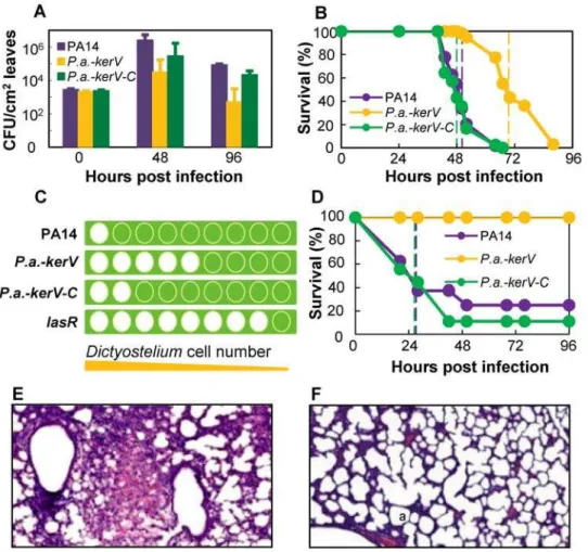
In a screen for novel evolutionarily conserved P. aeruginosa virulence factors using anArabidopsisinfiltration model [13,19], we identified a mutant with a TnphoA transposon insertion at gene PA14_41070 that exhibited decreased virulence compared to the parental strain PA14. This gene, annotated here askerV, encodes a hypothetical protein with 253 amino acids in length.
orders of magnitude less than those of PA14 (about 104.5 vs. 106.4CFU/ml at 48 h; about 102.7 vs. 104.9CFU/ml at 96 h; P,0.02); meanwhile the densities of CFUs of the complemented strainP.a.-kerV-Cwere similar to those of PA14 (P.0.1, Figure 1A). Accordingly the intensity of observed infection symptoms was reduced in leaves infected with theP.a.-kerVmutant, but restored in leaves infected withP.a.-kerV-C(data not shown).
The virulence of these strains was then examined in aDrosophila pricking model at 21uC in which about 100 bacterial cells were inoculated into the dorsal thorax of host flies. Flies infected with P.a.-kerVsurvived longer (n = 43, median survival 70 h) than those infected with PA14 (n = 47, 50 h) or P.a.-kerV-C (n = 42, 48 h) (P,0.0001, Figure 1B). These results are in agreement with our previous study [20], in which a kerV mutant (termed D12) was unable to evade host defense mechanisms in flies and therefore was deemed not proficient in infection.
Given the demonstrated involvement of KerV in both plant and fly pathogenesis, we asked whether KerV is indeed a key P. aeruginosavirulence factor important for infection of a broad range of hosts. The mutant’s virulence was tested in two additional eukaryotic hosts: amoeba and mouse. In the amoeba phagocytosis assay [21],Dictyostelium discoideumwith 2.5-fold sequential dilutions
starting with 2.06105cells were spotted on differentP. aeruginosa
lawns and the number of clear zones thatD. discoideummade by phagocytosingP. aeruginosawas recorded for each bacterial strain. In this assay, greater virulence is associated with a smaller number of clear zones and a bigger number of minimally required D. discoideum cells. Indeed, there was only one clear zone on the representative lawn of virulent PA14, indicating a minimum of 2.06105D. discoideumcells were needed for phagocytosis of PA14 (Figure 1C and Table S1). In contrast, eight clear zones (corresponding to about 3.36102 D. discoideumcells) were shown
on a representative lawn of the negative control lasR (a strain deficient in producing the master virulence regulator LasR [10]). Meanwhile, five clear zones (corresponding to about 5.16103D. discoideumcells) were shown on a representativeP. a.-kerVlawn and two clear zones (corresponding to about 8.06104 D. discoideum
cells) on a representativeP.a.-kerV-Clawn. Therefore our findings indicate that KerV plays an important role in P. aeruginosa resistance to amoeba phagocytosis.
In an acute mouse pulmonary infection model [22] in which 107 bacteria were administered intranasally,P.a.-kerVfailed to confer any mortality at 96 h post-infection, while the median survival of PA14 and P.a.-kerV-C infected mice were both 28 h (P,0.003,
Figure 1. KerV acts as a virulence determinant inP. aeruginosaagainst a range of eukaryotic hosts.(A) Bacteria quantities recovered from infected plant leaves. (B) Fly survival kinetics in a pricking infection model. Dotted lines are color-coded and represent survival medians for corresponding strains. (C) Amoeba phagocytosis ofP. aeruginosa. The graphic was produced from representative experimental observations. For detailed data see Table S1. Green square, bacterial lawn; green-filled circle, no phagocytosis of bacteria; white-filled circle, clear zone due to phagocytosis of bacteria. From left to right the numbers of laidDictyosteliumwere 2.06105, 8.06104, 3.26104, 1.36104, 5.16103, 2.06103, 8.26102,
3.36102and 1.36102. (D) Mouse survival kinetics in an acute lung infection model. (E and F) Histology of the infected neonatal mice lungs by PA14 (E)
Figure 1D). Histopathology of neonatal mouse lung tissues at 24 h post-infection showed that PA14 (Figure 1E) caused a typical lobar pneumonia characterized by pulmonary consolidation and intra-alveolar hemorrhage with massive inflammatory infiltrates. In contrast, neither inflammatory response nor hemorrhage was present inside the alveolar spaces of P.a.-kerV infected tissues (Figure 1F). These observations demonstrated that the virulence of thekerVmutant was attenuated in the mouse lung infection model and thus aroused much less inflammation. Interestingly, in a burn-mouse model [23], the mutant exhibited similar virulence compared to that of the parental strain in a 5-day experiment using 16105bacteria/mouse (P.0.05, Figure S2).
KerV is a hypothetical methyltransferase and is conserved in a defined genomic region in many Proteobacteria
A homology search in KEGG [24] revealed that KerV has putative methyltransferase type_11 motifs (Figure S3) and is annotated as a S-adenosylmethionine-depended methyltransferase (SAM-MT). SAM-MTs usually have three conserved amino acid motifs for SAM binding [25]. In KerV, the best match for motif I, usually the most conserved and most critical for binding SAM [26], is HAELPPSTG; however, the H and the first P are not present in other known methyltransferases to our knowledge. Secondary structure prediction by PROF algorithm [27] at PredictProtein site (http://www.predictprotein.org/) suggests it also lacks a classic seven-strandedbsheet [26]. Nevertheless, KerV possesses a fine post-I motif (LPGVD), a motif II (ADVVL), and a motif III (TVRPGGHLLL) with a commonly seen spacing of 11 residues between the putative motif I and post-I. In addition, the expected b strand for motif III is also suggested by PROF algorithm. These characteristics suggest that KerV encodes for either a novel methyltransferase with unique features or a protein with little familiarity.
A KerV ortholog search in KEGG returned 196 hits spanning mostly b- and c-Proteobacteria (Smith-Waterman (SW)-score
.100 and best-best search, Figure 2 and Table S2), most of which also possess the methyltransferase type_11 motif. These hits include well-characterized and emerging pathogens of humans, plants, insects and nematodes. Among the list are also bacterial strains that are known to have non-pathogenic interactions with hosts, including symbiotic and commensal associations (e.g., certainVibrio,Enterobacter,Sinorhizobiumstrains), and environmental species (e.g.,Methylobacillus,Nitrosospira,Chromobacterium).
Examination ofkerVand orthologs’ genomic context by KEGG [28] revealed that they are typically located within a defined neighborhood. In all 197 cases analyzed, 171 (87%) and 120 (61%) of the KerV orthologs appear next to a putative hydroxyacylglu-tathione hydrolase ortholog (gloB, PA14_41080) and a ribonucle-ase H ortholog (rnhA, PA14_41060), respectively. Furthermore, dnaQ (PA14_41050), which encodes the e subunit of DNA polymerase III, often co-localizes withkerV. In fact, 100 (51%) of the 197 genomes maintain the exact order of gene orthologs as gloB-kerV-rnhA-dnaQ, referred to as the ‘‘typical configuration’’ in this study (Figure 3A and 3B). In addition, 176 genomes (89%) share this conserved region. In these genomes, at least one of the gloBandrnhAorthologs is maintained in the typical configuration with respect to thekerVortholog and the remaining orthologs are present, if not nearby. A broader less-conserved area was observed beyond the immediatekerV-containing region discussed above, but it is not the focus in this study.
GloB, also annotated as glyoxalase II, is a component of the glyoxalase system. It is primarily involved in detoxification of endogenously formed reactive 2-oxoaldehyde species in various processes (e.g. glycolysis) in both prokaryotic and eukaryotic
Figure 2. KerV is conserved in Proteobacteria.Phylogenetic tree generated based on selected KerV orthologs using MAFFT in KEGG. The selection criteria were: 1) orthologs with a SW-score over 100; and 2) no more than two representative isolates per genus. * indicates that strains have known pathogenic interactions with eukaryotic hosts;+indicates that strains are known to interact with eukaryotic hosts, but have no known virulence properties. Strains without a label belong to environmental species and do not have substantial known associations with eukaryotes.
organisms [29,30]. RnhA specifically degrades RNA in DNA-RNA hybrids and is a key player in DNA replication [31,32]. Escherichia coli RnhA has been described as essential for growth [33]. DnaQ has 39–59 exonuclease activity that fixes DNA replication errors. Mutation of this gene can lead to a mutator phenotype [34]. Nevertheless, the mutation frequency ofP.a.-kerV was verified to be similar to that of the wild-type (data not shown). We tested aP. aeruginosa gloBmutant, retrieved from the PA14 transposon library [18], for virulence in a Drosophila pricking model. This mutant exhibited the parental virulence phenotype (data not shown). It is noteworthy thatrnhAanddnaQmutants are not available in both the PA14 and the PAO1 (another commonly used parental strain) transposon libraries [18,35], which strongly suggests the indispensability of these two genes for P. aeruginosa survival. Furthermore, although oriented in the same direction in
the chromosome, the dissimilar gene expression profiles ofkerV, rnhAanddnaQduring PA14 growth suggest that they may not be components of a shared operon in the P. aeruginosa genome (Figure 3C). The genesgloB,rnhAanddnaQwere relatively actively transcribed during early log phase but this transcription subse-quently subsided. On the contrary,kerVtranscription was found to be up-regulated as cells were approaching the late log phase and entering the early stationary phase. Nonetheless, the close and conserved physical association of kerV with these essential genes hints that the virulence mechanism of KerV may be related to fundamental bacterial physiology.
KerV orthologs mediate pathogenesis in other Proteobacteria
The presence of KerV orthologs in defined genomic context in other important pathogens suggested that KerV might represent a type of generic virulence determinant. We tested this hypothesis in a Drosophila feeding infection model employing kerV ortholog knockout mutants in twoc-Proteobacteria pathogens: V. cholerae andY. pseudotuberculosis.
V. cholerae, the aetiological agent of cholera, generally carries two major virulence factors encoded on a lysogenic filamentous bacteriophage (CTXW): cholerae toxin (CT) and a toxin co-regulated pilus (TCP) [36]. To test whether KerV ortholog is important for virulence in V. cholerae, three unique insertion mutants, labeledV.c.-kerV1, 2and3, and the wild-typeV. cholerae were retrieved from aV. choleraetransposon library [37] and fed to Drosophilato mimic the human disease cholera [38]. This library used a clinical isolate, C6706, which is a pandemic O1 El Tor isolate, as the wild-type. The KerV ortholog inV. choleraeO1 is encoded by VC2235 (Table S2). A ctxw mutant in the same parental background was also retrieved and used as a control for attenuated virulence. As expected, the flies infected with thectxw mutant had higher viability, with a median survival time of 64 h (n = 87) compared to 51 h (n = 91, P,0.0001) for flies infected with the parental strain (Figure 4A). Interestingly, all threekerV mutants exhibited a defect comparable to that of thectxwmutant, yielding a median survival time of 60 h (n$65, P,0.005). Since thekerVortholog and its adjacent genes are transcribed in opposite directions inV. cholerae(Figure 3B), the insertion of a transposon should not cause a polarity effect. Hence the phenotype observed forV.c.-kerVmutants can be attributed solely to disruption of the kerVortholog. We therefore conclude that the KerV ortholog is important forV. choleraeinfection.
Y. pseudotuberculosis is a gastrointestinal pathogen with several essential virulence factors on a 70-kb plasmid (pYV) [39]. In the virulentY. pseudotuberculosisparental strain YPIII, KerV ortholog is encoded by YPK_1107, which also does not form an operon with adjacent genes (Figure 3B). The kerV deletion mutant, Y.p.-kerV, was constructed and tested in an adaptedDrosophilafeeding assay. The assay was a slow killing model in which flies infected with the virulent YPIII had a median survival of 14 days (n = 45, Figure 4B). Flies infected with a control mutant lacking the pYV virulence plasmid, termed Y.p.-pYV in this study (YPIII pIB12 in [40]), exhibited slower mortality kinetics with a median survival of 17 days (n = 45, P,0.0001). TheY.p.-kerVstrain behaved essentially the same asY.p.-pYVwith the same median survival time (n = 40, P,0.0001). Our findings provide evidence that the KerV ortholog is a new virulence factor forY. pseudotuberculosis.
Discussion
In this study, we applied a combined approach of bench experiments and bioinformatics analyses to identify novel virulence
Figure 3. KerV orthologs are located in a conserved genomic neighborhood with typical configuration in many species.(A) Conservation of the typical configuration. Data are based on the 197 bacterial strains that have a KerV ortholog with SW-score.100. (B) The typical configuration in P. aeruginosa (P.a.), V. cholerae (V.c.) and Y. pseudotuberculosis(Y.p.). Green boxes represent the methyltransferase type_11 motif (Pfam08241) and are shown to scale with respective KerV orthologs. E, expectation value for homology with Pfam08241. SW, SW-score of ortholog if searched using PA14 KerV; I, identity of the ortholog. Arrows indicate the predicted transcriptional direction of each gene. (C) Transcriptional expression ofkerVin PA14 is distinctive from that ofgloB,
determinants. We discovered such a factor KerV inP. aeruginosa,V. cholera, and Y. pseudotuberculosis. Our results strongly suggest that KerV orthologs may be universal virulence determinants since they are conserved in many pathogenic Proteobacteria. The finding that KerV is important forP. aeruginosainfection of a broad range of hosts from amoebae to mice further strengthens this identity. The conservation of KerV in non-pathogenic bacteria also proposes that it is likely important in other ecological settings besides plant and animal infections, such as beneficial bacteria-host interactions and environmental processes. Interestingly, KerV orthologs are mostly well-conserved inb- andc-Proteobacteria with a few exceptions in
a-Proteobacteria by our criteria. The exclusiveness of KerV orthologs in Proteobacteria indicates that appearance of KerV is a late event in evolution and is unique to the physiology of this phylum. These analyses indicate that the natural reservoirs of most of the species that have KerV orthologs are not extreme in terms of temperature, pressure, oxygen and other nutrient levels, where higher organisms are easily found. In fact, a lot of these species are known to intimately associate with plants and animals symbiotically or pathogenically, although some have no known associations with eukaryotes. It is very likely that the essential function of KerV is for bacteria to interact with their environments, especially with other cohabitating organisms.
Co-localization of KerV with essential DNA replication and repair genes and reactive species-detoxification gene implies that its pro-virulence effect may not necessarily be related to a direct toxic enzymatic activity, but rather be attributed to a more generic role in fundamental bacterial physiology. Another hint may come from the distinct phenotypes ofP.a.-kerVmutant in an acute mouse pulmonary infection model and a burn-mouse model, where the mutant was defective in the former model while as fit as the
wild-type in the latter. Among the many dissimilarities between the two mouse models, different local host immune efficiency [41] and bacterial nutritional environments (rich environment with readily abundant supplies of proteins, amino acids, polysaccharides etc. at the burn site) are particularly notable. It is possible that KerV function is important for the bacterium to first establish a niche in the lung tissues, where nutrients are not as easily available and a competent host response is equipped, before launching its various known virulence attacks. Indeed, mechanisms involved in the maintenance of normal cell physiology can have profound pathogenic influences [42,43,44]. Irrespective of the details, KerV likely represents a not-yet-described virulence pathway. To our knowledge, KerV has never been linked to any knownP. aeruginosa virulence factors or their regulation systems, such as the las, rhl and mvfR quorum sensing systems, the GacS/GacA/rsmZ pathway, VfR, or the type III secretion system. A preliminary search of published transcriptional and proteomic studies of these systems did not reveal any associations with KerV. However, KerV may be involved with these systems post-transcriptionally or indirectly. Alternatively, KerV’s connection can be prominent in settings other than those employed in these studies (e.g., in vivo conditions).
KerV’s characteristics make it a promising anti-infective target for novel drug development. The fact that thekerVnull mutants did not exhibit growth defects in any of the three pathogens examined suggests that KerV is not specifically involved in bacterial replication and division. Targeting KerV pharmacologically may provide a strategy to impede virulence without directly interfering with bacterial cell viability. This strategy is therefore critically distinctive from traditional antibiotics as it should greatly reduce selection for drug resistance. Given the significance and conserva-tion of KerV, such an innovaconserva-tion would have broad applicaconserva-tions.
Materials and Methods
Ethics statement
All animals were handled in strict accordance with good animal practice as defined by the relevant national and local animal welfare bodies. All animal work was approved by the Massachu-setts General Hospital Institutional Animal Care and Use Committee.
Fly infection assays
A Drosophila melanogaster pricking assay using P. aeruginosa was employed as described by Apidianakis et al. [20]. Each inoculum included approximately 100 bacteria and the assay was carried out at 21uC. The Drosophila feeding assay using V. cholerae was adapted from a previous protocol [38] in which 5 ml of 1:50 water-diluted LB (Fisher Scientific) culture (original OD600nm= 3,
final CFU = 108/ml) supplemented with 1% sucrose was used for each replicate. The Drosophila feeding assay used to test Y. pseudotuberculosis virulence was developed specifically for this study where 5 ml of 1:1.25 water-diluted LB culture (original OD600nm= 2, final CFU = 10
9
/ml) supplemented with 4% sucrose was used. Five to nine day-old female flies were incubated at 29uC in both feeding assays. Fly survival was monitored until 100% mortality was achieved.
Plant infection assay
About 105bacterial cells/leaf were forced into the intercellular space of 5-week oldArabidopsis thalianaecotype Llagostera (Ll-O) leaves [13]. Throughout the course of the infection experiment, the plants were kept in a growth chamber at 30uC with a high relative humidity 80–90%. For each bacterial strain, cells were
recovered from 4 different leaves; two samples were collected from each specimen leaf. CFUs were counted at days 0, 2 and 4 post-infection by selecting on rifampicin (100mg/ml) LB agar plates.
Amoeba phagocytosis assay
In theD. discoideumphagocytosis model [21], nine dilutions of Dictyosteliumcells with 2.5-fold interval, starting from 2.06105cells, were spotted onto bacterial lawns formed by the testedP. aeruginosa strains. After 5 days of incubation at 25uC, the numbers of clear zones in the lawns were recorded.
Mouse acute lung infection assay
An acute lung infection model was used as described previously [22]. Briefly, P. aeruginosa cells were grown to OD600nm= 3,
harvested, and washed in saline. Twenty-microliter aliquots of bacterial solution containing 107 bacteria were administered intranasally to each mouse (N = 8 mice/experimental group). Mouse survival was monitored for 4 days.
Burn-mouse infection model
The assay followed a previously published protocol with modifica-tions [23]. Briefly, after mouse anesthetization, a full-thickness thermal burn injury was produced, which involved 5%–8% of the body surface area, on the dermis of the shaved abdomen. Subsequently an inoculum of 16105P. aeruginosacells was injected intradermally into the burn eschar. Mice survival was monitored for 5 days. Experiments were repeated twice with 8 mice/bacterial group for each set.
Infection assay statistical analysis
All of the infection assays were carried out at least 3 times unless otherwise noted. Each figure shows one representative dataset for each assay except Figure 1B, Figure 4 and Figure S2, which present compilations of multiple datasets. Survival curves were analyzed by Kaplan-Meier logrank test using MedCalc; CFUs were analyzed by student’s t-tests and error bars represent standard deviation in one dataset. A P-value below 0.05 was considered significant in all cases.
Bacterial mutant construction
TheP.a.-kerVmutant was made in PA14 background with the first 2 and last 7 codons intact, separated by a 6-nucleotide KpnI sequence, using a modified SOE-PCR protocol [45] and pEX18Ap [46]. Complementation of the P.a.-kerV mutants was achieved by replacing the mutated locus with the wild-type locus [46]. V.c.-kerV mutants were derived from a transposon library using a clinical isolate belonging to the O1 El Tor biotype as the wild-type [37]. The three V. cholerae kerV mutants, V.c.-kerV1 (EC1860),V.c.-kerV2(EC14225) andV.c.-kerV3(EC5287), had the transposon inserted into the gene ortholog at position 17.6%, 32.8% and 16.9%, respectively. The transposon and the gene were in the same orientation in all cases. Y.p.-kerVwas constructed to have a kanamycin cassette flanked by the first 21 and last 5 amino acids of YPK_1107 in virulent YPIII (pIB1+
) background [47].
PA14 gene expression profiling
PA14 was grown in LB broth at 37uC to OD600nm= 1, 2, 3 and
4. At each time point, cell aliquots were collected and processed for mRNA extraction, cDNA synthesis, and hybridization as described by De´ziel et al. [48]. Data presented were from one experiment where three replicates from cultures at OD600nm= 1, 2
and 3 and two replicates from cultures at OD600nm= 4 were
collected. The data were normalized using GeneSpring software (Agilent Technologies).
Bacterial growth curves
Growth curves were performed using SunriseTM microplate absorbance reader (Tecan) in triplicates at 37uC with shaking mode set at ‘‘Normal’’. Each curve was started by diluting overnight culture 1:100 in 200ml media. LB broth was used as rich
medium; M9 minimal salts (Sigma) supplemented with 0.4% glucose was used as minimal medium.
Supporting Information
Table S1 Raw data from Dictyostelium phagocytosis experi-ments.
Found at: doi:10.1371/journal.pone.0007167.s001 (0.01 MB PDF)
Table S2 List of P. aeruginosa kerV and its 196 orthologs analyzed in this manuscript.
Found at: doi:10.1371/journal.pone.0007167.s002 (0.34 MB DOC)
Figure S1 PA14, P.a.-kerV and P.a.-kerV-C growth curves in rich and minimal media.
Found at: doi:10.1371/journal.pone.0007167.s003 (1.33 MB TIF)
Figure S2 P.a.-kerV mutant exhibits similar virulence pheno-type as the parental strain in a burn-mouse model.
Found at: doi:10.1371/journal.pone.0007167.s004 (0.70 MB TIF)
Figure S3 Alignment of methyltransferase type_11 motifs in PA14 kerV gene and Pfam08241.
Found at: doi:10.1371/journal.pone.0007167.s005 (0.24 MB TIF)
Acknowledgments
We are grateful to Dr. Steven Clarke at UCLA for his insightful comments. We also thank Ewen Cameron and Dr. John J. Mekalanos at Harvard Medical School for providingV. choleraestrains; we thank Dr. Joan Mecsas, Dr. Molly Bergman and Dr. Ralph Isberg at Tufts University, Dr. Anne Derbise and Dr. Biliana Lesic at Pasteur Institute, forY. pseudotuberculosis strains and valuable technical support.
Author Contributions
Conceived and designed the experiments: DA YA RLB LR. Performed the experiments: DA YA ALB RLB BCG. Analyzed the data: DA YA ALB RLB BCG LR. Contributed reagents/materials/analysis tools: DA YA RLB BCG LR. Wrote the paper: DA YA RLB LR.
References
1. Williams P, Camara M, Hardman A, Swift S, Milton D, et al. (2000) Quorum sensing and the population-dependent control of virulence. Phil Tran Roy Soc London Ser B: Biol Sci 355: 667–680.
2. Rahme LG, Ausubel FM, Cao H, Drenkard E, Goumnerov BC, et al. (2000) Plants and animals share functionally common bacterial virulence factors. Proc Natl Acad Sci U S A 97: 8815–8821.
3. Braun V (2001) Iron uptake mechanisms and their regulation in pathogenic bacteria. Int J Med Micro 291: 67–79.
4. von Bodman SB, Bauer WD, Coplin DL (2003) Quorum sensing in plant-pathogenic bacteria. Ann Rev Phyt 41: 455.
5. Preston GM (2007) Metropolitan microbes: type III secretion in multihost symbionts. Cell Host & Microbe 2: 291–294.
6. Cegelski L, Marshall G, Eldridge G, Hultgren S (2008) The biology and future prospects of antivirulence therapies. Nat Rev Micro 6: 17–27.
7. Mahajan-Miklos S, Rahme LG, Ausubel FM (2000) Elucidating the molecular mechanisms of bacterial virulence using non-mammalian hosts. Mol Micro 37: 981–988.
9. Parsek MR, Greenberg EP (2000) Acyl-homoserine lactone quorum sensing in Gram-negative bacteria: A signaling mechanism involved in associations with higher organisms. Proc Natl Acad Sci U S A 97: 8789–8793.
10. Pukatzki S, Kessin RH, Mekalanos JJ (2002) The human pathogenPseudomonas aeruginosa utilizes conserved virulence pathways to infect the social amoeba
Dictyostelium discoideum. Proc Natl Acad Sci U S A 99: 3159–3164.
11. Hueck C (1998) Type III protein secretion systems in bacterial pathogens of animals and plants. Micro Mol Biol Rev 62: 379–433.
12. Hogan D, Kolter R (2002)Pseudomonas-Candidainteractions: an ecological role for virulence factors. Science 296: 2229–2232.
13. Rahme LG, Tan M-W, Le L, Wong SM, Tompkins RG, et al. (1997) Use of model plant hosts to identifyPseudomonas aeruginosavirulence factors. Proc Natl Acad Sci U S A 94: 13245–13250.
14. Tan M-W, Rahme LG, Sternberg JA, Tompkins RG, Ausubel FM (1999)
Pseudomonas aeruginosakilling ofCaenorhabditis elegansused to identifyP. aeruginosa
virulence factors. Proc Natl Acad Sci U S A 96: 2408–2413.
15. D’Argenio DA, Gallagher LA, Berg CA, Manoil C (2001)Drosophilaas a model host forPseudomonas aeruginosainfection. J Bact 183: 1466–1471.
16. Clatworthy A, Lee J, Leibman M, Kostun Z, Davidson A, et al. (2009)
Pseudomonas aeruginosainfection of zebrafish involves both host and pathogen determinants. Infect Immun 77: 1293–1303.
17. Fink R (1993)Pseudomonas aeruginosathe opportunist: pathogenesis and disease. Boca Raton, FL: CRC press.
18. Liberati N, Urbach J, Miyata S, Lee D, Drenkard E, et al. (2006) An ordered, nonredundant library ofPseudomonas aeruginosastrain PA14 transposon insertion mutants. Proc Natl Acad Sci U S A 103: 2833–2838.
19. Starkey M, Rahme LG (2009) ModelingPseudomonas aeruginosapathogenesis in plant hosts. Nat Protocols 4: 117–124.
20. Apidianakis Y, Mindrinos MN, Xiao W, Lau G, Baldini R, et al. (2005) Profiling early infection responses: Pseudomonas aeruginosa eludes host defenses by suppressing antimicrobial peptide gene expression. Proc Natl Acad Sci U S A 102: 2573–2578.
21. Cosson P, Zulianello L, Join-Lambert O, Faurisson F, Gebbie L, et al. (2002)
Pseudomonas aeruginosavirulence analyzed in aDictyostelium discoideumhost system. J Bact 184: 3027–3033.
22. Tang H, Kays M, Prince A (1995) Role ofPseudomonas aeruginosapili in acute pulmonary infection. Infect Immun 63: 1278–1285.
23. Stevens E, Ryan CM, Friedberg JS, Barnhill RL, Yarmush ML, et al. (1994) A quantitative model of invasivePseudomonasinfection in burn injury. J Burn Care Rehabil 15: 232–235.
24. Kanehisa M, Goto S (2000) KEGG: Kyoto encyclopedia of genes and genomes. Nucl Acids Res 28: 27–30.
25. Kagan R, Clarke S (1994) Widespread occurrence of three sequence motifs in diverse S-adenosylmethionine-dependent methyltransferases suggests a common structure for these enzymes. Arch Biochem Biophy 310: 417–427.
26. Martin J, McMillan F (2002) SAM (dependent) I AM: the S-adenosylmethio-nine-dependent methyltransferase fold. Curr Opin Stru Biol 12: 783–793. 27. Rost B, Sander C (1993) Prediction of Protein Secondary Structure at Better
than 70% Accuracy J Mol Bio 232: 584–599.
28. Kanehisa M, Goto S, Hattori M, Aoki-Kinoshita K, Itoh M, et al. (2006) From genomics to chemical genomics: new developments in KEGG. Nucl Acids Res 34: D354–357.
29. Cooper R (1984) Metabolism of Methylglyoxal in Microorganisms. Ann Rev Micro 38: 49–68.
30. Murata-Kamiya N, Kamiya H, Kaji H, Kasai H (2000) Methylglyoxal induces G:C to C:G and G:C to T:A transversions in the supF gene on a shuttle vector
plasmid replicated in mammalian cells. Mut Res/Gen Tox Env Mut 468: 173–182.
31. Itoh T, Tomizawa J (1980) Formation of an RNA primer for initiation of replication of ColE1 DNA by ribonuclease H. Proc Natl Acad Sci U S A 77: 2450–2454.
32. Ogawa T, Pickett GG, Kogoma T, Kornberg A (1984) RNase H Confers Specificity in thednaA-Dependent Initiation of Replication at the Unique Origin of theEscherichia coliChromosomein vivoandin vitro. Proc Natl Acad Sci U S A 81: 1040–1044.
33. Kanaya S, Crouch RJ (1984) Thernhgene is essential for growth ofEscherichia coli. Proc Natl Acad Sci U S A 81: 3447–3451.
34. Taft-Benz S, Schaaper RM (1998) Mutational analysis of the 39--.59 proofreading exonuclease ofEscherichia coliDNA polymerase III. Nucleic Acids Res 26: 4005–4011.
35. Jacobs M, Alwood A, Thaipisuttikul I, Spencer D, Haugen E, et al. (2003) Comprehensive transposon mutant library ofPseudomonas aeruginosa. Proc Natl Acad Sci U S A 100: 14339–14344.
36. Waldor M, Mekalanos J (1996)Vibrio cholerae: molecular pathogenesis, immune response, and vaccine development. In: Paradise L, ed. Ent Infec Immu. New York: Plenum Press. pp 37–55.
37. Cameron D, Urbach J, Mekalanos J (2008) A defined transposon mutant library and its use in identifying motility genes inVibrio cholerae. Proc Natl Acad Sci U S A 105: 8736–8741.
38. Blow NS, Salomon R, Garrity K, Reveillaud I, Kopin A, et al. (2005)Vibrio choleraeinfection ofDrosophila melanogastermimics the human disease cholera. PLoS Path 1: e8.
39. Cornelis G, Boland A, Boyd A, Geuijen C, Iriarte M, et al. (1998) The virulence plasmid ofYersinia, an antihost genome. Micro Mol Biol Rev 62: 1315–1352. 40. Bolin I, Norlander L, Wolf-Watz H (1982) Temperature-inducible outer
membrane protein of Yersinia pseudotuberculosis and Yersinia enterocolitica is associated with the virulence plasmid. Infect Immun 37: 506–512.
41. Robins EV (1989) Immunosuppression of the burned patient. Crit Care Nurs Clin North Am 1: 767–774.
42. Cai H, Strouse J, Dumlao D, Jung M, Clarke S (2001) Distinct reactions catalyzed by bacterial and yeast trans-aconitate methyltransferases. Biochem 40: 2210–2219.
43. Rietsch A, Mekalanos JJ (2006) Metabolic regulation of type III secretion gene expression inPseudomonas aeruginosa. Mol Micro 59: 807–820.
44. Wion D, Casadesus J (2006) N6-methyl-adenine: an epigenetic signal for DNA-protein interactions. Nat Rev Micro 4: 183–192.
45. Horton R, Hunt H, Ho S, Pullen J, Pease L (1989) Engineering hybrid genes without the use of restriction enzymes: gene splicing by overlap extension. Gene 77: 61–68.
46. Hoang T, Karkhoff-Schweizer RR, Kutchma AJ, Schweizer HP (1998) A broad-host-range Flp-FRT recombination system for site-specific excision of chromo-somally-located DNA sequences: application for isolation of unmarked
Pseudomonas aeruginosamutants. Gene 212 77-86.
47. Derbise A, Lesic B, Dacheux D, Ghigo JM, Carniel E (2003) A rapid and simple method for inactivating chromosomal genes inYersinia. FEMS Immu Med Micro 38: 113–116.