A novel
de novo COL1A1
mutation in a Thai boy with osteogenesis
imperfecta born to consanguineous parents
Siraprapa Tongkobpetch
1, 2, 3, Noppachart Limpaphayom
4, Apiruk Sangsin
1, 2, 3, 5, Thantrira Porntaveetus
6, 7,
Kanya Suphapeetiporn
2, 3and Vorasuk Shotelersuk
2, 31
Inter-Department Program of Biomedical Sciences, Faculty of Graduate School, Chulalongkorn University,
Bangkok, Thailand.
2
Center of Excellence for Medical Genetics, Department of Pediatrics, Faculty of Medicine, Chulalongkorn
University, Bangkok, Thailand.
3
Excellence Center for Medical Genetics, King Chulalongkorn Memorial Hospital, the Thai Red Cross
Society, Bangkok, Thailand.
4
Department of Orthopaedics, Faculty of Medicine, Chulalongkorn University, Bangkok, Thailand.
5
Department of Orthopedics, Faculty of Medicine, Chiang Mai University, Chiang Mai, Thailand.
6
Department of Physiology, Faculty of Dentistry, Chulalongkorn University, Bangkok, Thailand.
7
STAR on Craniofacial and Skeletal Disorders, Chulalongkorn University, Bangkok, Thailand.
Abstract
Osteogenesis imperfecta (OI) is genetically heterogeneous. Mutations inCOL1A1 and COL1A2 are responsible for at least 90% of the cases, which are transmitted in an autosomal dominant manner or arede novo events. We identi-fied a Thai boy with OI whose parents were first cousins. Because the proband was the product of a consanguineous marriage, we hypothesized that he might be homozygous for a mutation in a known gene causing a recessive form of OI. Using whole exome sequencing (WES), we did not find any pathogenic mutations in any known gene responsible for an autosomal recessive form of OI. Instead, we identified a COL1A1 frameshift mutation, c.1290delG (p.Gly431Valfs*110) in heterozygosis. By Sanger sequencing, the mutation was confirmed in the proband, and not detected in his parents, indicating that it was ade novo mutation. These findings had implication for genetic counsel-ing. In conclusion, we expanded the mutational spectrum ofCOL1A1 and provided another example of a de novo pathogenic mutation in heterozygosis in a patient born to consanguineous parents.
Keywords: osteogenesis imperfect,COL1A1, exome sequencing, next generation sequencing, Thai.
Received: February 16, 2016; Accepted: April 17, 2017.
Osteogenesis imperfecta (OI), or brittle bone disease, is a disorder characterized by low bone mass, bone fragility and often short stature. Extraskeletal manifestations may include blue sclerae, hearing loss and dentinogenesis imperfecta. The phenotypic severity of OI ranges from a very mild form without fractures to intrauterine fractures and perinatal lethality. The prevalence of OI is estimated to be about 7 per 100,000 births (Lindahlet al., 2015; Ste-vensonet al., 2012)
OI is genetically heterogeneous. About 90% of OI pa-tients carry mutations in theCOL1A1or COL1A2genes, which arede novoevents or transmitted in an autosomal dominant manner due to parental mosaicism. Other
inher-ited forms are rare and can be caused by mutations in differ-ent genes, including one gene responsible for the dominant form, IFITM5, 14 genes for the recessive form (BMP1, CRTAP, CREB3L1,FKBP10, LEPRE1, PLOD2, PPIB, SEC24D, SERPINF1, SERPINH1, SP7, SPARC, TMEM38BandWNT1(Forlino and Marini, 2016; Garbeset al., 2015; Mendoza-Londonoet al., 2015), and one gene for the X-linked form,MBTPS2(Lindertet al., 2016). These genes play an important role in the process of collagen type I synthesis or in the control of osteoblast differentiation or function. Notably, the causative mutation in some patients with OI remains unidentified. Identification of mutations is important for genetic counseling, risk assessment, and fa-cilitation of prenatal or preimplantation diagnosis.
Since OI can be caused by mutations in at least 18 genes, Sanger sequencing of all these genes would be time-consuming and prohibitively expensive. In 2010, whole exome sequencing (WES) was used to successfully
Send correspondence to Kanya Suphapeetiporn. Division of Medi-cal Genetics and Metabolism, Department of Pediatrics, Sor Kor Building, King Chulalongkorn Memorial Hospital, 10330 Bangkok, Thailand. E-mail: [email protected]
discover a new gene for a genetic disease for the first time (Nget al., 2010). Several novel causative genes were sub-sequently reported by next generation sequencing (NGS), including the most recently identified X-linked gene for OI,
MBTPS2(Lindertet al., 2016). NGS can analyze multiple regions in one reaction. It is therefore, a suitable tool for studying diseases with genetic heterogeneity, such as OI.
This study reports the clinical features and mutation analysis of a child with OI. He is the fourth child born to a healthy couple of first cousins. Their three older brothers are healthy, and there is no family history of OI or other bone diseases. He was born at full term and delivered by ce-sarean section, because of breech presentation. His birth weight was 2,950 g (25thcentile) and his birth length was 47 cm (3rdcentile). At birth, he had a closed fracture of the right femoral shaft, and was treated with a long leg cast for two weeks. His second fracture occurred at the age of one month at the left proximal femoral shaft. Serum calcium and alkaline phosphatase were within normal limit. DEXA scan showed markedly decreased bone density (lumbar spine 0.159 g/cm2, left hip 0.163 g/cm2, whole body 0.262 g/cm2). OI was then diagnosed clinically. Pamidronate was started at the age of five months.
From birth to the age of three years, his height was be-tween the 3rdand 10thcentiles, weight below the 3rdcentile, and head circumference around the 75thcentile, consistent with relative macrocephaly. By the age of four years, even under medication, he had six lower limb fractures caused by minor traumas. Bone deformities of lower extremities became notable. His cranium radiographs revealed Wor-mian bones, an important characteristic of OI. No clinical or radiographic evidence of spinal or upper extremity frac-ture was noted. From the age of four to eight years, he was free from fractures. However, at the age of eight years, he had a fracture of the right femoral shaft after a simple fall. His mental development was appropriate. At the last follow up, when he was 11 years old, his height was 123 cm (Z score of -3) and his weight was 27 kg (Z score of -1). He also had blue sclerae and pectus carinatum. Dentinogenesis imperfecta was not observed (Figure 1A). He had a limping gait due to leg length discrepancy. The standing antero-posteriorradiograph of the pelvis and femora revealed tilt-ing of pelvis and malunion at the right femoral shaft causing varus deformity (Figure 1B). There was a hypertro-phic nonunion of the shaft of the right femur (Figure 1B,C). Multiple dense metaphyseal lines caused by bisphospho-nate therapy were notable in both distal femoral and proxi-mal tibia metaphyses. He did not show platyspondyly or scoliosis. Based on clinical findings and follow-up, as well as radiological findings, the diagnosis of OI type IV was given to the patient.
After informed consent was obtained, genomic DNA was isolated from peripheral blood leukocytes using a Puregene Blood kit (Qiagen, Hilden, Germany). The geno-mic DNA was sent to Macrogen Inc. (Seoul, South Korea),
for whole-exome sequencing (WES). DNA was captured using the TruSeq DNA Sample Prep kit (Agilent Technol-ogies, Santa Clara, CA) and sequenced on a Hiseq2000 in-strument. Base calling was performed and quality scores analyzed using Real Time Analysis software version 1.7. Sequence reads were aligned against the University of Cali-fornia Santa Cruz human genome assembly hg19 using Burrows-Wheeler Alignment software (BWA). Single-nu-cleotide variants (SNVs) and insertions/deletions (Indels) were detected by Sequence Alignment/Map tools (SAMtools) and annotated against dbSNP & the 1000 Genomes Project. After quality filtering, we looked for variants located in the coding regions of known skeletal dysplasia genes for all potential pathogenic SNVs and Indels. Variant calling exclusion criteria were: (a) coverage < 10´, (b) quality score < 20, (c) minor allele frequency
³1% in the 1000 Genomes Project, and (d) non-coding ants and synonymous exonic variants. The remaining vari-ants were subsequently filtered out if they were present in our in-house database of 200 unrelated Thai exomes. Ex-isting SNVs or known pathogenic mutations were filtered out using the Human Gene Mutation Database (HGMD) and the Exome Aggregation Consortium database (ExAC). The pathogenic variant identified was confirmed by Sanger sequencing. DNA samples from the unaffected parents and one of the unaffected brother of the patient were also Sang-er sequenced to search for the mutation.
With a history of parental consanguinity, we hypothe-sized that our patient could be homozygous for a mutation in one of the 14 genes responsible for an autosomal reces-sive form of OI. A caveat is that he manifested blue sclerae. Patients with recessive forms of OI generally do not have blue sclerae or other secondary features. However, this is not absolute, as there have been reports of patients with
cessive forms of OI who had grayish sclerae (Beckeret al., 2011). Nonetheless, no mutations in any one of the known genes responsible for an autosomal recessive form of OI was identified in our patient.
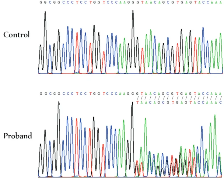
We then looked for a heterozygous mutation for the autosomal dominant form of OI, and found a c.1290delG mutation in exon 19 of theCOL1A1 gene. The deletion leads to a frameshift, resulting in a premature termination codon (PTC) at residue 540 (p.Gly431Valfs*110). The gene product, the alpha 1 chain of type I collagen, is ex-pected to be shortened from 1464 to 539 amino acid resi-dues. Sanger sequencing confirmed the presence of the
COL1A1c.1290delG mutation in heterozygosis (Figure 2). It was not present in neither of his parents nor the unaf-fected brother, indicating ade novomutation. This muta-tion was not present in the HGMD, the Osteogenesis Imperfecta Variant Database, or the in-house 200 Thai Exome Database.
Dentinogenesis imperfecta was not present in our pa-tient. It has been suggested that collagen plays
non-iden-tical roles in bone and dentin, since individuals with
COL1A1mutations have a wide variety of dentin and bone defects (O’Connell and Marini, 1999; Rauchet al., 2010).
Mutations in theCOL1A1gene can lead to OI by ei-ther haploinsufficiency or dominant negative effects. Haploinsufficiency is usually the consequence of splice site mutations, nonsense mutations, deletions or insertions. These mutations usually create a premature termination codon. These aberrant RNAs would usually be degraded by nonsense-mediated mRNA decay (NMD). With normal al-pha chains of type I collagen being produced from the wild-type allele, haploinsufficiency generally leads to a mild OI phenotype (Mariniet al., 2007). On the other hand, dominant negative effects result from missense mutations or from premature termination codon mutations that avoid NMD; the mutated alpha chain binding to normal alpha chains produced by the wild-type allele results in abnormal collagen type I. Classically known OI mutations with domi-nant-negative effect are the substitution of an amino acid for one of the obligatory glycine residues occurring in
ery third position along the chain of COL1A1, and other mutations, such as exon skipping defects or in-frame dele-tions. Most cases of OI with dominant negative effects are typically more severe than those with haploinsufficiency (Ben Amoret al., 2013).
Rauchet al. (2010) reported that mean Z-score for height was -1.3 in patients harboring mutations that lead to haploinsufficiency, and -5.5, for those with helical muta-tions, causing a dominant-negative effect. The patient pre-sented here had Z-scores of height and weight of£ -3.0, which were between these two groups. Since the cyclical intravenous pamidronate treatment was started in this pa-tient at the age of five months, the Z-score for height might have been less than -3 at the age of 11, if he had not received the medication. In fact, early treatment with intravenous di-sodium pamidronate may prevent scoliosis and basilar im-pression (Astromet al., 2007), and four years of cyclical intravenous pamidronate treatment was reported to lead to significant height gains in moderately to severely affected OI patients (Zeitlinet al., 2003). Taken together, the z-scores of our patient’s height and weight point to the form caused by a dominant negative effect (Rauchet al., 2010).
The severe phenotype of our patient suggested that theCOL1A1c.1290delG mutation led to a truncated alpha chain able to bind to the normal alpha chains, resulting in the production of defective collagen type I. The more se-vere phenotype of the patient reported could be explained by a possible abnormality in NMD efficiency. Although the mutation found in our patient is located in the NMD sensi-tive zone, 50-55 nucleotides upstream of the last exon-exon junction, the mutant RNA might still escape NMD. In 2014 the evidence pointing to the inter-individual variability in NMD efficiency and its correlation with clinical presenta-tions was reviewed, and it was proposed that it was a com-mon phenomenon in human populations (Nguyenet al., 2014). Therefore, it is possible that the mutant RNA es-capes NMD, at least partially, in our patient. There were previous reports of patients with severe OI who had prema-ture truncation mutations. A Korean patient with OI type IV had an eight-base pair deletion in exon 46 (Leeet al., 2006). Vietnamese patients with OI type III or IV were reported to have frameshift mutations located in the NMD sensitive zone, similar to our patient (Ho Duyet al., 2016). Taken to-gether, we hypothesize that variable NMD efficiency may be a cause of variable OI phenotypes in different patients with similar out-of-frame mutations.
Identification of this pathogenic mutation allowed more accurate genetic counseling, ruling out recessive in-heritance. Although the recurrence risk in this case is likely to be very low, the probability of parental mosaicism had to be considered, since no other tissue than blood was tested. An empirical recurrence risk of 27% for the perinatal lethal form of OI (type II) in the offspring of parents carrying mosaicism for collagen I mutations was reported (Pyottet
al., 2011). With mutation identification, prenatal or pre-implantation diagnosis can be provided.
In this study, using WES, we successfully identified a novelde novomutation in the COL1A1gene in a patient with OI, expanding its mutational spectrum. In addition, we provided another example of identification of a heterozy-gousde novomutation as the cause of a disease in a patient born to consanguineous parents, which allowed more accu-rate genetic counseling.
Acknowledgments
This study was supported by the Thailand Research Fund (RTA5680003, BRG5980001) through the Royal Golden Jubilee Ph.D. Program (Grant No. PHD/0160/2553) and the Chulalongkorn Academic Ad-vancement into Its 2ndCentury Project, the Ratchadapisek Sompoch Endowment Fund (2016) (59-006-HR, CU-59-064-AS), and Asia Research Center of the Korea Foun-dation for Advanced Studies at Chulalongkorn University.
References
Astrom E, Jorulf H and Soderhall S (2007) Intravenous pami-dronate treatment of infants with severe osteogenesis impe-rfecta. Arch Dis Child 92:332-338.
Becker J, Semler O, Gilissen C, Li Y, Bolz HJ, Giunta C, Ber-gmann C, Rohrbach M, Koerber F, Zimmermann K,et al.
(2011) Exome sequencing identifies truncating mutations in human SERPINF1 in autosomal-recessive osteogenesis imperfecta. Am J Hum Genet 88:362-371.
Ben Amor IM, Roughley P, Glorieux FH and Rauch F (2013) Skeletal clinical characteristics of osteogenesis imperfecta caused by haploinsufficiency mutations in COL1A1. J Bone Miner Res 28:2001-2007.
Forlino A and Marini JC (2016) Osteogenesis imperfecta. Lancet 387:1657-1671.
Garbes L, Kim K, Riess A, Hoyer-Kuhn H, Beleggia F, Bevot A, Kim MJ, Huh YH, Kweon HS, Savarirayan R,et al.(2015) Mutations in SEC24D, encoding a component of the COPII machinery, cause a syndromic form of osteogenesis imper-fecta. Am J Hum Genet 96:432-439.
Ho Duy B, Zhytnik L, Maasalu K, Kandla I, Prans E, Reimann E, Martson A and Koks S (2016) Mutation analysis of the COL1A1 and COL1A2 genes in Vietnamese patients with osteogenesis imperfecta. Hum Genomics 10:27.
Lee KS, Song HR, Cho TJ, Kim HJ, Lee TM, Jin HS, Park HY, Kang S, Jung SC and Koo SK (2006) Mutational spectrum of type I collagen genes in Korean patients with osteo-genesis imperfecta. Hum Mutat 27:599.
Lindahl K, Astrom E, Rubin CJ, Grigelioniene G, Malmgren B, Ljunggren O and Kindmark A (2015) Genetic epidemiol-ogy, prevalence, and genotype-phenotype correlations in the Swedish population with osteogenesis imperfecta. Eur J Hum Genet 23:1112.
Marini JC, Forlino A, Cabral WA, Barnes AM, San Antonio JD, Milgrom S, Hyland JC, Korkko J, Prockop DJ, De Paepe A,
et al.(2007) Consortium for osteogenesis imperfecta muta-tions in the helical domain of type I collagen: Regions rich in lethal mutations align with collagen binding sites for inte-grins and proteoglycans. Hum Mutat 28:209-221.
Mendoza-Londono R, Fahiminiya S, Majewski J, Care4Rare Can-ada, Tetreault M, Nadaf J, Kannu P, Sochett E, Howard A, Stimec J, et al.(2015) Recessive osteogenesis imperfecta caused by missense mutations in SPARC. Am J Hum Genet 96:979-985.
Ng SB, Buckingham KJ, Lee C, Bigham AW, Tabor HK, Dent KM, Huff CD, Shannon PT, Jabs EW, Nickerson DA,et al.
(2010) Exome sequencing identifies the cause of a mende-lian disorder. Nat Genet 42:30-35.
Nguyen LS, Wilkinson MF and Gecz J (2014) Nonsense-mediated mRNA decay: Inter-individual variability and hu-man disease. Neurosci Biobehav Rev 46:175-186.
O’Connell AC and Marini JC (1999) Evaluation of oral problems in an osteogenesis imperfecta population. Oral Surg Oral Med Oral Pathol Oral Radiol Endod 87:189-196.
Pyott SM, Pepin MG, Schwarze U, Yang K, Smith G and Byers PH (2011) Recurrence of perinatal lethal osteogenesis imperfecta in sibships: Parsing the risk between parental mosaicism for dominant mutations and autosomal recessive inheritance. Genet Med 13:125-130.
Rauch F, Lalic L, Roughley P and Glorieux FH (2010) Relation-ship between genotype and skeletal phenotype in children
and adolescents with osteogenesis imperfecta. J Bone Miner Res 25:1367-1374.
Stevenson DA, Carey JC, Byrne JL, Srisukhumbowornchai S and Feldkamp ML (2012) Analysis of skeletal dysplasias in the Utah population. Am J Med Genet A 158A:1046-1054. Zeitlin L, Rauch F, Plotkin H and Glorieux FH (2003) Height and
weight development during four years of therapy with cycli-cal intravenous pamidronate in children and adolescents with osteogenesis imperfecta types I, III, and IV. Pediatrics 111:1030-1036.
Internet Resources
Burrows-Wheeler Alignment (BWA), bio-bwa.sourceforge.net/ (accessed April 2015).
Sequence Alignment/Map tools (SAMtools), samtools.sourceforge.net/ (accessed April 2015).
The Human Gene Mutation Database at the Institute of Medical
Genetics in Cardiff (HGMD),
http://www.hgmd.cf.ac.uk/ac/index.php (accessed February 2016).
Exome Aggregation Consortium database (ExAC), exac.broadinstitute.org (accessed February 2016).
Osteogenesis Imperfecta Variant Database, https://oi.gene.le.ac.uk/home.php?select_db=COL1A1 (ac-cessed February 2016).
Associate Editor: Angela M. Vianna-Morgante