Mucin Secretion Induced by Titanium Dioxide
Nanoparticles
Eric Y. T. Chen., Maria Garnica., Yung-Chen Wang, Chi-Shuo Chen, Wei-Chun Chin*
Bioengineering, University of California Merced, Merced, California, United States of America
Abstract
Nanoparticle (NP) exposure has been closely associated with the exacerbation and pathophysiology of many respiratory diseases such as Chronic Obstructive Pulmonary Disease (COPD) and asthma. Mucus hypersecretion and accumulation in the airway are major clinical manifestations commonly found in these diseases. Among a broad spectrum of NPs, titanium dioxide (TiO2), one of the PM10 components, is widely utilized in the nanoindustry for manufacturing and processing of
various commercial products. Although TiO2NPs have been shown to induce cellular nanotoxicity and emphysema-like
symptoms, whether TiO2 NPs can directly induce mucus secretion from airway cells is currently unknown. Herein, we
showed that TiO2NPs (,75 nm) can directly stimulate mucin secretion from human bronchial ChaGo-K1 epithelial cells via a
Ca2+signaling mediated pathway. The amount of mucin secreted was quantified with enzyme-linked lectin assay (ELLA).
The corresponding changes in cytosolic Ca2+concentration were monitored with Rhod-2, a fluorescent Ca2+dye. We found
that TiO2 NP-evoked mucin secretion was a function of increasing intracellular Ca2+ concentration resulting from an
extracellular Ca2+influx via membrane Ca2+channels and cytosolic ER Ca2+release. The calcium-induced calcium release
(CICR) mechanism played a major role in further amplifying the intracellular Ca2+signal and in sustaining a cytosolic Ca2+
increase. This study provides a potential mechanistic link between airborne NPs and the pathoetiology of pulmonary diseases involving mucus hypersecretion.
Citation:Chen EYT, Garnica M, Wang Y-C, Chen C-S, Chin W-C (2011) Mucin Secretion Induced by Titanium Dioxide Nanoparticles. PLoS ONE 6(1): e16198. doi:10.1371/journal.pone.0016198
Editor:Meni Wanunu, University of Pennsylvania, United States of America
ReceivedSeptember 20, 2010;AcceptedDecember 7, 2010;PublishedJanuary 19, 2011
Copyright:ß2011 Chen et al. This is an open-access article distributed under the terms of the Creative Commons Attribution License, which permits unrestricted use, distribution, and reproduction in any medium, provided the original author and source are credited.
Funding:This study was supported by grants from NIH (1R15HL095039), NSF (CBET-0932404) and the UC CITRIS Program. CSC was supported by UC Merced Center of Excellence on Health Disparities (1P20MD005049-01 from the National Center on Minority Health and Health Disparities). EYTC was supported by GRC summer fellowship from UC Merced. The funders had no role in study design, data collection and analysis, decision to publish, or preparation of the manuscript.
Competing Interests:Dr. Chin is a member of the editorial board of PLoS ONE. This does not alter the authors’ adherence to all the PLoS ONE policies on sharing data and materials.
* E-mail: [email protected]
.These authors contributed equally to this work.
Introduction
Many published reports have demonstrated the association between NP exposure and pulmonary morbidity and mortality [1,2,3]. The adverse effects induced by NPs seem to exacerbate clinical symptoms of pre-existing respiratory illnesses such as asthma, COPD and Cystic Fibrosis (CF) [1,2,3,4,5,6]. During NP exposure, individuals with respiratory diseases showed more incidences of bronchoconstriction, medication use, bronchial hyperreactivity and lung fibrosis [2,7]. TiO2NPs are widely used in the nanotechnology
industry due to their vast array of applications that range from household commodities, such as components of paints and carpets, to personal products that include cosmetics, textiles, sunscreens and foods [8,9]. TiO2is also one of the PM10 components commonly
found in industries or manufacturing plants involved in processing mineral ore rutile [10]. It has been reported that.50% of TiO2NP
exposed workers had respiratory symptoms accompanied by reduction in pulmonary function [10,11]. Other reports have also indicated that inhalation of TiO2 NPs can induce pulmonary
inflammatory responses (characterized by neutrophil recruitment), epithelial cell death and increased permeability [2,9]. Furthermore, TiO2NPs have been shown to play a role in inducing epithelial
fibroproliferative changes, stimulating goblet cell hyperplasia and in instigating emphysema-like (such as alveolar enlargement) damages
in the lungs [2,10,12]. Overall, nanotoxicity induced by TiO2NP
exposure in both the occupational and ambient environment presents a significant and realistic health concern.
The harmful effects of NPs on the respiratory system not only encompass cellular apoptosis/necrosis, but also mucus hyperpro-duction which is closely associated with the pathogenesis of pulmonary diseases that include asthma, COPD and CF [2,10,13]. In these chronic pulmonary diseases, mucus hypersecretion and accumulation may lead to recurrent episodes of chronic bacterial infections, limited airflow and chronic inflammatory responses [2,14,15]. However, whether TiO2NPs can directly induce mucin
secretion has not been resolved.
Airway mucus plays a vital role in the constant clearance of inhaled pathogens and particulates. Mucus is a large, highly glycosylated protein consisting of an array of mucin peptides (apomucin) [14]. With their oligosaccharide sidegroups, such as sialic acid, sulfate, and carboxyl (COO2), mucins are usually polyanionic in nature [16]. Mucin secretion is closely regulated by cytosolic Ca2+
concentrations ([Ca2+
]C) in various epithelial cells
[17]. A rise in [Ca2+
]Cis crucial for initiating a cascade of down
stream events including the mobilization of granule-bound Ca2+,
Agonist-induced opening of various Ca2+
channels expressed on the cell membrane allows the influx of extracellular Ca2+
, which may serve as the external Ca2+source [19]. The initial upsurge in
the [Ca2+
]Cis usually relayed by triggering a secondary wave of
Ca2+
propagation from internal stores, such as the ER [19,20,21,22]. Ryanodine receptors (RYRs) on the ER have multiple allosteric Ca2+
binding sites responsible for triggering Ca2+
- induced Ca2+
release (CICR) into the cytosol [19,20,21,22]. The resultant increase in [Ca2+]
C could activate other cytosolic
proteins and modulate secretion of mucin, hormones or various neurotransmitters [17,23,24].
NPs have been shown to disturb cellular functions by elevating intracellular Ca2+
levels [25,26,27,28]. For example, ultrafine carbon black NPs can elicit Ca2+
-dependent secretion through the activation of L-type voltage-gated Ca2+
channels [25,26,28]. However, little is known regarding the intricate calcium signaling pathway regulating the exocytotic events of secretory products. In this study, we aim to investigate the mechanism through which TiO2NPs induce mucin secretion via a Ca
2+signaling mediated
pathway.
Materials and Methods
1. Culture of ChaGo-K1 cells
The human airway bronchial epithelial cell line ChaGo-K1, obtained from American Type Culture Collection (ATCC, Manassas, VA, USA), was used because it expresses MUC proteins and secretes mucin [29]. Cells were cultured in 15 cm cell culture plates (VWR, CA, USA) in RPMI 1640 medium (Invitrogen, CA, USA) supplemented with L-glutamine, 1% penicillin/streptomycin and 10% heat inactivated fetal bovine serum (FBS). Cultures were incubated in a humidified incubator at 37uC/5% CO2. Cell counts were performed using trypan blue
(Sigma-Aldrich, MO, USA) exclusion and a Bright-Line haemo-cytometer.
2. Nanoparticles and characterization
A mixture of anatase and rutile forms of ultrafine titanium (IV) dioxide (,75 nm) (Sigma-Aldrich, MO, USA) was used in this study because this form has been shown to result in more severe cellular injuries [30,31]. The TiO2 NPs have a surface area of
36 m2/g and the dispersion conductivity is 1040mS/cm (infor-mation from Sigma). All NP samples were sonicated before usage. The concentrations used were 1 mg/ml, 0.75 mg/ml, 0.5 mg/ml, 0.25 mg/ml, 0.1 mg/ml, and 0.05 mg/ml. The range of concen-trations used was consistent with the concenconcen-trations of TiO2NPs
found in previous reports [30]. The TiO2NPs were reconstituted
with Hanks’ solution (Invitrogen, CA, USA) before being tested. The size of NPs was independently confirmed using homodyne dynamics laser scattering (DLS) as described in previous studies [32,33].
3. Cell preparation
Cells were seeded at 26105cells per well in a 24-well plate, and
incubated for 24 hrs in RPMI 1640 supplemented with 10% FBS. Following 24 hr incubation, the RPMI medium was removed from the cells and the culture was rinsed with Hanks’ solution twice before use.
4. Measurements of cytosolic Ca2+concentrations induced by TiO2exposure
The cells were then loaded with a Rhod-2 AM dye (1mM)
(Kd= 570 nM, lEx= 552 nm and lEm= 581) (Invitrogen, CA,
USA) for 45 minutes. After the dye loading, the cells were rinsed,
incubated with either normal Hanks’ or Ca2+
-free Hanks’ solution, and treated with the appropriate TiO2concentrations. All calcium
signaling experiments were carried out on a thermoregulated stage at 37uC mounted on a Nikon microscope (Nikon Eclipse TE2000-U, Tokyo, Japan). ChaGo-K1 cells were incubated with cadmium chloride (200mM; Sigma-Aldrich, MO, USA) to block the membrane Ca2+
channels [34], followed by TiO2NP stimulation.
To investigate the interaction between TiO2and membrane Ca2+
channels, nifedipine (10mM; Sigma-Aldrich, MO, USA), an L-type Ca2+
channel blocker [27], was added to ChaGo-K1 cells prior to the exposure of TiO2. Antioxidant N-acetylcysteine (NAC,
250mM; Sigma-Aldrich, MO, USA) was also added to ChaGo-K1 cells to study the involvement of reactive oxygen species (ROS) [27,35], possibly generated as a result of TiO2stimulation, and the
activation of Ca2+
channels. Thapsigargin (100 nM; Sigma-Aldrich, MO, USA) [18] and ryanodine (100mM; Sigma-Aldrich, MO, USA) were added separately to deplete the ER Ca2+
content and to inhibit the CICR mechanism [20,21], correspondingly. These two blockers were utilized to investigate the contribution from the internal ER Ca2+
pool.
5. Calcein dye leakage measurements
ChaGo-K1 cells were seeded at the density of 26105cells per
well in a 24-well plate and cultured for 24 hrs. TiO2NP prepared
with calcein fluorescent dye (50mM) (Invitrogen, CA, USA) in
Hanks’ solution was incubated with the cells for 5 minutes at 37uC. Calcein is a biological inert green-fluorescent molecule of a molecular mass of 623 Da and an estimated molecular radius of 0.6 nm [36]. TiO2 NP solution containing the calcein dye was
then removed and cells were rinsed twice with PBS to remove possible remnants of calcein dye in the extracellular solution. The cells were subsequently stained with a fluorescent nucleus dye, hoechst (10mM) (Sigma-Aldrich, MO, USA), for 5 mintues at 37uC and thoroughly rinsed again [33]. Fresh Hanks’ solution was added into each well before taking fluorescent images of calcein and hoechst loaded cells with a Nikon fluorescence microscope. A percentage of calcein loaded cells against total number of cells, as indicated by hoechst fluorescence, was calculated for each of the TiO2NP concentrations used in the experiment.
6. Mucin secretion and ELLA Preparation
The cells were seeded at 26105cells per well in a 24-well plate
and cultured for 24 hrs. ChaGo-K1 cells were then rinsed with PBS and treated with BAPTA-AM (Invitrogen, CA, USA), thapsigargin Aldrich, MO, USA) or ryanodine (Sigma-Aldrich, MO, USA) for at least 30 minutes. Afterward the cells were stimulated for 15 minutes with the corresponding TiO2NP
concentrations (0.75 mg/ml, 0.5 mg/ml, 0.25 mg/ml, and 0.1 mg/ml) or ionomycin (1mM) (positive control) (Sigma-Aldrich,
MO, USA), both prepared in PBS. The supernatant containing secreted mucin was collected and briefly centrifuged at 8,000 rpm to remove the residual TiO2 NPs. The supernatant was then
incubated in a 96 well (Nunc MaxiSorp, VWR, CA, USA) plate overnight at 4uC. Afterward the 96-well plate was washed with PBST (PBS+0.05% Tween-20) and then blocked with 1% BSA. The 96 well plate was washed again with PBST and incubated with lectin (Wheat germ agglutinin, WGA) (Sigma-Aldrich, MO, USA), conjugated to horseradish peroxidase (HRP; 5mg/ml) (Sigma-Aldrich, MO, USA), at 37uC for 1 hr. The substrate, 3,39,5,59-Tetramethylbenzidine (TMB; Sigma-Aldrich, MO, USA), was added to each well at room temperature followed by H2SO4 (Sigma-Aldrich, MO, USA) in order to terminate the
7. Image Analysis
After staining the treated cells, image analysis was performed with an inverted Nikon Eclipse TE2000-U fluorescent microscope. Each photo was taken at a magnification of 2006and analyzed
using SimplePCI (Compix Inc., Imaging Systems, Sewickle, PA, USA). The data shown is a representative of Ca2+
signals of more than 200 cells.
8. Statistical Analysis
The data was presented as means6SD. Each experiment was performed independently at least three times. Statistical signifi-cance was determined using a Student’s t-test analysis with p values,0.05 (GraphPad Prism 4.0, GraphPad Software, Inc., San Diego, CA, USA).
Results
TiO2NP characterization
Dynamic laser scattering (DLS) was used to characterize the TiO2NPs. The particle size distribution ranged from,9 to 80 nm
due to minor aggregation or agglomeration while the predominant size is,50 nm (Fig. 1A).
TiO2NPs induce cytosolic Ca2+concentration increase
To investigate whether TiO2NPs could generate an increase in
[Ca2+
]C, ChaGo-K1 cells were loaded with Rhod-2 AM dye and
exposed to 0.05–1 mg/ml of TiO2NPs. The change in [Ca2+]C, as
represented by the fluorescence intensity within ChaGo-K1 cells, was monitored for 60 seconds. Figure 1B shows that 1 mg/ml of TiO2 NPs induced an approximate 150% increase, while lower
TiO2 concentrations (,0.1 mg/ml) caused a minor elevation
(,110%) in [Ca2+]C when compared with untreated cells. The
effect of TiO2 treatment on the [Ca2+]C of ChaGo-K1 cells
followed a concentration-dependent manner (Fig. 1B).
Extracellular source for Ca2+increase
To determine the main source of elevated [Ca2+
]C upon
stimulation, ChaGo-K1 cells were exposed to TiO2NPs in Ca2+
-free Hanks’ solution. EGTA (2 mM) was added in Hanks’ solution
to chelate possible traces of Ca2+
. TiO2 (0.05 mg/ml–1 mg/ml)
treatment under Ca2+
-free conditions failed to instigate a significant increase in [Ca2+
]C (Fig. 2A). Our data suggests that
the extracellular Ca2+
pool is the primary source of the observed cytosolic Ca2+
increase. We then tested whether TiO2NPs can
induce a Ca2+
influx via membrane channels. Blocking the channels with CdCl2(200mM) significantly inhibited an increase
in [Ca2+
]C (Fig. 2B). Co-treatment of cells with TiO2 NPs and
nifedipine greatly blocked the NP-induced [Ca2+
]C increase
(Fig. 2C). However, the incomplete blockage of extracellular Ca2+
influx via channels postulates additional Ca2+
leakage through perturbed cell membranes. To confirm whether TiO2
can instigate membrane disruption, thereby permitting unspecific extracellular Ca2+
entry, cytosolic leakage was assessed using the fluorescent calcein dye. It was found that the dye permeation ratio increased from approximately 4 to 13% with elevated TiO2
concentrations ranging from 0.1 to 1 mg/ml (Fig. 2D).
Oxidative stress induced Ca2+influx
To demonstrate that TiO2-evoked [Ca2+]C increase can be
associated with oxidative stress, cells were pretreated with an anti-oxidant, N-acetylcysteine (NAC) [27]. Pre-treatment with NAC was able to partially attenuate the increase in cytosolic Ca2+
level triggered by 1 mg/ml and 0.75 mg/ml TiO2exposure (Fig. 2E).
These results support the idea that oxidative stress, induced by TiO2 NPs, contributes to the observed [Ca2+]C increase and
promote Ca2+
-dependent mucin secretion.
The ER as an intracellular source of Ca2+
In order to determine the involvement of ER Ca2+
pool, it was depleted by pre-incubating the cells with thapsigargin. Pre-treatment with thapsigargin impeded TiO2 NPs from
triggering a sustained increase in the cytosolic Ca2+
level (Fig. 3A). We then investigated the role of the CICR mechanism by blocking RYRs (ryanodine receptors) [20]. Our results revealed that CICR was largely inhibited by ryanodine (a blocker for RYR associated with the CICR response) resulting in a significantly diminished [Ca2+
]C increase induced by NPs
(Fig. 3B).
Figure 1. TiO2NP characterization and resultant [Ca2+]Cchanges after NP treatment.A) DLS assessment of TiO2NPs in Hanks’ solution showed a size distribution of,9 to 80 nm. B) Cells were treated with TiO2NPs with concentrations of 0.05 mg/ml (yellow), 0.1 mg/ml (Light Blue), 0.25 mg/ml (Purple), 0.5 mg/ml (Green), 0.75 mg/ml (Red), and 1 mg/ml (Blue) in normal Hanks’ solution. Each line represents the average fluorescence intensity of approximately 200 cells per well.
Ca2+-dependency of TiO
2-induced mucin secretion
Enzyme-linked lectin assay (ELLA) was used to assess the amount of mucin secreted from ChaGo-K1 cells when stimulated with TiO2 NPs. When compared to the control, TiO2 NPs
increased mucin secretion by 113%, 125%, 133%, 137% and 150% at 0.05, 0.1, 0.25, 0.5 and 0.75 mg/ml, respectively (Fig. 4A). Chelating the intracellular Ca2+
with BAPTA-AM
yielded a significant reduction in mucin secretion (Fig 4B). Addition of thapsigargin (Fig. 4C) or ryanodine (Fig. 4D) also resulted in diminished mucin secretion induced by TiO2NPs. Our
data indicates that TiO2-induced mucin secretion is dependent on
the [Ca2+]
C, attributed to both external and internal Ca 2+pools
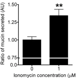
(Fig. 4A–D). Ionomycin (a Ca2+ ionophore) was used to elicit
mucin secretion as a positive control (Fig. 5).
Figure 2. Measurement of the [Ca2+]
C and calcein dye leakage after TiO2 NP treatment. Cells were treated with TiO2 NPs with concentrations ranging from 0.05 mg/ml–1 mg/ml, in A) Ca2+-free Hanks’ solution, B) in the presence of CdCl
2(200mM), C) nifedipine (10mM), D) calcein (50mM) (n = 12, **P,0.005), and E) NAC (250mM) (colors are as depicted in Figure 1B).
Figure 3. Measurement of [Ca2+]
Cafter stimulation by TiO2NPs.Cells were treated with TiO2NPs with concentrations ranging from 0.1 mg/ml –1 mg/ml, in the presence of A) thapsigargin (100 nM), and B) ryanodine (100mM) (colors used are consistent with Figure 1).
doi:10.1371/journal.pone.0016198.g003
Discussion
Recently, an increasing number of reports have shown that airborne particulate pollution found in both the ambient and working environments, particularly TiO2 NPs, can exacerbate
airway diseases [1,2,3,4,5,6,10,11,38]. Aggravated clinical mani-festations of COPD, CF and asthma may include intensified symptoms of mucociliary transport impairment and mucus hypersecretion [15,39]. The resultant accumulation of thick obstructive mucus usually occupies airway lumen, thereby limiting airflow and leading to morbidity [15,39]. Despite documentations of TiO2-induced cellular nanotoxicity effects, pulmonary
inflam-matory responses and emphysema-like pathology [12], whether TiO2 NPs can directly trigger mucin secretion has not been
resolved. In this study, we demonstrate that TiO2 NPs can
stimulate mucin secretion from bronchial epithelial ChaGo-K1 cells via a Ca2+
- dependent pathway.
Our study showed that TiO2NPs can induce mucin secretion
that increases as a function of TiO2NP concentration (Fig. 4A).
The TiO2concentration range used in our study is consistent with
previous reports representing the concentration found in ambience and nanotechnology industries [30,40,41,42]. While NP exposure has been long associated with increasing mucin synthesis due to goblet cell hyperplasia [13], our study indicates that TiO2NPs can
directly trigger mucin secretion in the airway.
It has been well established that intracellular Ca2+plays a vital role
in stimulus-secretion coupling [43]. Previous reports have docu-mented that an elevated [Ca2+
]Cprecedes mucin granule exocytosis
[17]. NP exposure has been shown to trigger an intracellular Ca2+
increase in various cells; therefore, we examined the cellular Ca2+
signaling pathway involved during TiO2stimulation [25,28,44]. At
TiO2 concentrations of 0.5, 0.75, and 1 mg/ml, there was a
sustained elevation in [Ca2+
]C. At lower doses (0.05, 0.1 and
0.25 mg/ml), the [Ca2+
]Cincreased gradually within the 1stminute
(Fig. 1B). Our data demonstrated that TiO2 NPs induced a
concentration dependent increase in [Ca2+]
C, which is consistent
with results from the mucin secretion measurements (Fig. 4A). The stimulus-induced intracellular Ca2+
signal can be evoked by the entry of Ca2+
through voltage-gated Ca2+
channels, or by the
release of Ca2+
from intracellular Ca2+
stores [43,45,46]. Previous researches have suggested that extracellular Ca2+
influx plays an important role in the elevated [Ca2+
]C during NP stimulation
[25,27,28,47]. Data from experiments performed in Ca2+
-free Hanks’ solution confirmed that [Ca2+]
C failed to increase when
treated with TiO2NPs (Fig. 2A). To characterize the nature of the
Ca2+
influx induced by TiO2NPs, we first evaluated the effect of
cadmium chloride (CdCl2), a general Ca2+ channel blocker
[34,48]. Figure 2B shows that the [Ca2+]
C remained low and
relatively unchanged with CdCl2. Secondly, nifedipine, a widely
used L-type Ca2+
channel blocker, markedly diminished the increase in [Ca2+
]C(Fig. 2C). The effect of nifedipine implies that
TiO2 NPs can activate L-type voltage gated Ca2+ channels,
allowing extracellular Ca2+influx into the cytosol. This
observa-tion is consistent with previous reports showing that ultrafine carbon black and ZnO NP-induced [Ca2+
]Celevation can also be
attenuated by nifedipine [27,28]. In addition, several reports have suggested that oxidative stress induced by NPs can exert an impact on the intracellular Ca2+signaling pathway and that the activity of
Ca2+
channels may be altered by ROS [27,28,44]. Results from Figure 2E showed that NAC significantly reduced the rising [Ca2+
]C generated by TiO2 NPs. Huang et al, has also
demonstrated that NAC can attenuate the intracellular Ca2+level
when challenged with ZnO NPs [27]. Our results support the idea that NAC and other antioxidants may be effective in reducing NP-instigated mucin hypersecretion. NPs such as TiO2can damage
cell membrane integrity by possible lipid peroxidation [27,31], thereby creating pores on the lipid bilayer [49] that may allow the transient influx of extracellular Ca2+. Our data further
demon-strated that co-adminstration of TiO2NPs and fluorescent calcein
dye lead to intracellular leakage and the permeation efficiency increased in a TiO2concentration dependent manner (Fig. 2D).
Calcein has also been previously utilized to evaluate the efficacy of peptides in causing membrane perturbation [50]. Our result suggests that the possible membrane perturbation/transient pore formation induced by TiO2 NPs allows an extracellular Ca2+
influx and may account for the portion of Ca2+
that can not be completely abolished by blocking L-type Ca2+ channels with
nifedipine.
Increasing the [Ca2+]
Cof human goblet cells has been shown to
trigger degranulation [17]. We used BAPTA (cytosolic Ca2+
chelator) to test whether the increase in Ca2+
induced by TiO2
NPs could stimulate mucin exocytosis. It is evident that BAPTA significantly inhibited mucin exocytosis (Fig. 4B), indicating that TiO2NPs can elicit a [Ca
2+]
Cincrease, thereby leading to mucin
secretion.
Besides the external Ca2+source (Hanks’ solution), the ER is
one of the major internal Ca2+
stores. Figures 3A and 4C revealed that when the ER Ca2+
had been depleted by pretreatment with thapsigargin, the TiO2 NP-induced [Ca2+]C failed to increase
significantly, and the subsequent mucin secretion was abolished. Our data indicates that the ER plays a critical role in relaying TiO2-induced Ca2+ signaling. CICR is a positive feedback
mechanism where the ER amplifies a small increase in [Ca2+
]C,
(e.g. due to voltage-gated Ca2+
influx [22]), with the activation of RYRs that will lead to the release of more Ca2+ from the ER
[19,20]. Previous studies have shown that through activation of RYRs with Ca2+
, CICR can generate an overall increase in [Ca2+
]C[20,21,22]. Our data showed that ryanodine inhibited a
continual rise in [Ca2+
]C when applying TiO2 NPs (Fig. 3B).
Therefore, it is indicative that the TiO2-instigated increase in
[Ca2+]
Cwas also CICR dependent. The effect of ryanodine was
further demonstrated by the lack of mucin secretion under TiO2
NP stimulation (Fig. 4D).
In summary, our study indicates that cellular exposure to TiO2
NPs can activate membrane L-type Ca2+
channels, induce ROS production and possibly disrupt the cellular membrane. Influx of extracellular Ca2+
into the cytoplasm raises [Ca2+
]C, which in turn
can trigger ryanodine receptors on the ER to release ER resident Ca2+ via the CICR mechanism. A sufficient increase in the
cytosolic Ca2+
level results in subsequent mucin secretion. More importantly, our results provide a direct link between airborne particulate matters and the pathogenesis of chronic airway diseases involving mucus hypersecretion and airway obstruction. In addition, we demonstrate that once thought inert and harmless TiO2NPs can indeed interfere with intracellular Ca
2+signaling,
possibly leading to pathological states.
Acknowledgments
The authors thank Profs. Pedro Verdugo and Paul Quinton for their support and encouragement during the preparation of this manuscript.
Author Contributions
Conceived and designed the experiments: EYTC MG YCW CSC WCC. Performed the experiments: EYTC MG YCW. Analyzed the data: EYTC MG YCW. Contributed reagents/materials/analysis tools: EYTC MG YCW CSC. Wrote the paper: EYTC WCC.
References
1. Alfaro-Moreno E, Nawrot TS, Nemmar A, Nemery B (2007) Particulate matter in the environment: pulmonary and cardiovascular effects. Curr Opin Pulm Med 13: 98–106.
2. Gwinn MR, Vallyathan V (2006) Nanoparticles: health effects–pros and cons. Environ Health Perspect 114: 1818–1825.
3. Sethi S (2004) New developments in the pathogenesis of acute exacerbations of chronic obstructive pulmonary disease. Curr Opin Infect Dis 17: 113–119. 4. Atkinson RW, Anderson HR, Sunyer J, Ayres J, Baccini M, et al. (2001) Acute
effects of particulate air pollution on respiratory admissions: results from APHEA 2 project. Air Pollution and Health: a European Approach. Am J Respir Crit Care Med 164: 1860–1866.
5. Ling SH, van Eeden SF (2009) Particulate matter air pollution exposure: role in the development and exacerbation of chronic obstructive pulmonary disease. Int J Chron Obstruct Pulmon Dis 4: 233–243.
6. Stone V (2000) Environmental air pollution. Am J Respir Crit Care Med 162: S44–47.
7. Boezen M, Schouten J, Rijcken B, Vonk J, Gerritsen J, et al. (1998) Peak expiratory flow variability, bronchial responsiveness, and susceptibility to ambient air pollution in adults. Am J Respir Crit Care Med 158: 1848–1854. 8. Card JW, Zeldin DC, Bonner JC, Nestmann ER (2008) Pulmonary applications
and toxicity of engineered nanoparticles. Am J Physiol Lung Cell Mol Physiol 295: L400–411.
9. Johnston HJ, Hutchison GR, Christensen FM, Peters S, Hankin S, et al. (2009) Identification of the mechanisms that drive the toxicity of TiO(2) particulates: the contribution of physicochemical characteristics. Part Fibre Toxicol 6: 33. 10. Ahn MH, Kang CM, Park CS, Park SJ, Rhim T, et al. (2005) Titanium dioxide
particle-induced goblet cell hyperplasia: association with mast cells and IL-13. Respir Res 6: 34.
11. Garabrant DH, Fine LJ, Oliver C, Bernstein L, Peters JM (1987) Abnormalities of pulmonary function and pleural disease among titanium metal production workers. Scand J Work Environ Health 13: 47–51.
12. Chen HW, Su SF, Chien CT, Lin WH, Yu SL, et al. (2006) Titanium dioxide nanoparticles induce emphysema-like lung injury in mice. FASEB J 20: 2393–2395.
13. Hyun JS, Lee BS, Ryu HY, Sung JH, Chung KH, et al. (2008) Effects of repeated silver nanoparticles exposure on the histological structure and mucins of nasal respiratory mucosa in rats. Toxicol Lett 182: 24–28.
14. Voynow JA, Rubin BK (2009) Mucins, mucus, and sputum. Chest 135: 505–512.
15. Rogers DF (2007) Physiology of airway mucus secretion and pathophysiology of hypersecretion. Respir Care 52: 1134–1146; discussion 1146-1139.
16. Verdugo P (1990) Goblet cells secretion and mucogenesis. Ann Rev Physiol 52: 157–176.
17. Abdullah LH, Conway JD, Cohn JA, Davis CW (1997) Protein kinase C and Ca2+activation of mucin secretion in airway goblet cells. Am J Physiol 273: L201–210.
18. Nguyen T, Chin WC, Verdugo P (1998) Role of Ca2+/K+ion exchange in intracellular storage and release of Ca2+. Nature 395: 908–912.
19. Berridge MJ, Bootman MD, Roderick HL (2003) Calcium signalling: dynamics, homeostasis and remodelling. Nat Rev Mol Cell Biol 4: 517–529.
20. Ashby MC, Craske M, Park MK, Gerasimenko OV, Burgoyne RD, et al. (2002) Localized Ca2+uncaging reveals polarized distribution of Ca2+-sensitive Ca2+ release sites: mechanism of unidirectional Ca2+waves. J Cell Biol 158: 283–292. 21. Meissner G (1994) Ryanodine receptor/Ca2+ release channels and their
regulation by endogenous effectors. Annu Rev Physiol 56: 485–508. 22. Solovyova N, Veselovsky N, Toescu EC, Verkhratsky A (2002) Ca(2+) dynamics
in the lumen of the endoplasmic reticulum in sensory neurons: direct visualization of Ca(2+)-induced Ca(2+) release triggered by physiological Ca(2+) entry. Embo J 21: 622–630.
23. Mogami H, Zhang H, Suzuki Y, Urano T, Saito N, et al. (2003) Decoding of short-lived Ca2+influx signals into long term substrate phosphorylation through activation of two distinct classes of protein kinase C. J Biol Chem 278: 9896–9904.
24. Zhu H, Hille B, Xu T (2002) Sensitization of regulated exocytosis by protein kinase C. Proc Natl Acad Sci U S A 99: 17055–17059.
25. Brown DM, Donaldson K, Borm PJ, Schins RP, Dehnhardt M, et al. (2004) Calcium and ROS-mediated activation of transcription factors and TNF-alpha cytokine gene expression in macrophages exposed to ultrafine particles. Am J Physiol Lung Cell Mol Physiol 286: L344–353.
26. Brown DM, Hutchison L, Donaldson K, Stone V (2007) The effects of PM10 particles and oxidative stress on macrophages and lung epithelial cells: modulating effects of calcium-signaling antagonists. Am J Physiol Lung Cell Mol Physiol 292: L1444–1451.
27. Huang CC, Aronstam RS, Chen DR, Huang YW (2009) Oxidative stress, calcium homeostasis, and altered gene expression in human lung epithelial cells exposed to ZnO nanoparticles. Toxicol In Vitro 24: 45–55.
28. Stone V, Tuinman M, Vamvakopoulos JE, Shaw J, Brown D, et al. (2000) Increased calcium influx in a monocytic cell line on exposure to ultrafine carbon black. Eur Respir J 15: 297–303.
29. Dahiya R, Kwak KS, Byrd JC, Ho S, Yoon WH, et al. (1993) Mucin synthesis and secretion in various human epithelial cancer cell lines that express the MUC-1 mucin gene. Cancer Res 53: 1437–1443.
30. Chen E, Ruvalcaba M, Araujo L, Chapman R, Chin WC (2008) Ultrafine titanium dioxide nanoparticles induce cell death in human bronchial epithelial cells. Journal of Experimental Nanoscience 3: 171–183.
31. Gurr JR, Wang AS, Chen CH, Jan KY (2005) Ultrafine titanium dioxide particles in the absence of photoactivation can induce oxidative damage to human bronchial epithelial cells. Toxicology 213: 66–73.
32. Chen EY, Wang YC, Chen CS, Chin WC (2010) Functionalized Positive Nanoparticles Reduce Mucin Swelling and Dispersion. PLoS One 5: e15434. 33. Chen EY, Yang N, Quinton PM, Chin WC (2010) A New Role for Bicarbonate
in Mucus Formation. Am J Physiol Lung Cell Mol Physiol.
34. Nguyen T, Chin WC, O’Brien JA, Verdugo P, Berger AJ (2001) Intracellular pathways regulating ciliary beating of rat brain ependymal cells. J Physiol 531: 131–140.
35. Kawasaki S, Takizawa H, Takami K, Desaki M, Okazaki H, et al. (2001) Benzene-extracted components are important for the major activity of diesel exhaust particles: effect on interleukin-8 gene expression in human bronchial epithelial cells. Am J Respir Cell Mol Biol 24: 419–426.
36. Edwards DA, Prausnitz MR, Langer R, Weaver JC (1995) Analysis of Enhanced Transdermal Transport by Skin Electroporation. Journal of Controlled Release 34: 211–221.
37. Kemp PA, Sugar RA, Jackson AD (2004) Nucleotide-mediated mucin secretion from differentiated human bronchial epithelial cells. Am J Respir Cell Mol Biol 31: 446–455.
38. Stone V, Johnston H, Clift MJ (2007) Air pollution, ultrafine and nanoparticle toxicology: cellular and molecular interactions. IEEE Trans Nanobioscience 6: 331–340.
39. Randell SH, Boucher RC, Grp UNCVL (2006) Effective mucus clearance is essential for respiratory health. Am J Respir Cell Mol Biol 35: 20–28. 40. Barlow PG, Clouter-Baker A, Donaldson K, Maccallum J, Stone V (2005)
Carbon black nanoparticles induce type II epithelial cells to release chemotaxins for alveolar macrophages. Part Fibre Toxicol 2: 11.
41. Sayes CM, Wahi R, Kurian PA, Liu Y, West JL, et al. (2006) Correlating nanoscale titania structure with toxicity: a cytotoxicity and inflammatory response study with human dermal fibroblasts and human lung epithelial cells. Toxicol Sci 92: 174–185.
42. Zhang AP, Sun YP (2004) Photocatalytic killing effect of TiO2 nanoparticles on Ls-174-t human colon carcinoma cells. World J Gastroenterol 10: 3191–3193. 43. Petersen CC, Toescu EC, Petersen OH (1991) Different patterns of
receptor-activated cytoplasmic Ca2+ oscillations in single pancreatic acinar cells: dependence on receptor type, agonist concentration and intracellular Ca2+ buffering. Embo J 10: 527–533.
45. Berridge MJ, Irvine RF (1984) Inositol trisphosphate, a novel second messenger in cellular signal transduction. Nature 312: 315–321.
46. Berridge MJ, Irvine RF (1989) Inositol phosphates and cell signalling. Nature 341: 197–205.
47. Brown DM, Hutchison L, Donaldson K, MacKenzie SJ, Dick CA, et al. (2007) The effect of oxidative stress on macrophages and lung epithelial cells: the role of phosphodiesterases 1 and 4. Toxicol Lett 168: 1–6.
48. Boulton CL, O’Shaughnessy CT (1991) The Effect of Calcium Channel Antagonists on Spontaneous and Evoked Epileptiform Activity in the Rat Neocortex In Vitro. Eur J Neurosci 3: 992–1000.
49. Kelly CV, Leroueil PR, Orr BG, Banaszak Holl MM, Andricioaei I (2008) Poly(amidoamine) dendrimers on lipid bilayers II: Effects of bilayer phase and dendrimer termination. J Phys Chem B 112: 9346–9353.
![Figure 1. TiO 2 NP characterization and resultant [Ca 2+ ] C changes after NP treatment](https://thumb-eu.123doks.com/thumbv2/123dok_br/18192726.332515/3.918.97.627.751.1016/figure-tio-np-characterization-resultant-ca-changes-treatment.webp)
![Figure 2. Measurement of the [Ca 2+ ] C and calcein dye leakage after TiO 2 NP treatment](https://thumb-eu.123doks.com/thumbv2/123dok_br/18192726.332515/4.918.90.655.89.897/figure-measurement-ca-calcein-dye-leakage-tio-treatment.webp)
![Figure 3. Measurement of [Ca 2+ ] C after stimulation by TiO 2 NPs. Cells were treated with TiO 2 NPs with concentrations ranging from 0.1 mg/ml –1 mg/ml, in the presence of A) thapsigargin (100 nM), and B) ryanodine (100 mM) (colors used are consistent wi](https://thumb-eu.123doks.com/thumbv2/123dok_br/18192726.332515/5.918.90.655.93.366/measurement-stimulation-concentrations-ranging-presence-thapsigargin-ryanodine-consistent.webp)