Lipases Belonging to Families II and V from
Anaerovibrio
lipolyticus
5ST
Florence Prive´, Naheed N. Kaderbhai, Susan Girdwood, Hilary J. Worgan, Eric Pinloche, Nigel D. Scollan,
Sharon A. Huws, C. Jamie Newbold*
Institute of Biological, Environmental and Rural Sciences, Aberystwyth University, Aberystwyth, Ceredigion, United Kingdom
Abstract
Following the isolation, cultivation and characterization of the rumen bacteriumAnaerovibrio lipolyticusin the 1960s, it has been recognized as one of the major species involved in lipid hydrolysis in ruminant animals. However, there has been limited characterization of the lipases from the bacterium, despite the importance of understanding lipolysis and its impact on subsequent biohydrogenation of polyunsaturated fatty acids by rumen microbes. This study describes the draft genome of Anaerovibrio lipolytica 5ST, and the characterization of three lipolytic genes and their translated protein. The uncompleted draft genome was 2.83 Mbp and comprised of 2,673 coding sequences with a G+C content of 43.3%. Three
putative lipase genes,alipA,alipBandalipC, encoding 492-, 438- and 248- amino acid peptides respectively, were identified using RAST. Phylogenetic analysis indicated that alipA and alipB clustered with the GDSL/SGNH family II, and alipC clustered with lipolytic enzymes from family V. Subsequent expression and purification of the enzymes showed that they were thermally unstable and had higher activities at neutral to alkaline pH. Substrate specificity assays indicated that the enzymes had higher hydrolytic activity against caprylate (C8), laurate (C12) and myristate (C14).
Citation:Prive´ F, Kaderbhai NN, Girdwood S, Worgan HJ, Pinloche E, et al. (2013) Identification and Characterization of Three Novel Lipases Belonging to Families II and V fromAnaerovibrio lipolyticus5ST. PLoS ONE 8(8): e69076. doi:10.1371/journal.pone.0069076
Editor:Paul J. Planet, Columbia University, United States of America
ReceivedFebruary 11, 2013;AcceptedJune 4, 2013;PublishedAugust 12, 2013
Copyright:ß2013 Prive´ et al. This is an open-access article distributed under the terms of the Creative Commons Attribution License, which permits unrestricted use, distribution, and reproduction in any medium, provided the original author and source are credited.
Funding:The authors acknowledge funding from the Biotechnology and Biological Sciences Research Council (UK), Department of Environment Food and Rural Affairs, English Beef and Lamb Executive, Hybu Cig Cymru, Quality Meat Scotland, the Welsh Government (BEACON: 80561) and European Union Prosafebeef (FOOD-CT-2006-36241). The funders had no role in study design, data collection and analysis, decision to publish, or preparation of the manuscript.
Competing Interests:The authors received funding from a commercial source (Quality Meat Scotland). The authors confirm that this does not alter their adherence to all the PLOS ONE policies on sharing data and materials.
* E-mail: cjn@aber.ac.uk
Introduction
Hobson and Mann [1] isolated a bacterium from the sheep rumen able to hydrolyze linseed oil triglycerides to glycerol and fatty acids, using anaerobic techniques and a combination of differential and selective media [2]. It was named Anaerovibrio lipolytica [3], since changed to A. lipolyticus [4]. The growth characteristics of strain 5ST were described in continuous culture. Ribose, fructose and D-lactate were used as growth substrates and glycerol was fermented to propionate, lactate and succinate [5,6,7]. Ruminal lipase activity in animals receiving mainly concentrate feeds is thought to be accomplished mainly by A. lipolyticus, although other lipolytic species might be expected to predominate in grazing animals asA. lipolyticuslacks the ability to hydrolyze galacto- and phospholipids [8]. These latter lipids are known to be hydrolyzed in vitro byButyrivibrio fibrisolvensstrains S2 and LM8/1B [9]. Culture studies have shownA. lipolyticusto be present at around 107/ml in rumen [10] and molecular studies based on the concentration 16S rDNA have tended to support this [11,12,13] suggesting that a major role for A. lipolyticus in the rumen. Despite the possible importance ofA. lipolyticusin ruminal lipid metabolism its lipase activity remains relatively unstudied.A. lipolyticusextracellular lipase activity was characterized in cell free-medium and after purification by chromatography on Sephadex columns; the lipases were most active at pH 7.4 and 20 to 22uC,
and diglycerides were hydrolyzed more rapidly than triglycerides [8,14,15]. However no recent studies have been undertaken to enhance our knowledge of the lipases inA. lipolyticus. This study describes a genomic analysis of A. lipolyticus5ST using the 454 pyrosequencing technology (Roche, Life Sciences) and the identification of three lipolytic genes in this important rumen organism; their expression and the subsequent purification and characterization of the protein products from these genes.
Materials and Methods
Preparation of Anaerovibrio lipolyticus 5ST genomic DNA
gsAssembler v2.5.3 software (Roche, Life Sciences), using the default parameters.
Annotation and sequence analysis
The contigs were submitted for genome annotation to the RAST server at http://rast.nmpdr.org [16], tRNAscan-SE 1.23 [17] and RNAmmer 1.2 [18].
The predicted lipase genes and amino acid sequences were compared for similarity to known sequences using BLASTN and BLASTP search. Their signal sequences for peptide cleavage were predicted using SignalP 4.0 [19]. CD search [20], the Pfam database (version 25.0, available at http://pfam.sanger.ac.uk/) and ClustalW [21] were used to search for conserved domains in the predicted amino acid sequences and to execute multiple alignments to find potential gene products relatedness to known families of lipolytic enzymes. The theoretical molecular mass and isoelectric point of the deduced lipolytic protein sequences were calculated using the Compute pI/Mw tool on the ExPASy proteomics server (available at http://expasy.org/tools/pi_tool. html, May 2011).
Expression and purification of recombinant lipases
Primers for the amplification of the lipase genes were designed with FastPCR 6.1 [22], with and without the N-terminal signal sequence where one could be identified (Table S2). The PCR reaction was set up in a total volume of 25ml as follows: 2mL of
template (,100 ng), 1ml of forward and reverse primer (10 pM), 8.5ml of molecular water and 12.5ml of PCR mastermix (ImmoMixTM, Bioline UK Ltd., London, UK). Initial activation of the Taq was performed for 10 min at 95uC, followed by 25 cycles as follows: 95uC for 30 s, 50uC for 30 s, 72u for 2 min, followed by a final extension at 72uC for 8 min and holding of samples at 4uC. After PCR, the products were verified by electrophoresis on a 1% agarose gel using a 1 kb ladder. The band of interest was cut out with a sterile razor blade and the DNA eluted using the MinElute Gel Extraction kit (Qiagen, Crawley, UK).
The expression of the lipolytic genes was then undertaken using the pTrcHis TOPOH TA Expression kit (Invitrogen, Carlsbad, CA, USA) following the supplier’s protocol. The PCR product was ligated to the pTrcHis TOPO vector and introduced intoE. coli
TOP10 cells. Twelve colonies for each transformation were picked for secondary screening and their insert was analysed for size and orientation by tip-dip PCR using the gene specific forward primer and the vector specific pTrcHis reverse primer (59-GAT TTA ATC TGT ATC AGG-39). Protein expression was accomplished by growing and inducing 50 ml of cells as follows: 2 ml of LB broth containing 50mg/ml ampicillin were inoculated with a single colony and grown overnight at 37uC with shaking. Subsequently, 50 ml of LB broth containing 50mg?ml21
ampicil-lin were inoculated with 1 ml of the overnight culture and grown until mid-log. The culture was then induced with IPTG to a final concentration of 1 mM and the culture grown at 37uC with shaking at 100 rpm for 5 h. The cells were then harvested by centrifugation at 3000g, 10 min, 4uC, and the pellets stored at 280uC before proceeding to protein purification. Purification of the proteins was carried out in native conditions using the ProBondTMPurification System (Invitrogen, Carlsbad, CA, USA).
Bradford reagent in a microplate, the plate was shaken for 30 s and incubated at room temperature for 20 min. The formation of the blue-coloured Coomassie-Blue G-250 complex was then monitored at 595 nm on a PowerWave XS microplate reader (BioTek Instruments Inc., Potton, UK).
Phylogenetic placement
Predicted protein sequences were aligned using the built-in ClustalW (default parameters), and a phylogenetic tree built using the Maximum Parsimony method with default parameters and 500 bootstrap replications with the MEGA5 software [24].
Enzymatic assays
Enzyme activity was quantified on a temperature-controlled Powerwave XS microplate reader (BioTek Instruments Inc., Potton, UK) based on the level ofr-nitrophenol released following the hydrolysis of r-nitrophenyl ester substrates by the enzyme [25,26]. The production of r-nitrophenol was monitored in triplicate every minute for 10 min at 410 nm, and data were collected with the software Gen5 v1.10 (BioTek Instruments Inc., Potton, UK). Unless otherwise described, enzyme activity was measured by a standard assay at 39uC, with 1 mMr-nitrophenyl ester substrates in 50 mM morpholineethanesulfonic acid (MES, pH 6.5) containing 1% acetonitrile. The substrate used in standard conditions wasr-nitrophenyl caprylate (C8) for alipA, alipBss and alipC. After pre-incubation for 3 min, the reaction was started by the addition of 2ml of the eluted fraction of purified
enzyme (,0.4 mg?ml21). Blank reactions were performed with every measurement to subtract appropriate values for nonenzy-matic hydrolysis of the substrate. One unit of enzyme activity was defined as the amount of activity required to release 1mmol ofr -nitrophenol?min21fromr-nitrophenyl ester.
Substrate specificity
The following r-nitrophenyl esters with different chain length were purchased from Sigma-Aldrich (Dorset, UK) or TCI Europe (Zwijndrecht, Belgium) and used at 1 mM final concentration for assaying substrate specificities: r-nitrophenyl butyrate (C4), r -nitrophenyl caproate (C6), r-nitrophenyl caprylate (C8), r -nitrophenyl caprate (C10), r-nitrophenyl laurate (C12), r -nitrophenyl myristate (C14), r-nitrophenyl palmitate (C16) and r-nitrophenyl stearate (C18). The r-nitrophenyl ester substrates with C4 to C10 acyl chains were dissolved in acetonitrile at a concentration of 100 mM.r-nitrophenyl ester substrates with C12 to C18 acyl chains were dissolved in a 1:4 mixture of acetonitrile and 2-propanol in order to solubilise the substrate, and reactions were performed with final concentrations of 1% acetonitrile and 4% 2-propanol.
Effect of pH on enzyme activity
Figure 1. Maximum Parsimony tree of lipases.Protein sequences were aligned using the built-in ClustalW (default parameters), the tree was built using the Maximum Parsimony method with default parameters and 500 bootstrap replications.
doi:10.1371/journal.pone.0069076.g001
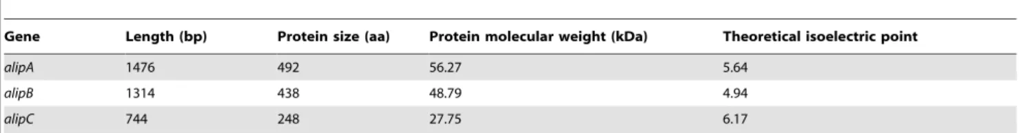
Table 1.Putative lipase/esterase genes identified fromAnaerovibrio lipolyticus5ST using the RAST annotation and features of the encoded proteins.
Gene Length (bp) Protein size (aa) Protein molecular weight (kDa) Theoretical isoelectric point
alipA 1476 492 56.27 5.64
alipB 1314 438 48.79 4.94
alipC 744 248 27.75 6.17
photometrically at 348 nm as it is the pH-independent isobestic wavelength ofr-nitrophenoxide andr-nitrophenol [28].
Effect of temperature on enzyme stability and thermostability
The effect of temperature on the activity of enzyme activity was examined across the range 25–70uC under standard assay conditions. The pH of the MES buffer was adjusted to 6.5 at respective temperatures. The thermostability of the enzymes was analysed by measuring the residual activity after incubating the enzyme (2ml in 50 mM MES, pH 6.5) for 1 h at 50, 60 and 70uC.
Effect of metal ions
The effect of metal ions on the activity of the enzymes were investigated by incubating the enzymes with various metal chloride salts (Na+
, K+
, NH4+, Mg2+, Ca2+, Mn2+, Zn2+, Co2+) at final concentrations of 5 mM in 50 mM MES (pH 6.5) for 30 min at room temperature. The remaining activity was then measured under standard assay conditions.
Nucleotide sequence accession numbers
The draft genome was deposited with NCBI BioProject Accession: PRJNA187036 The nucleotide sequences of the genes reported here are available in the GenBank database under accession numbers KC579357–KC579359.
Results
Pyrosequencing results and identification of three lipolytic genes in the draft genome
Pyrosequencing generated 340,862 high quality reads with an average length of 425 bp, representing 144,706,594 bp of total information. These data represented 366 coverage for an
estimated bacterial genome size of 4 Mbp. The assembly of the uncompleted draft genome resulted in 285 contigs with 2,830,874 bp total sequence information, comprising 247 large contigs (.5000 bp) with a total size of 2,816,384 bases. The RAST annotation identified 2,673 coding sequences and the G+C content was 43.3%. Copies of the 5ST and 23S rRNA genes (6 and 1 respectively) and 60 predicted tRNA genes were identified
within the genome. There were 268 subsystems represented in the genome, however 63% of the predicted genes could not be assigned to a subsystem. Two genes annotated as ‘‘GDSL family lipolytic enzyme’’ and one gene annotated as ‘‘carboxylesterase’’ were named alipA, alipB and alipC respectively. Phylogenetic analysis indicated that alipA and alipB clustered with the GDSL/ SGNH family II, and alipC clustered with lipolytic enzymes from family V (Figure 1).
The lipase genes alipA, alipB and alipC identified as novel members of family II and V lipases
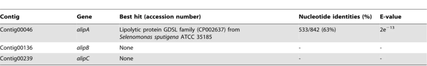
Gene length varied from 744 to 1,476 bp; and features of the encoded proteins are presented in Table 1. Tables 2 and 3 present the results of the BLASTN and BLASTP analysis of the identified putative lipase genes. GenealipAmatched with a gene coding for a lipolytic protein from Selenomonas sputigena, with 63% identity, whereas no homologous sequences were found for genesalipBand
alipCin the Genbank database. However, the proteins were found to match with proteins from variousVeillonellaceae, and the best hits were with GDSL lipolytic proteins fromSelenomonasspecies for the proteins alipA and alipB (56 and 41% identity respectively) and with a lipase/esterase from Mitsuokella multiacidafor alipC (51% identity). AlipC also shared 42% amino acid identity (e-value 8e250) with a lipase from a rumen metagenome RlipE2 [29].
Domain analysis (Table S1) revealed that alipA and alipC did not contain a signal peptide; alipB contained a putative 24-residual signal peptide at the N-terminus. AlipA and alipB contained a unique SGNH/GDSL hydrolase superfamily domain (c|01053) at amino acid residues 306–482 and 242–413 respectively. No conserved domain could be identified on half of the protein sequences, on the N side. AlipC contained a COG1647 domain (esterase/lipase function prediction) at amino acid residues 1–244 and an esterase/lipase superfamily domain (c|12031) at amino acid residues 85–227.
The proteins alipA and alipB contained, respectively, the lipase-conserved catalytic triad residues Asp466/Asp405 and His469/ His408 and the catalytic nucleophile Ser309/Ser249 in a GDS(L) motif (Figure 2). These indicated that alipA and alipB were related to enzymes from family II as defined by Arpigny and Jaeger [30]. The protein alipC contained the catalytic triad Asp194, His224
Selenomonas sputigenaATCC 35185
Contig00136 alipB None -
-Contig00239 alipC None -
-doi:10.1371/journal.pone.0069076.t002
Table 3.Best matches using BLASTP for the predicted amino acid sequence of lipolytic genes identified inAnaerovibrio lipolyticus 5ST.
Protein Putative function (accession number) Most similar homolog (e-value) Identity (overlapped aa)
alipA Lipolytic protein GDSL family (AEC00120) Selenomonas sputigenaATCC 35185 (0.0) 278/495 (56%)
alipB GDSL-like protein (EFR39963) Selenomonassp.oraltaxon 137 str. F0430 (5e2108) 169/411 (41%)
alipC Putative esterase/lipase (EEX68534) Mitsuokella multiacidaDSM 20544 (3e280) 126/249 (51%)
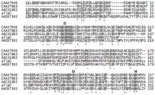
and Ser90 in the pentapeptide motif GQSMG, and multiple amino acid alignments indicated alipC would be a new member of family V (Figure 3).
Expression and purification of alipA, alipB and alipC
In order to investigate the biochemical properties of the enzymes, they were expressed in the pTrcHis TOPO vector in
Escherichia coli. AlipB without its signal sequence (alipBss) was chosen as alipB was either not expressed, degraded or insoluble during expression as it could not be detected on SDS-PAGE (data not shown). Figure 4 illustrates the purification process, with the total lysates ofE. coliTOP10 expressing the recombinant proteins before and after induction with 1 mM IPTG, and the purified fraction after elution from the nickel resin. The purification protocol routinely yielded 0.2 to 0.4 mg?ml21of purified protein from 50 ml cultures grown for 5 h after induction.
Substrate specificity
To examine substrate specificity, activity was tested against variousr-nitrophenyl esters with different acyl chain lengths. The results under standard assay conditions of pH 6.5 and 39uC are presented in Table 4. AlipA and alipBss showed a narrow chain length specificity, with the highest specific activity against r
-nitrophenyl laurate (640 U?mg21) and myristate (157 U?mg21) respectively, and lower specific activity against r-nitrophenyl caproate (33 and 43 U?mg21 respectively). AlipC showed a broader range of activity with higher specific activities against short to medium acyl chain length: the activities were 187 U?mg21 againstr-nitrophenyl butyrate, 270 U?mg21againstr-nitrophenyl caprylate, 118 and 242 U?mg21againstr-nitrophenyl laurate.
Effect of pH and temperature on enzyme activity
The effects of pH and temperature on the activity of the enzymes were determined (Figure 5). AlipA and alipC had maximal activity at pH 8.5 and 9.0 respectively, and presented .50% activity in alkaline pH ranges, respectively 7.5–9.5 and 9.0–10.0. AlipBss showed.50% of maximum activity in the pH range 6.0–8.0, with maximal activity at pH 7.5. The optimum temperatures were determined as 40uC (alipA, alipC) and 55uC (alipBss). The temperature range where the enzyme retained more than 50% activity was 40–50uC for alipA, 35–55uC for alipBss, and 35–50uC for alipC. The temperature stability of the proteins was examined by measuring its residual activity after incubating the purified enzymes for 1 h at 50, 60 or 70uC (Table 5) and thus represent both temperature stability including the protein unfold-ing and refoldunfold-ing potential of the proteins followunfold-ing thermal shock.
Figure 2. Conserved sequence motifs of alipA and alipB and lipolytic enzymes from the GDSL family.Alignment of Pfam conserved domains. The accession numbers of the aligned sequences are for the following organisms: CAA47020, triacylglycerol lipase fromPhotorhabdus luminescens; AAC38796, outer membrane esterase fromSalmonella typhimurium; AAB61674, lipase/esterase fromPseudomonas aeruginosaPAO1. Conserved motifs are highlighted. The possible catalytic triad (Serine (S), Aspartic acid (D), Histidine (H)) is shown at the top of the alignment whenever necessary.
doi:10.1371/journal.pone.0069076.g002
Figure 3. Conserved sequence motifs of alipC and lipolytic enzymes from family V.Alignment of Pfam conserved domains. The accession numbers of the aligned sequences are for the following organisms: CAA47949, triacylglycerol lipase fromPsychrobacter immobilis; CAA37863, triacylglycerol lipase fromMoraxellasp.; AAC21862, putative esterase/lipase fromHaemophilus influenzae; AAC67392, lipolytic enzyme fromSulfolobus acidocaldarius. Conserved motifs are highlighted. The possible catalytic triad (Serine (S), Aspartic acid (D), Histidine (H)) is shown at the top of the alignment whenever necessary.
The proteins alipBss and alipC appeared to be temperature sensitive as less than 50% of activity was measured after 1 h incubation at 50uC. Activities ranged from 8 to 45% after incubating at 60 or 70uC. AlipA appeared to have some thermostability: it retained around 50% activity after incubation at 60 and 70uC.
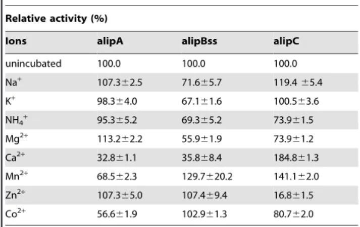
Effect of ions on enzyme activity
The effects of different metal cations at a concentration of 5 mM on the activities of the enzyme was assayed, the results are presented in Table 6. Ca2+, Mn2+ and Co2+ inhibited alipA activity(,68% activity), K+and NH
4+did not modify the activity (98–99% activity), while Na+
, Mg2+
and Zn2+
activated alipA. AlipBss activity was strongly inhibited by Ca2+
and Mg2+ (residual activities, 33 and 56% respectively) and moderately inhibited by Na+
, K+
and NH4+ (residual activities, 72, 67 and 69% respectively), whereas Mn2+
, Zn2+
and Co2+
activated alipBss (residual activities, 130, 107, and 103% respectively). Zn2+
strongly inhibited alipC activity (17% residual activity); NH4+, Mg
2+and Co2+
had more moderate inhibitory effects (residual activities from 74 to 81%). K+
had no effect, while Na+ , Ca2+
and Mn2+
activated alipC (residual activities, 119, 185, and 141% respectively).
Discussion
Next-generation sequencing has provided scientists with quick and increasingly affordable methods to access genomic data. The 454 technology has been used successfully in a number of studies to establish draft and complete bacterial genomes and establish their gene catalogues, for example for the bacteria Leuconostoc argentinum, Lactobacillus animalis [31,32], Staphylococcus epidermidis
A487 [33],Acinetobacter baumannii[34]; and, combined with other sequencing platforms, the genomes of the rumen bacteria
Megasphaera elsdenii [35], Ruminococcus albus [36] and Fibrobacter succinogenes[37].
The genus Anaerovibrio currently includes a single species, A. lipolyticus. The 454 technology was used in this study to establish the draft genome ofA. lipolyticus5ST, and identify putative lipase genes.
The draft genome was annotated using the Rapid Annotation using Subsystem Technology (RAST) server [16]. However, the accuracy of the annotation also relies on the automated pipeline
used [38], some predicted genes could be dissimilar to anything in the reference databases as they could have evolved extensively, represent uncharacterized sequences, or be misidentified [39]. Reference databases and computational methods constituting annotation pipelines are constantly developed, and there is hence a need to reprocess genome annotations on a regular basis to improve their quality and completeness [39,40]. As it was not the primary objective in this study, the genomic sequence of the bacterium remains as draft. However it would be valuable to complete the draft sequence, and subsequently annotate the complete genome, as the presence of other lipolytic genes may have been overlooked. In addition a closed genome would be useful to better understandA. lipolyticus’s role in the rumen and to share its genome in public databases for future use in molecular studies.
AlipA,alipBandalipCexhibited very low nucleotide as well as amino acid sequence similarity to previously available sequences, except for a rather low match with some amino acid sequences from the Veillonellaceae generaSelenomas,MitsuokellaandCentipeda. It is therefore proposed that they represent novel esterases/lipases, and that they have not been isolated yet from previous metagenomic studies in the rumen [29,41].
AlipA and alipB exhibited the distinct GDSL sequence motif located at the N-terminal part and Ser, Asp and His residues as part of the active site. They also contained a SGNH hydrolase superfamily domain, thus classifying them as belonging to the subfamily of GDSL/SGNH enzymes [42,43,44]. The GDSL family of enzymes was first identified by Upton and Buckley [42], these enzymes contain a GDS(L) motif located in the N-terminal part of the protein instead of the conserved lipase motif GXSXG [30]. The SGNH hydrolase subfamily was proposed from the crystal structures of Aspergillus aculeatus rhamnogalacturonan acetylesterase, Streptomyces scabies and influenza C virus esterases and an acetylhydrolase isolated fromBos taurus[45,46]; where four amino acids were found to be essential for catalysis, namely serine, glycine, asparagine, and histidine. The catalytic serine is located in a GDS(L) motif, while the Gly and Asn serve as proton donors to the oxyanion hole, and the His helps increasing the nucleophilicity of the Ser by deprotonating its hydroxyl group [45]. However sequence analysis showed that only the second half of alipA and alipB amino acid sequences were predicted for esterase/lipase function, as the conserved domains were present on amino acid residues 306–482 and 242–413 on the native sequence of alipA
Figure 4. Analysis of the proteins expressed inEscherichia coli
TOP10 cells following purification on a 15% denaturing polyacrylamide gel.Lanes M, protein standards; 1, before induction with 1 mM IPTG; 2, after cells were induced with IPTG and grown at 37uC for 5 h; 3, recombinant protein purified under native conditions with the ProBondTMpurification system. Arrows designate the positions
of the proteins on the gels.
doi:10.1371/journal.pone.0069076.g004
alipA alipBss alipC
pNP-acylesters
Butyrate (C4) 139.4617.4 91.8622.9 186.7641.5
Caproate (C6) 73.8619.0 48.6611.5 76.0648.9
Caprylate (C8) 98.4617.4 59.4621.8 269.6671.7
Caprate (C10) 32.869.5 43.2611.4 117.565.3
Laurate (C12) 639.6624.0 97.2624.7 242.0638.4
Myristate (C14) 172.2617.4 156.6611.5 193.6634.2
Palmitate (C16) 123.0619.1 64.8627.6 83.0637.5
Stearate (C18) 73.8617.3 ND 34.6620.8
ND. Not detected.
(492 aa total) and alipB (438 aa total) respectively. No conserved domain or homologies could be detected on the first part of the protein sequences; thus further studies would be needed to assess whether these parts of the proteins would contain other catalytic domains or serve protein structural functions such as autotran-sport. For example, GDSL hydrolases fromPseudomonas aeruginosa,
S. typhimuriumandPhotobacterium luminescens harbour a C-terminus domain encompassing approximately one third of the entire protein. This domain is composed of 12b-sheets which form ab -barrel inserted into the bacterial outer membrane and forms a structure similar to porines [47,48,49]. The N-terminal part of the protein carrying the catalytic activity is exported through theb -barrel and often cleaved off after translocation [50,51,52,53,54,55]. Enzymes belonging to the GDSL family were also described as exhibiting broad substrate specificity, due to a flexible active site that changes conformation with the binding of different substrates [43], and regioselectivity. In contrast, alipA had a very high specific activity againstr-nitrophenyl laurate, and
Figure 5. The effect of pH (A) and temperature (B) on the activity of proteins alipA, alipBss and alipC.The pH assays were determined againstr-nitrophenyl caprylate (C8) at a constant temperature of 39uC in a wide-range pH buffer set at the indicated pH values. Temperature assays were determined against the same substrate at a constant pH of 6.5 in 50 mM MES.
doi:10.1371/journal.pone.0069076.g005
Table 5.Relative activity of lipases cloned fromAnaerovibrio lipolyticus5ST after incubation for one hour at 50, 60 or 70uC.
Relative activity (%) Temperature of incubation
alipA alipBss alipC
40uC 100.0 100.0 100.0
50uC 16.869.8 33.6611.0 32.8612.4
60uC 45.3612.5 24.066.1 8.265.8 70uC 49.268.4 15.666.5 9.863.2
The enzymes were incubated 1 h at 50, 60 and 70uC in 50 mM MES buffer (pH 6.5); the residual activities were measured with a standard assay againstr -nitrophenyl caprylate (C8). The activity of the enzyme at 40uC was defined as 100%. Control activities at 40uC were 794, 564 and 793 U?mg21protein for
alipBss against r-nitrophenyl myristate. Assessment of their activity towards a broader range of substrate would be needed to further characterize these enzymes.
Analysis of the alipC-encoded protein showed that the protein belongs to the serine hydrolase group and has common features to basic lipases [30], as well as the conserved sequence motifs of family V bacterial lipases in its deduced amino acid sequence. It is therefore proposed that alipC could be grouped in this family. Lipases from family V have predicted molecular masses averaging 30 kDa [44] ; likewise alipC was a small, single-domain protein with a molecular mass of 27.75 kDa and a pI of 6.17. Comparison of the biochemical properties of alipC with those of other lipases is however difficult since very little information about the family V of lipases is available. Members of family V are proteins sharing significant homology with other bacterial enzymes such as epoxide hydrolases, dehalogenases and haloperoxidases, which also possess the a/b hydrolase fold [30,44]. Only four enzymes from this family have been cloned and characterized to date: the carbox-ylesterase Est2 from Acetobacter pasteurianus showing maximum activity at 78uC and pH 7.8 [56,57], the carboxylesterase EstV fromHelicobacter pyloriwith an optimum temperature of 50uC and pH of 10 [58], the lipase Lip1 isolated from the thermophilic bacterium Fervidobacterium changbaicumshowing maximum activity at 78uC and pH 7.8 [27] and the lipase RlipE2 from a metagenomic library of bovine rumen with maximum activity at 30uC and pH 7.5 [29]. Both Est2 and EstV presented typical characteristics of carboxylesterases, with EstV showing preference
(C8) [27], while RlipE2 showed high hydrolytic activity against longer chain ester substrates (C12, C16, C18) and triolein (C18) [29]. The characterization of alipC therefore gives new insights into lipase family V.
A. lipolyticus 5ST lipase activity was investigated by methods available in the 1970s [8,15]. An extracellular lipase being associated with cell surface or extracellular membranous structures was described. Activity was measured in the pH range 6.6–7.8 and temperature range 15–35uC. Optimum activity was observed at pH 7.4 and temperature 20 to 22uC, activity was enhanced by CaCl2 and BaCl2 while ZnCl2 and HgCl2 were inhibitory. Diglycerides were hydrolyzed more rapidly than triglycerides. AlipA, alipB and alipC do not match this description, though enzyme activities with different buffers and substrates cannot be directly compared [59]. Only alipB had a signal peptide suggesting it might be secreted from the cell and its activity was somewhat inhibited by Ca2+and slightly enhanced by Zn2+. AlipC activity was enhanced by Ca2+ and strongly inhibited by Zn2+, but maximal activity was observed at pH 9. Fay and colleagues [60] also observed that pure cultures of A. lipolyticus 5ST could not efficiently catalyze r-nitrophenyl palmitate and concluded this substrate was more likely to indicate esterase activity rather than lipase activity.
So far, only six pure cultures of obligate anaerobic and lipolytic bacteria have been isolated from the rumen of sheep, cattle and deer [1,10,61,62,63,64] (. It was estimated thatA. lipolyticusalone could account for the total rates of esterified fatty acid production in the rumen, based on lipid hydrolysis rates and population density studies [65]. Three lipase/esterase genes from the draft genome ofA. lipolyticus5ST were isolated, and their characteriza-tion is an important step in increasing our knowledge on the lipase activity within the rumen.
Supporting Information
Table S1 Conserved domains and predicted signal sequences in the proteins alipA, alipB and alipC.
(DOCX)
Table S2 Primers used for amplification of the lipolytic genes inAnaerovibrio lipolytica5S.
(DOCX)
Author Contributions
Conceived and designed the experiments: FP NS SH CJN. Performed the experiments: FP NK SG HW EP. Analyzed the data: FP NK SG CJN. Contributed reagents/materials/analysis tools: NK SH EP. Wrote the paper: FP SH CJN.
References
1. Hobson PN, Mann SO (1961) The isolation of glycerol fermenting and lipolytic bacteria from the rumen of the sheep. J Gen Microbiol 25:227–240. 2. Stewart CS, Flint HJ, Bryant MP (1997) The rumen bacteria. In The rumen
microbial ecosystem. 2nded. Hobson PN, and Stewart C S. (eds). Blackie Academic and Professional Publishers London, pp. 10–72.
3. Hungate RE (1966) The rumen and its microbes. Academic Press, New York, USA.
4. Stro¨mpl C, Tindall BJ, Jarvis GN, Lu¨nsdorf H, Moore ERB, et al. (1999) A re-evaluation of the taxonomy of the genusAnaerovibrio, with the reclassification of
Anaerovibrio glyceriniasAnaerosinus glycerinigen. nov., comb. nov., andAnaerovibrio
burkinabensisasAnaeroarcus burkinabensis[corrig.]. gen. nov., comb. nov. Int J Syst Bacteriol 49(4):1861–1872.
5. Hobson PN, Summers R (1967) The continuous culture of anaerobic bacteria. J Gen Microbiol 47:53–65.
6. Hobson PN, Summers R (1966) Effect of growth rate on the lipase activity of a rumen bacterium. Nature 209:736–737.
7. Henderson C, Hobson PN, Summers R (1969) The production of amylase, protease and lipolytic enzymes by two species of anaerobic rumen bacteria. In Proceedings of the Fourth Symposium on Continuous Cultivation of Micro-organisms, Prague. Academic Press London, pp. 89–204.
Relative activity (%)
Ions alipA alipBss alipC
unincubated 100.0 100.0 100.0
Na+ 107.3
62.5 71.665.7 119.465.4
K+ 98.3
64.0 67.161.6 100.563.6
NH4+ 95.365.2 69.365.2 73.961.5
Mg2+ 113.2
62.2 55.961.9 73.961.2
Ca2+ 32.8
61.1 35.868.4 184.861.3
Mn2+
68.562.3 129.7620.2 141.162.0
Zn2+
107.365.0 107.469.4 16.861.5
Co2+ 56.6
61.9 102.961.3 80.762.0
The enzymes were incubated 30 min in 50 mM MES buffer (pH 6.5) with the metal ions at 5 mM final concentration; the residual activities were measured with a standard assay againstr-nitrophenyl caprylate (C8). The activity of the enzyme not incubated with ions was defined as 100%. Control activities (inincubated) were 337, 296 and 270 U?mg21protein for alipA, alipBss and
alipC respectively).
8. Henderson C (1971) A study of the lipase produced byAnaerovibrio lipolytica, a rumen bacterium. J Gen Microbiol 65:81–89.
9. Harfoot CG, Hazlewood GP (1997) Lipid metabolism in the rumen. In The rumen microbial ecosystem. Hobson PN, and Stewart CS. (eds). Blackie Academic and Professional Publishers, pp. 382–426.
10. Prins RA, Lankhorst,A, Van der Meer P, Van Nevel CJ (1975) Some characteristics ofAnaerovibrio lipolytica, a rumen lipolytic organism. Antonie van Leeuwenhoek 41:1–11.
11. Tajima K, Aminov RI, Nagamine T, Matsui H, Nakamura M, et al. (2001) Diet-dependent shifts in the bacterial population of the rumen revealed with real-time PCR. Appl Environ Microbiol 67:2766–2774.
12. Koike S, Yabuki H, Kobayashi Y (2007) Validation and application of real-time polymerase chain reaction assays for representative rumen bacteria. Anim Sci J 78:135–141.
13. Huws SA, Lee MR, Muetzel SM, Scott MB, Wallace RJ, et al. (2010) Forage type and fish oil cause shifts in rumen bacterial diversity. FEMS Microbiol Ecol 73(2):396–407.
14. Henderson C (1970) The lipases produced byAnaerovibrio lipolyticain continuous culture. Biochem J 119(3):5P–6P.
15. Henderson C, Hodgkiss W (1973) An electron microscopic study ofAnaerovibrio lipolytica(strain 5S) and its lipolytic enzyme. J Gen Microbiol 76:389–393. 16. Aziz RK, Bartels D, Best AA, DeJongh M, Disz T, et al. (2008) The RAST
Server: Rapid Annotations using Subsystems Technology. BMC Genomics 9:75. 17. Lowe TM, Eddy SR (1997) tRNAscan-SE: a program for improved detection of
transfer RNA genes in genomic sequences. Nucleic Acids Res 25:955–964. 18. Lagesen K, Hallin P, Rødland EA, Staerfeldt HH, Rognes T, et al. (2007)
RNAmmer: consistent and rapid annotation of ribosomal RNA genes. Nucleic Acids Res 35:3100–3108.
19. Petersen TN, Brunak S, von Heijne G, Nielsen H (2011) SignalP 4.0: discriminating signal peptides from transmembrane regions. Nature Methods 8:785–786.
20. Marchler-Bauer A, Lu S, Anderson JB, Chitsaz F, Derbyshire MK, et al. (2011) CDD: a Conserved Domain Database for the functional annotation of proteins. Nucleic Acids Res 39(D):225–229.
21. Thompson JD, Higgins DG, Gibson TJ (1994) CLUSTAL W: improving the sensitivity of progressive multiple sequence alignment through sequence weighing, position specific gap penalties and weight matrix choice. Nucleic Acids Res 22:4673–4680.
22. Kalendar R, Lee D, Schulman AH (2009) FastPCR software for PCR primer and probe design and repeat search. Genes, Genomes, Genomics 3(1):1–14. 23. Bradford MM (1976) A rapid and sensitive method for the quantitation of
microgram quantities of protein utilizing the principle of protein dye binding. Anal Biochem 72:248–254.
24. Tamura K, Peterson D, Peterson N, Stecher G, Nei M, et al. (2011) MEGA5: Molecular Evolutionary Genetics Analysis using Maximum Likelihood, Evolutionary Distance, and Maximum Parsimony Methods. Molecular Biology and Evolution 28: 2731–2739.
25. Lee YP, Chung GH, Rhee JS (1993) Purification and characterisation of
Pseudomonas fluorescens SIK W1 lipase expressed in Escherichia coli. Biochim Biophys Acta 1169:156–164.
26. Pinsirodom P, Parkin KL (2001) Lipase assays. Curr Prot Food Anal Chem C3.1.1–C3.1.13.
27. Cai J, Xie Y, Song B, Wang Y, Zhang Z, et al. (2011)Fervidobacterium changbaicum
Lip1: identification, cloning, and characterization of the thermophilic lipase as a new member of bacterial lipase family V. Appl Microbiol and Biotechnol 89:1463–1473.
28. Hotta Y, Ezaki S, Atomi A, Imanaka T.(2002) Extremely stable and versatile carboxylesterase from a hyperthermophilic archaeon. Appl Environ Microbiol 68:3925–3931.
29. Liu K, Wang J, Bu D, Zhao S, McSweeney C, et al. (2009) Isolation and biochemical characterization of two lipases from a metagenomic library of China Holstein cow rumen. Bioche Biophys Res Commun 385:605–611.
30. Arpigny JL, Jaeger KE (1999) Bacterial lipolytic enzymes: classification and properties. Biochem J 343:177–183.
31. Nam SH, Choi SH, Kang A, Kim DW, Kim RN, et al. (2010) Genome sequence ofLeuconostoc argentinumKCTC 3773. J Bacteriol 192(24):6094–6491. 32. Nam SH, Choi SH, Kang A, Kim DW, Kim RN, et al. (2011) Genome
sequence ofLactobacillus animalisKCTC 3501. J Bacteriol 193(5):1280–1281. 33. Al-Marhous MM, Jack RW, Sandiford SK, Tagg JR, Beatson SA, et al. (2011)
Identification of a haemolysin-like peptide with antibacterial activity using the draft genome sequence ofStaphylococcus epidermidisstrain A487. FEMS Immunol Med Microbiol 62(3):273–282.
34. Gao F, Wang Y, Liu YJ, Wu XM, Lv X, et al. (2011) Genome sequence of
Acinetobacter baumanniiMDR-TJ. J Bacteriol 193(9):2365–2366.
35. Marx H, Graf AB, Tatto NE, Thallinger GG, Mattanovich D, et al (2011) Genome sequence of the ruminal bacteriumMegasphaera elsdenii. J Bacteriol 193(19):5578–5579.
36. Suen G, Stevenson DM, Bruce DC, Chertkov O, Copeland A, et al. (2011a) Complete genome of the cellulolytic ruminal bacteriumRuminococcus albus7. J Bacteriol 193(19): 5574–5575.
37. Suen G, Weimer PJ, Stevenson DM, Aylward FO, Boyum J, et al. (2011b) The complete genome sequence ofFibrobacter succinogenesS85 reveals a cellulolytic and metabolic specialist. PLoS ONE 6(4):e18814.
38. Bakke P, Carney C, DeLoache W, Gearing M, Ingvorsen K, et al. (2009) Evaluation of three automated genome annotations forHalorhabdus utahensis. PLoS One 4(7):e6291.
39. Stothard P, Wishart DS (2006) Automated bacterial genome analysis and annotation. Curr Opin Microbiol 9:505–510.
40. Madupu R, Brinkac LM, Harrow J, Wilming LG, Bohme U, et al. (2010) Meeting report: a workshop on best practices in genome annotation. Database 2010 doi:10.1093/database/baq001.
41. Ferrer M, Golyshina OV, Chernikova TN, Khachane AN, Reyes-Duarte D, et al. . (2005) Novel hydrolase diversity retrieved from a metagenome library of bovine rumen microflora. Environ Microbiol 7(12):1996–2010
42. Upton C, Buckley JT (1995) A new family of lipolytic enzymes? Trends Biochem Sci 20(5):178–179.
43. Akoh CC, Lee GC, Liaw YC, Huang TH, Shaw JF (2004) GDSL family of serine esterases/lipases. Prog Lipid Res 43:534–552.
44. Hausmann S, Jaeger KE (2010) Lipolytic enzymes from bacteria. In Handbook of hydrocarbon and lipid microbiology. Timmis KN (ed). Springer-Verlag, pp.1099–1126.
45. Mølgaard A (2002) Rhamnogalacturonan acetylesterase, a member of the SGNH-hydrolase family. In Advances in Pectin and Pectinase Research. Voragen, F., Schols, H., and Visser, R.G.F. (eds). Kluwer Academic Publishers, pp. 299–313.
46. Mølgaard A, Kauppinen S, Larsen S (2000) Rhamnogalacturonan acetylesterase elucidates the structure and function of a new family of hydrolases. Structure 8(4):373–383
47. Henderson IR, Navarro-Garcia F, Nataro JP (1998) The great escape: structure and function of the autotransporter proteins. Trends Microbiol 6:370–378. 48. Tamm LK, Arora A, Kleinschmidt JH (2001) Structure and assembly of
beta-barrel membrane proteins. J Biol Chem 276:32399–32402.
49. Oomen CJ, van Ulsen P, van Gelder P, Feijen M, Tommassen J, et al. (2004) Structure of the translocator domain of a bacterial autotransporter. EMBO J 23:1257–1266.
50. Pohlner J, Halter R, Beyreuther K, Meyer TF (1987) Gene structure and extracellular secretion ofNeisseria gonorrhoeaeIgA protease. Nature 325:458–462. 51. O’Toole PW, Austin JW, Trust TJ (1994) Identification and molecular characterization of a major ringforming surface protein from the gastric pathogenHelicobacter mustelae. Mol Microbiol 11:349–361.
52. Suhr M, Benz I, Schmidt MA (1996) Processing of the AIDA-I precursor: removal of AIDAc and evidence for the outer membrane anchoring as a beta-barrel structure. Mol Microbiol 22:31–42.
53. Steinhauer J, Agha R, Pham T, Varga AW, Goldberg MB (1999) The unipolar
Shigellasurface protein IcsA is targeted directly to the bacterial old pole: IcsP cleavage of IcsA occurs over the entire bacterial surface. Mol Microbiol 32:367– 377.
54. St Geme JW III, Cutter D (2000) TheHaemophilus influenzaeHia adhesin is an autotransporter protein that remains uncleaved at the C terminus and fully cell associated. J Bacteriol 182:6005–6013.
55. Dautin N, Bernstein HD (2007) Protein secretion in gram-negative bacteria via the autotransporter pathway. Annu Rev Microbiol 61:89–112.
56. Kashima Y, Ijima M, Okamoto A, Koizumi Y, Udaka S, et al. (1998) Purification and characterization of intracellular esterases related to ethylacetate formation inAcetobacter pasteurianus. J Ferm Bioeng 85:584–588.
57. Kashima Y, Nakajima Y, Nakano K, Tayama K, Koizumi Y, et al. (1999) Cloning and regulation of ethanol-regulated esterase genes in Acetobacter pasteurianus. J Biosci Bioeng 87:9–27.
58. Ruiz C, Falcocchio S, Pastor FIJ, Saso L, Diaz P (2007)Helicobacter pyloriEstV: identification, cloning and characterization of the first lipase isolated from an epsilon-proteobacterium. Appl Environ Microbiol 73(8):2423–2431. 59. Reynolds RD (1982) Buffer-dependent variations in assay of tyrosine
aminotransferase holoenzyme. Arch Biochem Biophys 219(1):140–148. 60. Fay JP, Jakober KD, Cheng KJ, Costerton JW (1990) Esterase activity of pure
cultures of rumen bacteria as expressed by the hydrolysis ofr -nitrophenylpal-mitate. Can J Microbiol 36:585–589.
61. Hazlewood GP, Dawson RMC (1975) Isolation and properties of a phospholipid hydrolysing bacterium from ovine rumen fluid. J Gen Microbiol 89:163–174. 62. Hazlewood GP, Dawson RMC (1979) Characteristics of a lipolytic and fatty acid
requiring
63. Jarvis GN, Stro¨mpl C, Moore ERB, Thiele JH (1998) Isolation and characterisation of obligately anaerobic, lipolytic bacteria from the rumen of red deer. Syst Appl Microbiol 21:135–143.
64. Cirne DG, Delgado OD, Marichamy S, Mattiasson B (2006)Clostridium lundense
sp. nov., a novel anaerobic lipolytic bacterium isolated from bovine rumen. Int J Syst Evol Microbiol56:625–628.