A co m parative study o f aging o f the
e lastic fibe r syste m o f the diaphragm
and the re ctus abdo m inis m uscle s in
rats
Laboratório de Anatomia Médico-Cirúrgica, Departamento de Cirurgia, Faculdade de Medicina, Universidade de São Paulo, São Paulo, SP, Brasil C.J. Rodrigues and
A.J. Rodrigues Junior
Abstract
In the present study the age-related changes of the striated muscle elastic fiber system were investigated in the diaphragm and rectus abdominis muscles of 1-, 4-, 8- and 18-month-old rats. The activation patterns of these muscles differ in that the diaphragm is regularly mobilized tens of times every minute during the entire life of the animal whereas the rectus abdominis, although mobilized in respira-tion, is much less and more irregularly activated. The elastic fibers were stained by the Verhoeff technique for mature elastic fibers. Weigert stain was used to stain mature and elaunin elastic fibers, and Weigert-oxone to stain mature, elaunin and oxytalan elastic fibers. The density of mature and elaunin elastic fibers showed a progressive increase with age, whereas the amount of oxytalan elastic fibers decreased in both diaphragm and rectus abdominis muscles and their muscular fascias. These age-related quantitative and structural changes of the elastic fiber system may reduce the viscoelastic properties of the diaphragm and rectus abdominis muscles, which may compromise the transmission of tensile muscle strength to the tendons and may affect maximum total strength.
Co rre spo nde nce
C.J. Rodrigues Disciplina de Topografia Estrutural Humana, FM, USP Av. Dr. Arnaldo, 455, Sala 1304 01246-903 São Paulo, SP Brasil
Fax: + 55-11-852-4877 E-mail: aldojunq@ mandic.com.br
Research supported by the Sandoz Foundation for Gerontological Research (047/89). Publication supported by FAPESP.
Received November 18, 1999 Accepted August 22, 2000
Ke y wo rds
·Aging
·Elastin
·Extracellular matrix
·Diaphragm
·Rectus abdominis
Intro ductio n
Structural changes in the diaphragm due to the aging process, mainly in the muscle fibers, have been described (1-3) but few studies have assessed alterations of the interstitial connective tissue, which main-tains muscle-tendon integrity and is involved in the transmission of muscular forces. Dur-ing agDur-ing the extracellular matrix of the skel-etal muscle undergoes remodeling to accom-modate the growth of the muscle fibers (4). In a previous report we demonstrated a
de-crease in the collagen concentration and muscle fiber size, accompanied by a rear-rangement of the distribution of the types of muscle fibers in both diaphragm (DIA) and rectus abdominis (RA) muscles during aging (5).
present study was performed to evaluate the age-related changes of the elastic fiber sys-tem of two muscles that are activated differ-ently, the diaphragm, which contracts regu-larly tens of times per minute and during the entire life of the animal, and the rectus abdominis, whose activation is much less frequent.
Mate rial and Me tho ds
Thirty-two male Wistar rats were used in this investigation. Groups of 8 animals were sacrificed 1, 4, 8 or 18 months after birth and are referred to in this paper as young, young adult, adult and aged or older, respectively. Mean weights (± SD) were 68 ± 10.06, 319 ± 33.4, 420 ± 37.15 and 458 ± 43.34 g, respec-tively. The animals received water and stan-dard chow food ad libitum.
Tissue iso latio n
The animals were killed by ether inhala-tion. After death, a cross-section of the RA and of the entire right coastal DIA was ob-tained. One representative strip from both DIA and RA was fixed in a 10% formalde-hyde solution, dehydrated, and embedded in paraffin. The paraffin blocks were sectioned, providing 5-µm thick serial sections used for quantitative assessment of elastic fiber con-tent.
Staining pro ce dure
Three adjacent sections were submitted to one of the following selective methods for staining elastic fibers: Verhoeffs (V) io-dine-iron hematoxylin method (8) to stain only the mature elastic fibers; Weigerts (W) resorcin-fuchsin method (9) to stain mature and elaunin elastic fibers, and Weigerts tech-nique preceded by oxidation performed through oxone (WO), as previously described (10) to stain the oxytalan, elaunin and ma-ture elastic fibers.
Mo rpho m e tric e valuatio n
The linear density (LV) of the elastic fiber system was determined in 25 random microscopic fields per histologic section stained by the Verhoeff (LVV), Weigert (LVW) and Weigert-oxone (LVWO) tech-niques and all measurements were performed by the same observer. The sections were scanned randomly at 1,000X magnification in a continuous line from edge to edge, em-ploying a test eyepiece reticule with 10 par-allel lines and 100 points that contains a simple square lattice test system of 10,500 µm2. Each elastic fiber completely inter-sected by any one of the test lines was counted. The intersections of fibers with a test reticule are related to the length of those fibers per unit area, according to the equa-tion LV = 2Na, where Na is the total length of fibers per unit area (11). The area of the tissue examined was determined by count-ing the number of points of intersection in tissue sample.
Ele ctro n micro sco py
Small pieces of tissue were fixed in a solution containing 0.1% tannic acid and 3% buffered glutaraldehyde, followed by post-fixation in 1% osmium tetroxide for 1 h. The fixed material was stained in 0.5% aqueous uranyl acetate overnight and rou-tinely embedded. The sections were cut with an ultratome apparatus and double-stained with uranyl acetate and lead citrate. Thin sections (70 nm) were subsequently analyzed with a Zeiss 9S2 electron micro-scope.
Statistical analysis
Re sults
Body weight and cross-sectional area of muscle fibers from both DIA and RA in-creased significantly as a function of age following a second degree curve, as previ-ously reported (5). The usual three compo-nents of the elastic fiber system in the extra-cellular matrix were found in both muscles. In cross-sections, the elastic fibers of young rats were thin, straight and oriented perpen-dicularly to the long axis of the muscle cells (Figure 1). In the older animals, however, the elastic fibers were thickened and curled (Figure 2). These aging changes of the elas-tic fibers were easily demonstrated in 18-month-old rat muscle.
At the ultrastructural level the mature
Figure 1 - Diaphragm (A) and rectus abdominis muscles (B) from a young (1 month) rat. Thin and straight elastic fibers (ar-row s) surrounding the muscle cells. W eigert -oxone st ain (800X).
Figure 2 - Diaphragm (A) and rec-tus abdominis muscles (B) from an aged (18 months) rat. Thick and curled elastic fibers (arrow s) surrounding the muscle cells. Weigert-oxone stain (800X).
elastic fibers showed a central core of elastin surrounded by some microfibrils, while the oxytalan fibers were composed only of bundles of ordered microfibrils. The elaunin fibers exhibited a low amount of elastin (Fig-ure 3). In the DIA and RA of aged rats we found foci of oxytalan fibers that were ran-domly distributed in an oval-shaped arrange-ment corresponding to abnormal oxytalan fibers (Figure 4).
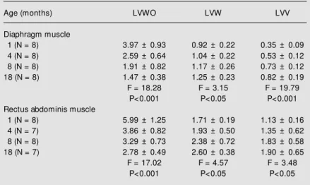
The LV of elastic fibers in both muscles was quantified on selectively stained sec-tions. LV values represented the total length of mature, elaunin and oxytalan fibers per unit area in WO-stained tissue, mature and elaunin fibers in W-stained tissue, and ma-ture fibers in V-stained tissue. The mean values of LVV-, LVW- and LVWO-stained
A
B
10 µm
A
B
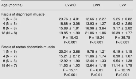
and LVWO in both DIA and RA and their muscular fascia.
D iscussio n
There was a progressive increase of the linear density of mature and elaunin elastic fibers and a decrease of the oxytalan fibers with age, as assessed in the DIA and RA muscles of the rat. Considering that the ex-tracellular matrix of skeletal muscle has the function to maintain muscle-tendon integ-rity and the transmission of muscle tension, it is obvious that by altering the composition of collagen and elastin or changing their architectural arrangement, these properties may be compromised.
The tissue elasticity is primarily depend-ent upon the concdepend-entration of the mature and elaunin elastic fibers and the tissue resis-tance is related to the oxytalan elastic fibers (6). The high linear density of oxytalan fi-bers observed in both DIA and RA from young rats is a marker of a strong local mechanical stress (12). This result may be explained by the fact that in this phase of development the respiratory frequency is higher than in the adult, thus requiring a large overall mechanical work exerted by both the DIA and the RA, which is closely associated with respiration (13). If this hy-pothesis is correct, the abrupt decline in the quantity of oxytalanic fibers in both muscles in adult rats indicates a lower degree of mechanical load upon the DIA and RA. It is interesting that a reduction of oxytalan elas-tic fibers as a function of age was described for the pulmonary parenchyma of man with an abrupt decline between one and twenty years of age (11).
It is known that the concentration and the phenotypes of collagen may influence the muscular passive and/or active length-force and the viscoelastic properties (14). Further-more, it has been demonstrated that slow-twitch muscles have a greater amount of collagen than fast muscles (15). In a previ-fibers for each age group are indicated in
Table 1 for the muscles, and in Table 2 for their muscular fascia. There was a positive correlation between LVV and LVW with age, and a negative correlation between age
Table 1 - Linear density (LV) of the elastic fiber system.
Values are reported as the mean ± SD (x 10-3 µm-2); N, number of rats. WO,
Weigert-oxone; W, Weigert; V, Verhoeff: methods for staining elastic fibers. The data on LV of elastic fiber concentration in the tissue w ere analyzed by ANOVA correlating them w ith age.
Age (months) LVWO LVW LVV
Diaphragm muscle
1 (N = 8) 3.97 ± 0.93 0.92 ± 0.22 0.35 ± 0.09
4 (N = 8) 2.59 ± 0.64 1.04 ± 0.22 0.53 ± 0.12
8 (N = 8) 1.91 ± 0.82 1.17 ± 0.26 0.73 ± 0.12
18 (N = 8) 1.47 ± 0.38 1.25 ± 0.23 0.82 ± 0.19
F = 18.28 F = 3.15 F = 19.79
P<0.001 P<0.05 P<0.001
Rectus abdominis muscle
1 (N = 8) 5.99 ± 1.25 1.71 ± 0.19 1.13 ± 0.16
4 (N = 7) 3.86 ± 0.82 1.93 ± 0.50 1.35 ± 0.62
8 (N = 8) 3.29 ± 0.73 2.38 ± 0.72 1.83 ± 0.58
18 (N = 7) 2.78 ± 0.49 2.60 ± 0.38 1.90 ± 0.65
F = 17.02 F = 4.57 F = 3.48
P<0.001 P<0.05 P<0.05
Figure 3 - Diaphragm muscle from an adult (8 months) rat. Elaunin elastic fibers (EL) run perpendicular to the long axis of the muscle cell (28,500X).
Figure 4 - Diaphragm muscle from an aged (18 months) rat. An abnormal oxytalan fiber (row ) show ing haphazardly ar-ranged microfibrils (28,500X).
1 µm
ous report we demonstrated a smaller amount of collagen in the RA muscle than in the DIA (5). In the present study we demonstrated a higher amount of elastic fibers in these rat RA muscles than in the DIA. These data were compatible with the function of RA muscle, which is faster and stronger.
With aging the DIA and RA muscles showed a smaller amount of oxytalan fibers, which promote resistance, and an increased amount of mature and elaunin fibers, which are the principal structural elements of elas-ticity (6,10). However, these mature and elaunin elastic fibers undergo distortion of their normal architecture, becoming thicker, tortuous and fragmented. These morphologi-cal changes of the elastic fibers reduce their original elasticity because their total stretch is compromised and there are reduced breaks of hydrophobic links, with a consequent re-duced storage of free and elastic energy (16). These structural alterations seem to be pro-moted by an increased and ordered arrange-ment of collagen (17), which compresses the elastic fibers, and by the action of tissue elastase, that causes degenerative changes in these fibers. Cannon and Read (18) showed evidence of increased elastolytic plasma ac-tivity in patients around 60 years of age with direct inguinal hernia when compared to the plasma activity of patients within the same age range but without hernias.
These age-related elastic fiber changes explain the skin looseness and wrinkling (19,20), the weakness of the transversalis fascia, which promotes the high prevalence of direct inguinal hernia in the elderly (21), as well as the weakness of the fascia lata (22) and of the splenic capsule (23).
The decrease of oxytalanic fibers and the structural and architectural alterations in the elastic fibers that occur with aging in both DIA and RA would expose the interstitial collagen of skeletal muscle to deformation tensions higher than it normally endures. As a consequence, collagen fibers would be submitted to greater deformation, and since they represent a resistance element, they would end up by rupturing, promoting re-duced muscle viscoelastic properties, which may compromise the transmission of tensile muscle strength to the tendons and may af-fect maximum total strength.
Ackno wle dgm e nts
We thank Prof. Dr. Cesar Timo-Iaria for helpful comments.
Table 2 - Linear density (LV) of the elastic fiber system.
Values of LV are reported as the mean ± SD (x 10-3 µm-2); N, number of rats. For
abbreviations, see legend to Table 1. The data on LV of elastic fiber concentration in the tissue w ere analyzed by ANOVA correlating them w ith age.
Age (months) LVWO LVW LVV
Fascia of diaphragm muscle
1 (N = 8) 23.76 ± 4.01 12.66 ± 2.27 5.25 ± 0.82
4 (N = 8) 18.88 ± 3.08 13.93 ± 1.27 8.42 ± 2.50
8 (N = 8) 15.89 ± 1.81 16.96 ± 3.64 9.17 ± 2.82
18 (N = 8) 19.85 ± 1.90 21.06 ± 1.86 16.39 ± 1.77
F = 10.43 F = 18.24 F = 39.78
P<0.001 P<0.001 P<0.001
Fascia of rectus abdominis muscle
1 (N = 8) 20.24 ± 3.66 9.78 ± 1.21 6.19 ± 1.15
4 (N = 7) 15.21 ± 2.12 11.86 ± 1.74 8.30 ± 1.87
8 (N = 8) 12.92 ± 1.90 12.44 ± 1.33 9.54 ± 1.38
18 (N = 7) 11.53 ± 1.03 12.64 ± 1.18 11.14 ± 1.75
F = 15.11 F = 6.01 F = 12.19
Re fe re nce s
1. Daw CK, Starnes JW & White TP (1988). M uscle atrophy and hypoplasia w ith ag-ing: impact of training and food restric-tion. Journal of Applied Physiology, 64: 2428-2432.
2. Eddinger TJ, Cassens RG & M oss RL (1986). M echanical and histochemical characterization of skeletal muscles from senescent rats. AmericanJournal of Phys-iology, 251: C421-C430.
3. Zhang Y & Kelsen SG (1990). Effects of aging on diaphragm contractile function in golden hamsters. American Review of Respiratory Disease, 142: 1396-1401. 4. Gosselin LE, M artinez DA, Vailas AC &
Sieck GC (1993). Interstitial space and col-lagen alterations of the developing rat dia-phragm. Journal of Applied Physiology, 74: 2450-2455.
5. Rodrigues CJ, Rodrigues Junior AJ & Bohm GM (1996). The effects of aging on the muscle fibers and collagen content of the diaphragm: A comparison w ith the rectus abdominis muscle. Gerontology, 42: 218-228.
6. Ross R (1973). The elastic fiber. A review .
Journal of Histochemistry and Cytochem-istry, 21: 199-208.
7. Dieler R & Schroder JM (1990). Increase of elastic fibers in muscle spindles of rats follow ing single or repeated denervation w ith or w ithout reinnervation. Virchow s Archiv. A, Pathological Anatomy and His-topathology, 417: 213-221.
8. Verhoeff FH (1908). Some new staining
methods of w ide applicability including a rapid differential stain for elastic tissue.
Journal of the American M edical Associa-tion, 50: 876-877.
9. Weigert C (1898). Über eine M ethode zur Färbung elastischer Fasern. Zentralblatt für Allgemeine Pathologie, und Pathologi-sche Anatomie, 9: 289-292.
10. Fullmer HM & Lillie RD (1958). The oxytalan fiber: a previously undescribed connective tissue fiber. Journal of His-tochemistry and CyHis-tochemistry, 6: 425-430.
11. Niew oehner DE & Kleinerman J (1977). M orphometric study of elastic fibers in norm al and em physem at ous hum an lungs. American Review of Respiratory Disease, 115: 15-21.
12. Cleary EG & Gibson M A (1983). Elastin-associated microfibrils and microfibrillar proteins. International Review of Connec-tive Tissue Research, 10: 97-209. 13. Crosfilt M & Widdicombe J (1961).
Physi-cal characteristics of the chest and lungs and the w ork of breathing in different mammalian species. Journal of Physiolo-gy, 158: 1-14.
14. M ays PK, Bishop JE & Laurent GJ (1988). Age-related changes in the proportion of types I and III collagen. M echanisms of Ageing and Development, 45: 203-212. 15. Kovanen V, Suominen H & Heikkinen E
(1984). Collagen of slow tw itch and fast tw itch muscle fibers in different types of rat skeletal muscle. European Journal of
Applied Physiology, 52: 235-242. 16. Gosline JM (1978). Hydrophobic
interac-tion and a model for the elasticity of elas-tin. Biopolymers, 17: 677-695.
17. Danielsen L & Kobayashi T (1972). Degen-eration of dermal elastic fibers in relation to age and light exposure. Preliminary re-port on electron microscopic studies. Acta Dermato-Venereologica, 52: 1-10. 18. Cannon DR & Read RC (1981). M etastatic
emphysema. M echanism for acquiring in-guinal herniation. Annals of Surgery, 194: 270-277.
19. Imayama S & Braverman IM (1989). A hypothetical explanation for the aging of skin. American Journal of Pathology, 134: 1019-1025.
20. Tsuji T & Hamada T (1981). Age-related changes in human dermal elastic fibers.
British Journal of Dermatology, 105: 57-63.
21. Rodrigues Junior AJ, Tolosa EM C & Carvalho CAF (1990). Electron microscop-ic study on the elastmicroscop-ic and elastmicroscop-ic related fibers in the human fascia transversalis at different ages. Gegenbaurs M orphologi-sches Jahrbuch, 136: 645-652.
22. Bick EM (1961). Aging in the connective tissues of the human musculoskeletal system. Geriatrics, 16: 448-453. 23. Rodrigues CJ, Sacchetti JC & Rodrigues