Fo o d re strictio n induce s
in vivo
ve ntricular dysfunctio n in
spo ntane o usly hype rte nsive rats
witho ut im pairm e nt o f
in vitro
m yo cardial co ntractility
Departamentos de 1Clínica Médica and 2Pediatria, Faculdade de Medicina de Botucatu,
Universidade Estadual Paulista, Botucatu, SP, Brasil K. O koshi1, J.R. Fioretto2,
M.P. O koshi1, A.C. Cicogna1, F.F. Aragon1, L.S. Matsubara1 and B.B. Matsubara1
Abstract
Cardiac structures, function, and myocardial contractility are affected by food restriction (FR). There are few experiments associating under-nutrition with hypertension. The aim of the present study was to analyze the effects of FR on the cardiac response to hypertension in a genetic model of hypertension, the spontaneously hypertensive rat (SHR). Five-month-old SHR were fed a control or a calorie-restricted diet for 90 days. Global left ventricle (LV) systolic function was evaluated in vivo by transthoracic echocardiogram and myocardial contractility and diastolic function were assessed in vitro in an isovolumetrically beating isolated heart (Langendorff preparation). FR reduced LV systolic function (control (mean ± SD): 58.9 ± 8.2; FR: 50.8 ± 4.8%, N = 14, P < 0.05). Myocardial contractility was preserved when assessed by the +dP/dt (control: 3493 ± 379; FR: 3555 ± 211 mmHg/s, P > 0.05), and developed pressure (in vitro) at diastolic pressure of zero (control: 152 ± 16; FR: 149 ± 15 mmHg, N = 9, P > 0.05) and 25 mmHg (control: 155 ± 9; FR: 150 ± 10 mmHg, N = 9, P > 0.05). FR also induced eccentric ventricular remodeling, and re-duced myocardial elasticity (control: 10.9 ± 1.6; FR: 9.2 ± 0.9%, N = 9, P < 0.05) and LV compliance (control: 82.6 ± 16.5; FR: 68.2 ± 9.1%, N = 9, P < 0.05). We conclude that FR causes systolic ventricular dysfunction without in vitro change in myocardial contractility and diastolic dysfunction probably due to a reduction in myocardial elas-ticity.
Co rre spo nde nce
J.R. Fioretto
Departamento de Pediatria Faculdade de Medicina, UNESP Caixa Postal 530
18618-970 Botucatu, SP Brasil
Fax: + 55-14-6822-0421 E-mail: jrf@ fmb.unesp.br
Research supported by FAPESP (No. 95/4318-5).
Received May 8, 2003 Accepted January 22, 2004
Ke y words •Undernutrition
•Isolated heart
•Echocardiogram
•Langendorff preparation
•Ventricular remodeling
Intro ductio n
Researchers have long believed that the heart is spared in the presence of undernutri-tion and have focused their studies on other organs and systems that are strongly affected
by food restriction (FR).
(1-6), with important ultrastructural changes occurring in the rat myocardium (4,6). We have previously demonstrated that FR in-creases myocardial hydroxyproline concen-tration and causes left ventricular (LV) ec-centric remodeling and diastolic dysfunc-tion (7). We have also observed that FR induces major ultrastructural changes in the myocardium of normotensive and spontane-ously hypertensive rats (SHR) (4,8). If these pathological conditions, hypertension and undernutrition, coexist in the same individual, we may assume that ventricular function is markedly affected. However, there are few experiments associating undernutrition with systemic arterial hypertension (4,9,10). This association is very interesting because un-dernutrition is a low nutrient supply condi-tion, while systemic arterial hypertension causes further energy consumption by in-creasing myocardial protein synthesis (11).
The aim of the present study was to ana-lyze the effects of FR on in vivo LV systolic function and on in vitro LV contractility and diastolic function in a genetic model of hy-pertension, the SHR. To the best of our knowledge, this is the first study of SHR with FR in which the echocardiogram and isolated heart technique are used in the same animal.
Mate rial and Me thods
Study groups
All investigations were performed ac-cording to the Guide for the Care and Use of Laboratory Animals published by the U.S. National Institutes of Health and were ap-proved by the Animal Research Committee of the Medical School of Botucatu, São Paulo, Brazil.
Five-month-old male SHR were fed a control or a restricted diet for 90 days. The control group (N = 23) had free access to regular rat chow (Purina Labina®, São Paulo, SP, Brazil), and its consumption was
meas-ured daily. The animals subjected to FR (N = 23) received 50% of the amount of chow consumed by the control group on the previ-ous day. Water was provided ad libitum. The rats were housed in individual cages at room temperature (23ºC) on a 12-h light/dark cycle. Body weight was measured once a week and blood pressure was recorded at the begin-ning of the protocol and before sacrifice using the indirect tail-cuff technique (12).
Echocardiographic study
frac-tional shortening index ((LVDD-LVSD)/ LVDD x 100).
Intra-observer (K.O.) variability was cal-culated by reading the M-mode tracings twice in a blind fashion (mean ± SD; LVDD: 1.31 ± 1.55; LVSD: 2.49 ± 1.91; LVWT: 2.60 ± 1.56%).
Le ft ve ntricular study - Lange ndo rff
pre paratio n
The in vitro study was performed on 9 animals from each group after the echocardi-ogram. Hearts from four animals were fro-zen and stored for later morphological study. One heart was eliminated from the in vitro
study because it did not reach stability. The hearts were studied using a modified Lan-gendorff preparation procedure as previously described (7). Briefly, rats were anesthetized with sodium thiopental (50 mg/kg, ip) and heparinized (1,000 IU). The chest was en-tered by median sternotomy under artificial ventilation. The ascending aorta was iso-lated and cannuiso-lated for retrograde perfu-sion with filtered oxygenated Krebs-Henseleit solution maintained at constant temperature and perfusion pressure, 37ºC and 75 mmHg, respectively. The Krebs-Henseleit solution, gassed with 95% oxygen-5% carbon diox-ide, pH 7.3-7.4, had the following composi-tion: 115 mM NaCl, 5.4 mM KCl, 1.2 mM MgSO4, 2.5 mM CaCl2, 1.15 mM NaH2PO4,
25 mM NaHCO3, 11 mM glucose, and 8 mM
mannitol. The heart was removed quickly from the chest and attached to the perfusion apparatus (model 830; Hugo Sachs Elektronic, Grunstasse, Germany). The pul-monary artery was cut to vent the right ven-tricle during systole, the left atrial append-age was removed, and a latex balloon (7 mm in length) was placed inside the LV via the mitral valve orifice. The balloon was already attached to a plastic cannula connected to a three-way stopcock through which the bal-loon was filled with saline solution or emp-tied; ventricular pressure was measured
us-ing a P23XL transducer and a polygraph (model 40-9800-20 Windograph; Gould, Valleyview, OH, USA). Once the heart de-veloped stable isovolumetric contractions (maintenance of systolic and diastolic pres-sure at a certain intraventricular volume), the balloon volume was increased in 20-µl increments over an end-diastolic pressure range of 0-25 mmHg. Pressure and volume within the balloon were recorded after each increase and corresponded to the LV pres-sure and volume, respectively. The volume at zero end-diastolic pressure reflects un-stressed ventricular volume (V0), which was used as an index of chamber size. To ensure stability of the preparation, two or three data sets were recorded. All hearts were paced from the right atrium at 230 beats/min using an artificial pacer (model 79232; Hugo Sachs Elektronic).
Diastolic function was analyzed by meas-uring or calculating the following variables: maximal rate of decrease in LV pressure (-dP/dt, index of myocardial relaxation), per-cent variation in LV volume required to increase diastolic pressure from 0 to 25 mmHg (∆V25, index of LV compliance), and percent myocardial strain caused by a dia-stolic stress of 25 g/cm2 (ε
25, index of myo-cardial elasticity). Myomyo-cardial elasticity was calculated as follows (14,15):
stress = (1.36 x LVP x V2/3)/[(V + 0.943 x LVW)2/3 - V2/3]
strain = {[V1/3 + (V + 0.943 x LVW)1/3]/ [V01/3 + (V0 + 0.943 x LVW)1/3] - 1} x 100
where LVP is LV pressure (mmHg), V is chamber volume (ml), LVW is left ventricu-lar weight (g), and V0 is volume (ml) at a diastolic pressure of 0 mmHg.
Re sults
There were no differences in initial body weight (BWi) between the experimental groups. FR decreased body weight (BW) with no effect on tail-cuff systolic blood pressure or in vivo heart rate (Table 1).
The echocardiographic study (Table 1) showed that FR did not change LVDD, in-creased LVSD, and reduced LVWT. Nor-malization of LVDD to BW showed that FR increased this ratio. Undernutrition decreased the LVWT/LVDD ratio and LV systolic per-formance assessed by the fractional shorten-ing index.
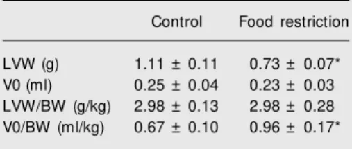
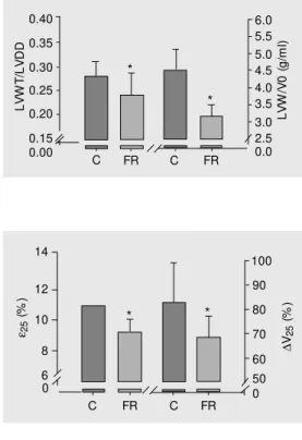
The in vitro study (Table 2) showed that FR reduced LVW in proportion to BW re-duction. The volume at zero mmHg LV dia-stolic pressure (V0) was not changed after FR. Undernutrition reduced both the LVW/ V0 and LVWT/LVDD ratios, indicating the occurrence of eccentric ventricular remodel-ing (Figure 1). FR did not change +dP/dt, Piso 0 and Piso 25 (Table 3). Food-restricted ani-mals showed increased developed stress in both diastolic pressures (zero and 25 mmHg). LV relaxation, evaluated by the -dP/dt index, was not affected by FR (Table 3). However, the ε25 and ∆V25 values respectively showed that FR reduced myocardial elasticity and LV compliance (Figure 2).
D iscussio n
The undernutrition protocol used in this experiment was based on the fact that human food deprivation is generally the result of a deficiency of all diet components (16). Inves-tigators have been using different reduction levels of total chow amount without specific deficiencies (6,17-19). Our undernutrition protocol was effective by causing a marked body weight reduction. This study did not include a control group, preventing conclu-sions about the impact of hypertension on cardiac remodeling in normotensive animals. FR did not change tail-cuff systolic blood
Table 1. General characteristics of rats and echo-cardiographic data.
Control Food restriction BWi (g) 350 ± 20 345 ± 14 BW (g) 380 ± 33 258 ± 22*
HR (in vivo, bpm) 268 ± 43 240 ± 24
SBP (mmHg) 190 ± 24 196 ± 21 LVDD (mm) 7.03 ± 0.46 6.96 ± 0.45 LVSD (mm) 2.91 ± 0.70 3.44 ± 0.47* LVWT (mm) 1.97 ± 0.14 1.64 ± 0.17* LVDD/BW (mm/kg) 18.7 ± 1.2 27.2 ± 3.2* FS (% ) 58.9 ± 8.2 50.8 ± 4.8* Data are reported as means ± SD for 14 animals in each group. BWi = body w eight at the begin-ning of the food restriction (FR) protocol; BW = body w eight at the end of the FR protocol; HR = heart rate; SBP = tail-cuff systolic blood pressure; LVDD = left ventricular diastolic diameter; LVSD = left ventricular systolic diameter; LVWT = left ventricular w all thickness; FS = left ventricular fractional shortening.
* P < 0.05 vs control (Student t-test).
Table 2. Structural study of the left ventricle in an isolated Langendorff heart preparation.
Control Food restriction LVW (g) 1.11 ± 0.11 0.73 ± 0.07* V0 (ml) 0.25 ± 0.04 0.23 ± 0.03 LVW/BW (g/kg) 2.98 ± 0.13 2.98 ± 0.28 V0/BW (ml/kg) 0.67 ± 0.10 0.96 ± 0.17* Data are reported as means ± SD for 9 animals in each group. LVW = left ventricular w eight; V0 = left ventricular unstressed volume; BW = body w eight.
* P < 0.05 vs control (Student t-test).
0 and 25 mmHg (DS-0 and DS-25, respec-tively). Developed stress was calculated us-ing the same formula as described above.
Statistical analysis
pressure, in agreement with data reported by Gradin and Persson in a study on SHR (17). However, Overton et al. (20) found a reduc-tion in blood pressure which was explained by diminished sympathetic support. As was also the case in the present study, these investigators did not observe FR interfer-ence with heart rate. Since we found no changes in tail-cuff systolic blood pressure or heart rate, we may assume that these parameters had no influence on the in vivo
results.
FR did not alter LVDD but decreased LVWT; consequently, the LVWT/LVDD ratio was decreased in FR rats. In undernour-ished children and patients with anorexia nervosa, echocardiographic studies have al-ways shown lower LVDD and LVWT (1,2,21,22). In our laboratory, LVDD and LVWT reduction was seen in normotensive rats submitted to the same FR protocol. This highlights the question concerning the influ-ence of hypertension on ventricular cavity remodeling in rats submitted to FR. The absence of a reduction in LV chamber size suggests an inability to adapt to hemody-namic load when undernutrition restricts myocardial growth. This was confirmed by the decrease in ejection index found in the in vivo study. The lower LVWT/LVDD ratio indicates that FR caused a change in the shape of the ventricular cavity, showing an eccentric remodeling.
The in vitro study confirmed the struc-tural findings obtained in vivo; FR did not alter V0, but decreased LVW. Consequently, the LVW/V0 ratio was lower in FR rats, indicating eccentric remodeling.
FR decreased the ability of the LV to eject, which may have been caused by the reduction in myocardial contractility and/or cardiac load changes. The M-mode echocar-diogram is an efficient method for in vivo
studies, which, however, can be used only in the study of myocardial contractility. The study of the isolated heart from the same animal allowed us to gain a better
under-Figure 1. Ratio of left ventricular w all thickness (LVWT) to dia-stolic diameter (LVDD) and ratio of left ventricular w eight (LVW) to volum e (V0). Data are re-ported as means ± SD for 9 ani-mals in each group. C = control group; FR = f ood-rest rict ed group. Note that both ordinates and abscissa are discontinuous. * P < 0.05 compared to control animals (Student t-test).
ε25
( % ) 14 100 12 10 8 6
C FR C FR 90 80 70 60 50 0 0 * * ∆ V2 5 ( % )
Figure 2. Left ventricular dia-stolic function evaluated in the isolated heart. Data are reported as means ± SD for 9 animals in each group. C = control group; FR = food-restricted group; ε25 = percent age of m yocardial st rain caused by a diast olic stress of 25 g/cm2; ∆V25 =
per-centage of variation in ventricu-lar volume required to increase diastolic pressure from 0 to 25 mmHg. Note that both ordinates and abscissa are discontinuous. * P < 0.05 compared to control animals (Student t-test). Table 3. Functional study of the left ventricle in an
isolated Langendorff heart preparation.
Control Food restriction Systole
+dP/dt (mmHg/s) 3493 ± 379 3555 ± 211 Piso 0 (mmHg) 152 ± 16 149 ± 15
Piso 25 (mmHg) 155 ± 9 150 ± 10
DS-0 (g/cm2) 104 ± 19 136 ± 19*
DS-25 (g/cm2) 174 ± 19 217 ± 22*
Diastole
-dP/dt (mmHg/s) 2090 ± 234 1986 ± 246 Data are reported as means ± SD for 9 animals in each group. +dP/dt = rate of rise of ventricular pressure; Piso 0 and Piso 25 = developed pressure
at diastolic pressure of zero and 25 mmHg, re-spectively; DS-0 and DS-25 = developed stress at diastolic pressure of zero and 25 mmHg, respec-tively; -dP/dt = rate of decrease of left ventricular pressure.
* P < 0.05 vs control (Student t-test).
L V W T /L V D D 0.40 L V W /V 0 ( g /m l) 6.0 0.35 0.30 0.25 0.20 0.15
C FR C FR 5.5 5.0 4.5 4.0 3.5 3.0 2.5 0.0 0.00 * *
and heart rate were controlled in the study of the isolated isovolumetrically beating heart. We do not think that the increase of devel-oped stress in FR rats is an indication of improved systolic performance because this index is inappropriate for the evaluation of myocardial contractility when the LV shows eccentric remodeling (7). However, an in-creased developed stress with unchanged systolic pressure may help understand the mechanism by which ventricular perfor-mance was impaired in the in vivo study on FR rats. Since both groups had the same in vivo arterial systolic pressure and undernu-trition caused eccentric remodeling, we as-sumed that the FR rats presented a higher in vivo afterload. This increased afterload may explain the ventricular ejection impairment even when the inotropic state is unchanged. One might ask if the in vivo preload condi-tion would favorably affect LV performance of FR rats, i.e., if for a given diastolic pres-sure the preload is higher in an eccentrically shaped ventricle. Consequently, preload re-serve would be recruited in these hearts, helping systolic function. However, we be-lieve that this was not the case in our study. We analyzed all hearts in the same diastolic pressure range, and systolic performance was the same in both groups.
FR caused a decrease in myocardial elas-ticity and ventricular compliance. In our pre-vious studies, we observed that FR increased myocardial hydroxyproline concentration in adult and young normotensive rats (3,7), possibly explaining the alterations found in the diastolic property. It was interesting to observe that the increase in myocardial stiff-ness and the consequent reduction in ven-tricular compliance occurred in the presence of eccentric remodeling. This remodeling, caused by a disproportional reduction in LV weight (or wall thickness) compared to LV volume (or diastolic diameter), might have led to increased ventricular compliance (25). In summary, SHR submitted to FR for 90 days showed a reduction in LV wall thick-ness, eccentric remodeling, in vivo systolic performance depression, and changes in pas-sive diastolic properties, with no changes in tail-cuff systolic blood pressure, heart rate, LV diastolic diameter, or myocardial con-tractility.
Ackno wle dgm e nts
We thank J.C. Georgette, V.M. Souza, and M.A. Dallaqua for expert technical as-sistance and Colin E. Knaggs for revising the English text.
Re fe re nce s
1. Simone G, Scalfi L & Galderisi M (1994). Cardiac abnormalities in young w omen w ith anorexia nervosa. British Heart Journal, 71: 278-292.
2. St. John Sutton M G, Plappert T, Crosby L, Douglas P, M ullen J & Reichek N (1985). Effects of reduced left ventricular mass on cham-ber architecture, load, and function: a study of anorexia nervosa.
Circulation, 72: 991-1000.
3. Cicogna AC, Padovani CR, Okoshi K, M atsubara LS, Aragon FF & Okoshi M P (2001). The influence of temporal food restriction on the performance of isolated cardiac muscle. Nutrition Research, 21: 639-648.
4. Okoshi M P, Okoshi K, Dal Pai V, Dal Pai-Silva M , M atsubara LS & Cicogna AC (2001). M echanical, biochemical and morphological changes in the heart from chronic food restricted rats. Canadian Journal of Physiology and Pharmacology, 79: 754-760.
5. M cKnight KA, Rupp H, Dhalla KS, Beamish RE & Dhalla NS (1999). Biphasic changes in heart performance w ith food restriction in rats.
Journal of Applied Physiology, 87: 1909-1913.
6. Rossi M A & Zucoloto S (1982). Ultrastructural changes in nutritional cardiomyopathy of protein-calorie malnourished rats. British Journal of Experimental Pathology, 63: 242-253.
7. Fioretto JR, Queiroz SS, Padovani CR, M atsubara LS, Okoshi K & M atsubara BB (2002). Ventricular remodeling and diastolic myocar-dial dysfunction in rats submitted to protein-calorie malnutrition.
American Journal of Physiology,282: H1327-H1333.
8. Okoshi M P, Pai VD, M eyer M M , Pai-Silva M D & Cicogna AC (1998). M orphological alterations in heart muscle tissue from undernour-ished hypertensive rats. Journal of the American College of Cardiol-ogy, 31: 338C.
Balti-more, M D, USA.
10. Sw oap SJ, Boddell P & Baldw in KM (1995). Interaction of hyperten-sion and caloric restriction on cardiac mass and isomyosin expres-sion. American Journal of Physiology, 268: R33-R39.
11. Katz AM (1994). The cardiomyopathy of overload: an unnatural grow th response in the hypertrophied heart. Annals of Internal M edicine, 121: 363-371.
12. Pfeffer JM , Pfeffer M A & Frohlich ED (1971). Validity of an indirect tail-cuff method for determining systolic arterial pressure in unanes-thetized normotensive and spontaneously hypertensive rats. Jour-nal of Laboratory and Clinical M edicine,78: 957-962.
13. Sahn DJ, DeM aria A, Kisslo J & Weyman AE (1978). The Committee on M -M ode Standardization of the American Society of Echocardi-ography. Recommendations regarding quantitation in M -mode echo-cardiography: results of a survey of echocardiographic measure-ments. Circulation, 58: 1072-1083.
14. Doering CW, Jalil JE, Janicki JS, Pick R, Shahriar A, Abrahams C & Weber KT (1988). Collagen netw ork remodeling and diastolic stiff-ness of the rat left ventricle w ith pressure overload hypertrophy.
Cardiovascular Research, 22: 686-695.
15. Jalil JE, Doering CW, Janicki JS, Pick R, Shroff SG & Weber KT (1989). Fibrillar collagen and myocardial stiffness in the intact hyper-trophied rat left ventricle. Circulation Research, 64: 1041-1050. 16. Hoffer LJ (1994). Starvation. In: Shils M E, Olson JA & Shike M
(Editors), M odern Nutrition in Health and Disease. Lea & Febiger, Philadelphia, PA, USA.
17. Gradin K & Persson B (1990). Blood pressure and sympathetic activity in spontaneously hypertensive rats during food restriction.
Journal of Neural Transmission,79: 183-191.
18. Hilderman T, M cKnight K, Dhalla KS, Rupp H & Dhalla NS (1996). Effects of long-term dietary restriction on cardiovascular function and plasma catecholamines in the rat. Cardiovascular Drugs and Therapy,10: 247-250.
19. Rupp H, M aisch B & Brilla CG (1997). Schedule-induced psychologi-cal stress and molecular structures of cardiomyocytes. American Journal of Physiology, 272: R776-R782.
20. Overton JM , VanNess JM & Castro RM (1997). Food restriction reduces sympathetic support of blood pressure in spontaneously hypertensive rats. Journal of Nutrition,127: 655-660.
21. Bergman JW, Human DG, M oor M M A & Schulz JM (1988). Effect of kw ashiorkor on the cardiovascular system. Archives of Disease in Childhood,63: 1359-1362.
22. Kothari SS, Patel TM , Shetalw ad AN & Patel TK (1992). Left ventric-ular mass and function in children w ith severe protein energy mal-nutrition. International Journal of Cardiology,35: 19-25.
23. Döring HJ & Dehnert H (1988). The isolated perfused w arm-blooded heart according to Langendorff. In: DöringC (Editor), M ethods in Experimental Physiology and Pharmacology. Biological M easure-ment Techniques. Biomesstechnik-Verlag, Berlin, Germany. 24. Little WC (2001). Assessment of normal and abnormal cardiac
function. In: Braunw ald E, Zipes DP & Libby P (Editors), Heart Disease. A Textbook of Cardiovascular M edicine. W.B. Saunders Company, Philadelphia, PA, USA.