Me thylm e rcury into xicatio n and
histo che m ical de m o nstratio n o f
NAD PH-diapho rase activity in the
striate co rte x o f adult cats
1Laboratório de Biologia Ambiental, Universidade Federal do Pará,
Santarém, PA, Brasil
Laboratórios de 2Biofísica Celular and 3Neuroquímica Celular e Molecular,
Departamento de Fisiologia, Centro de Ciências Biológicas, Universidade Federal do Pará, Belém, PA, Brasil
R.B. O liveira1,
W. Gomes-Leal2,
J.L.M. do-Nascimento3
and C.W. Picanço-Diniz2
Abstract
The effects of methylmercury (MeHg) on histochemical demonstra-tion of the NADPH-diaphorase (NADPH-d) activity in the striate cortex were studied in 4 adult cats. Two animals were used as control. The contaminated animals received 50 ml milk containing 0.42 µg MeHg and 100 g fish containing 0.03 µg MeHg daily for 2 months. The level of MeHg in area 17 of intoxicated animals was 3.2 µg/g wet weight brain tissue. Two cats were perfused 24 h after the last dose (group 1) and the other animals were perfused 6 months later (group 2). After microtomy, sections were processed for NADPHd his-tochemistry procedures using the malic enzyme method. Dendritic branch counts were performed from camera lucida drawings for control and intoxicated animals (N = 80). Average, standard deviation and Student t-test were calculated for each data group. The
concentra-tions of mercury (Hg) in milk, fish and brain tissue were measured by acid digestion of samples, followed by reduction of total Hg in the digested sample to metallic Hg using stannous chloride followed by atomic fluorescence analysis. Only group 2 revealed a reduction of the neuropil enzyme activity and morphometric analysis showed a reduc-tion in dendritic field area and in the number of distal dendrite branches of the NADPHd neurons in the white matter (P<0.05). These results suggest that NADPHd neurons in the white matter are more vulnerable to the long-term effects of MeHg than NADPHd neurons in the gray matter.
Co rre spo nde nce
C.W. Picanço-Diniz Passagem São Pedro, 99 Rodovia do Coqueiro
67113-000 Ananindeua, PA Brasil
E-mail: leal@ marajo.ufpa.br
Presented at the XIII Annual Meeting of the Federação de Sociedades de Biologia Experimental, Caxambu, MG, Brasil, August 26-29, 1998.
Research supported by FNMA,
FINEP, CNPq, CAPES, PRO PESP/ UFPa. W. Gomes-Leal was the recipient of a PIBIC-CNPq fellowship.
Received April 13, 1998 Accepted June 29, 1998
Ke y wo rds
•NADPH-dehydrogenase
•Visual cortex
•Neurotoxicity
•Neuropil
•Mercury
Mercury (Hg) in both organic and inor-ganic forms is a potent neurotoxic chemical. Methylmercury (MeHg), the organic form, is a poison which can affect different organs and systems. However, in all species the main target is the nervous system. In the central nervous system (CNS), the visual cortex and granular layer of the cerebellum are the major targets for the effects of MeHg.
neu-ronal lesion (2-4). Indeed, a number of pa-pers suggest that glial cells have an impor-tant role in neurotoxicity (4). Nevertheless, MeHg inhibits several mitochondrial en-zymes, interferes with protein synthesis and alters the activity and transport of enzymes such as glutathione peroxidase, adenyl cy-clase and dehydrogenases (1,5,6). Inhibition of protein synthesis is observed after in vivo
or in vitro exposure to MeHg and may be an
early effect of MeHg (5). Thus, MeHg may alter the synthesis and activity of key en-zymes in cell metabolism. These effects may extend to neurons that contain NADPH-dia-phorase (NADPH-d), an NADPH-depend-ent enzyme that has been idNADPH-depend-entified to be resistant to several neuropathological condi-tions such as NMDA neurotoxicity, stroke and some neurodegenerative disorders, such as Huntington, Parkinson and Alzheimer dis-eases (7,8).
Mapping of NADPH-d throughout the nervous system became possible due to the pioneering work of Thomas and Pearse (7) who described “active solitary neurons” in non-fixed matter in the cerebral cortex and basal ganglia of various species. These cells were shown to be resistant to metabolic poi-soning by substances such as carbon monox-ide, sulfanylammonox-ide, thalidomide and tetra-chloromethane vapor. Studies on these neu-rons were intensified after the discovery by Scherer-Singler et al. (9) that the enzyme activity was preserved in matter fixed with aldehydes. The work of Hope et al. (10) demonstrated that neuronal NADPH-d is a nitric oxide synthase (NOS), the enzyme that synthesizes nitric oxide (NO), a novel and unusual gaseous messenger molecule in mammalian tissues (11). Throughout the brain and peripheral paraformaldehyde-fixed tissues, all NOS-staining neurons also stain for NADPH-d (12). Paraformaldehyde fixa-tion presumably inactivates virtually all NADPH-dependent oxidative enzymes ex-cept NOS, supporting the idea that the NADPH-d stain labels NOS neurons
selec-tively (11). Therefore, NADPH-d histochem-istry provides a simple method for the local-ization of this novel messenger system in the CNS. NO has been implicated in several physiological as well as pathological roles in the CNS which have been recently reviewed (13).
stan-dard deviation and Student t-test were
calcu-lated for each data group. The concentra-tions of mercury in milk, fish and brain tissue were measured by acid digestion of samples, followed by reduction of total Hg in the digested sample to metallic Hg using stannous chloride followed by atomic fluo-rescence analysis.
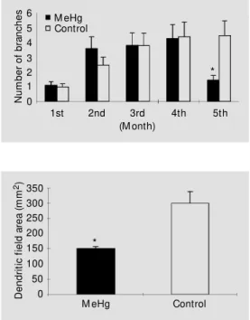
The morphologic pattern of NADPHdn in the control animals was similar to that already described elsewhere (14). The final concentration of MeHg in the visual cortex was 3.2 µg/g wet weight brain tissue. No differences in NADPHdn in gray matter were found between groups 1 and 2 for all param-eters analyzed (P>0.05). Analysis of NADPHdn in the white matter showed a reduction of the number of distal branches (Figure 1) in group 2 (P<0.05) and of the dendritic area field (Figure 2). However, morphological alterations in NADPHdn in the white and gray matter were virtually impossible to distinguish by qualitative light microscopy analysis. The enzyme activity in the neuropil was considerably less intense in the brains of intoxicated animals belonging to group 2 when compared to control (not illustrated). No alterations were observed in group 1.
Different results for NADPHdn were observed in adult cat striate cortex after a short and long time of severe intoxication. The long-term effects of MeHg seem to af-fect mainly NADPH-diaphorase neurons in the white matter and the neuropil activity in the gray matter. Neurons in the gray matter did not seem to be altered by the schedule of treatment with MeHg. Our results for the gray matter are similar to those reported by other authors who suggest a resistance of NADPH-positive neurons in gray matter to pathologic conditions like neurotoxicity me-diated by NMDA receptors underlying stroke and neurodegenerative disorders, such as Huntington, Parkinson and Alzheimer dis-eases (7-8). It is unknown why these cells are resistant. This phenomenon may be a
char-acteristic of gabaergic neurons, which have been shown to be resistant to toxic levels of NMDA in mouse cell culture (15). Cortical NADPHdn seem to synthesize GABA in the cerebral cortex (16). These inhibitory cells may present some protective “intrinsic safety factor” due to their important role in regulat-ing cerebral excitability (15). Teleologically, this may denote that critical neuronal sys-tems may be less vulnerable to pathological conditions in the CNS. Inhibitory cells con-stitute a critical neuronal system in cerebral cortex in that, despite their small number (only 20 to 25% of the total neurons in the cerebral cortex), they control the excitability of the entire brain. The resistance of NADPHdn to pathological insults is likely to be due to a number of functional adaptations including perhaps the major characteristic -an unusual metabolism.
The fact that the effects of MeHg were observed only after a long survival time of 6 months (group 2) may indicate that a long silent period is necessary for the effects of MeHg to appear. Indeed, it has been reported that some patients with Minamata disease developed the clinical features of severe poi-soning after they had stopped eating the
N
u
m
b
e
r
o
f
b
ra
n
c
h
e
s 6 5 4 3 2 1 0
1st 2nd 3rd 4th 5th
M eHg Control
(M onth)
*
Figure 1 - Number of dendritic branches of w hit e m at t er NADPHd-positive neurons in the striate cortex of the adult cat 6 months after intoxication. Note that the number of more distal dendritic branches is significant-ly decreased in the treated ani-mal compared to the control one (* P<0.05).
Figure 2 - Dendritic field area of w hite matter NADPHd-positive neurons in the striate cortex of the adult cat, 6 months after in-toxication. Note the significant reduction of the values in the treated animal compared to the control one (* P<0.05).
D
e
n
d
ri
ti
c
f
ie
ld
a
re
a
(
m
m
2)350 300 250 200 150 100 50 0
*
polluted fish (1). The silent period may rep-resent the time necessary for MeHg to over-come the compensatory function of the CNS. The decreased number of more distal branches in the white matter NADPH-dia-phorase neurons matched the decreased den-dritic field area. However, extensive quanti-fication was necessary to show these results, indicating that NADPH-diaphorase neurons are affected only by high MeHg doses. Other authors have demonstrated a similar fact for NMDA excitoxicity in culture (15). Differ-ences in neuronal vulnerability to pathologi-cal insults between white and gray matter may be related to glial function. A number of recent papers have suggested that astrocytes may play a major role in MeHg neurotoxicity (2-4). Astrocytes show basal levels of metallothioneins (MTs), metal-binding pro-teins whose biosynthesis is greatly enhanced by various factors including heavy metals such as Zn, Co, Cd and Hg (2). By virtue of their high thiol group (-SH) content, MTs have a very high affinity for MeHg. Indeed, wherever an MeHg compound was identi-fied in biological fluids, it was complexed with SH-containing ligands (2-3). The MeHg-MT complex may keep MeHg in a relatively nontoxic form in astrocytes, thereby protect-ing both astrocytes and juxtaposed neurons from the cytotoxic effects of the metal (3). Thus, MT induction may cause CNS toler-ance to MeHg, at least during the early stages of intoxication. A recent report showed that in the human brain MT immunoreactivity was limited to a subpopulation that probably represented protoplasmic astrocytes (17). This astrocyte type is more characteristic in the gray matter, a possible reason for its lower vulnerability.
We found a striking decrease in diapho-rase neuropil reactivity in group 2, six months after MeHg intake. The anatomical substrate for diaphorase neuropil reactivity has not been fully determined. There is evidence from electron microscopy studies that the NADPHd reactivity in lamina 4C of
pri-mates is mainly due to presynaptic axon terminals both from intra- and extracortical projections (18). The alterations of the inten-sity in NADPH-d activity in neuropil could suggest changes in transport processes of NADPH-diaphorase/NOS enzymes to distal branch regions of dendrites and axon termi-nals. This possibility agrees with the evi-dence that MeHg acts by altering both pro-tein synthesis and transport (1,5,6). The spe-cific mechanism for these effects is unknown. Alterations in the integrity of microtubules has been reported in a variety of experimen-tal systems (1). The decreased NADPHd neuropil reactivity may also be related to some type of astrocyte dysfunction. The as-trocyte-mediated tolerance to MeHg (dis-cussed above) may be an important factor to be overcome during the silent period of MeHg intoxication. Direct lesion of NADPHd ax-onal terminals may then occur. This possi-bility is under investigation by our group. Axonal terminals stained by iontophoretic injection of biocytin into the striate of the cats belonging to group 1 did not display qualitatively perceptible morphological al-terations. However, we cannot rule out some subtle alterations such as a reduced number of axonal boutons (Gomes-Leal W, Jesus-Silva SG, Oliveira RB and Picanço-Diniz CW, unpublished results).
expo-sure. Astroglial glutamate transporters carry out most of the functional glutamate trans-port and are essential for maintaining low extracellular glutamate (19). Higher doses and longer periods of exposure to MeHg may impair astrocyte function in terms of excitatory transmitter uptake (5). Thus, MeHg may damage the CNS through excitotoxic mechanisms like those mediated by NMDA. Indeed, ligand and voltage-gated ion chan-nels represent a plausible early target for the action of MeHg (20). Since a large battery of events are mediated by ion channels, it fol-lows that their disruption by mercurials could lead to potentially deleterious consequences for the cell. Thus, studies on mercury and other metal effects on the ion channels of
neurons (including the NADPHdn subpopu-lation) and astrocytes represent a more sen-sitive method than histochemistry to study the chronic neurotoxic effects of metals on the nervous system. Such studies are now required to reveal the mechanisms that en-sure protection of NADPHdn in the gray matter described in the present study and to determine why NADPHdn in the white mat-ter and neuropil seem to be more vulnerable.
Ackno wle dgm e nts
The authors are indebted to Erinaldo J. da Silva and Silmara S. Morais for preparing part of the drawings utilized for morphomet-ric analysis in the present paper.
Re fe re nce s
1. WHO (1991). IPCS, Enviromental Health
Criteria118. World Health Organization,
Geneva.
2. Aschner M (1996). M ethylmercury in as-trocytes – What possible significance?
Neurotoxicology, 17: 93-106.
3. Aschner M , Cherian M G, Klaassen CD, Palmiter RD, Erickson JC & Bush AI (1997). M etallothioneins in brain – The role in physiology and pathology.
Toxicol-ogy and Applied PharmacolToxicol-ogy, 142:
229-242.
4. Aschner M & Kimelberg HK (1996). The
Role of Glia in Neurotoxicity. CRC Press,
Times M irror International Publishers Ltd., New York.
5. Clarkson TW (1997). The toxicology of mercury. Critical Review s in Clinical
Labo-ratory Sciences, 34: 369-403.
6. M ottet NK, Vahter M E, Charleston JS & Friberg LT (1997). M etabolism of methyl-mercury in the brain and its toxicological significance. M etal Ions in Biological
Sys-tems, 34: 371-403.
7. Thomas E & Pearse AGE (1964). The soli-tary active cells. Histochemical demon-stration of damage-resistant nerve cells w ith a TPH-diaphorase reaction. Acta
Neuropathologica, 3: 238-249.
8. Ferrant e RJ, Kow all NW , Beal M F, Richardson Jr EP, Bird ED & M artin JB (1985). Selective sparing of a class of stri-atal neurons in Huntington’s disease.
Sci-ence, 230: 561-563.
9. Scherer-Singler U, Vincent SR, Kimura H & M cGeer FG (1983). Demonstration of a unique populat ion of neurons w it h NADPH-diaphorase histochemistry.
Jour-nal of Neuroscience M ethods, 9: 229-234.
10. Hope BT, M ichael GJ, Knigge KM & Vincent SR (1991). Neuronal NADPH-dia-phorase is a nitric oxide synthase. Pro-ceedings of the National Academy of
Sci-ences, USA, 88: 2811-2814.
11. Daw son TM & Snyder SH (1994). Gases as biological messengers: Nitric oxide and carbon monoxide in the brain. Journal of
Neuroscience, 14: 5147-5159.
12. Daw son TM , Bredt DS, Fotuhi M , Hw ang P & Snyder SH (1991). Nitric oxide syn-thase and neuronal NADPH-diaphorase are identical in brain and peripheral
tis-sues. Proceedings of the National
Acade-my of Sciences, USA, 88: 7797-7801.
13. Wolf G (1997). Nitric oxide and nitric ox-ide synthase: biology, pathology, localiza-tion. Histology and Histopathology, 12: 251-261.
14. M izukaua K, M cGeer PL, Vincent SR & M cGeer EG (1989). Distribution of re-duced nicotinamide adenine dinucleotide phosphate (NADPH) diaphorase-positive cells and fibers in the cat central nervous system. Journal of Comparative Neurol-ogy, 279: 281-311.
15. Tecoma ES & Choi W (1989). GABAergic
neocort ical neurons are resist ant t o NM DA receptor-mediated injury. Neurol-ogy, 39: 676-682.
16. Valtschanoff JG, Weinberg RJ, Kharazia VN & Schmidt HHHW (1993). Neurons in rat cerebral cortex that synthesize nitric oxide: NADPH-diaphorase histochemistry, NOS immunocytochemistry, and colocali-zation w ith GABA. Neuroscience Letters, 157: 157-161.
17. Blauw geers HG, Sillevis-Smitt PA, De Jong JA & Troost D (1993). Distribution of metallothionein in the human central ner-vous system. Glia, 8: 62-70.
18. Aoki C, Fenstemaker S, Lubin M & Go GG (1993). Nitric oxide synthase in the visual cortex of monocular monkeys as revealed by light and electron microscopic immu-nocytochemistry. Brain Research, 620: 97-113.
19. Rothstein JD, Dykes-Hoberg M , Pardo CA, Bristol LA, Jin L, Kunci RW, Kanai Y, Hediger M A, Wang Y, Schielk JP & Welty DF (1996). Knockout of glutamate trans-porters reveals a major role for astroglial transport in excitoxicity and clearance of glutamate. Neuron, 16: 675-686. 20. Sorois JE & Atchison WD (1996). Effects