Ro le o f Tamm-Ho rsfall pro te in
and uro mo dulin in calcium o xalate
crystallizatio n
1Kidney Stone Program, Division of Biological Sciences and the
Pritzker School of Medicine, University of Chicago, Chicago, IL, USA
2Departamento de Clínica Médica, Hospital de Clínicas,
Universidade Federal do Paraná, Curitiba, PR, Brasil M. Carvalho1,2,
R.A. Mulinari2 and
Y. Nakagawa1
Abstract
One of the defenses against nephrolithiasis is provided by macromol-ecules that modulate the nucleation, growth, aggregation and retention of crystals in the kidneys. The aim of the present study was to determine the behavior of two of these proteins, Tamm-Horsfall and uromodulin, in calcium oxalate crystallization in vitro. We studied a group of 10 male stone formers who had formed at least one kidney stone composed of calcium oxalate. They were classified as having idiopathic nephrolithiasis and had no well-known metabolic risk factors involved in kidney stone pathogenesis. Ten normal men were used as controls, as was a group consisting of five normal women and another consisting of five pregnant women. Crystallization was in-duced by a fixed supersaturation of calcium oxalate and measured with a Coulter Counter. All findings were confirmed by light and scanning electron microscopy. The number of particulate material deposited from patients with Tamm-Horsfall protein was higher than that of the controls (P<0.001). However, Tamm-Horsfall protein decreased the particle diameter of the stone formers when analyzed by the mode of the volume distribution curve (P<0.002) (5.64 ± 0.55 µm compared to 11.41 ± 0.48 µm of uromodulin; 15.94 ± 3.93 µm and 12.45 ± 0.97 µm of normal men Tamm-Horsfall protein and uromodu-lin, respectively; 8.17 ± 1.57 µm and 9.82 ± 0.95 µm of normal women Tamm-Horsfall protein and uromodulin, respectively; 12.17 ± 1.41 µm and 12.99 ± 0.51 µm of pregnant Tamm-Horsfall protein and uromodulin, respectively). Uromodulin produced fewer particles than Tamm-Horsfall protein in all groups. Nonetheless, the total volume of the crystals produced by uromodulin was higher than that produced by Tamm-Horsfall protein. Our results indicate a different effect of Tamm-Horsfall protein and uromodulin. This dual behavior suggests different functions. Tamm-Horsfall protein may act on nucleation and inhibit crystal aggregation, while uromodulin may promote aggrega-tion of calcium oxalate crystals.
Co rre spo nde nce
M. Carvalho
Rua Chichorro Jr., 144, Apto. 142 80035-040 Curitiba, PR Brasil
Fax: + 55-41-262-7658 E-mail: carvalho@ mais.sul.com.br
Received December 14, 2001 Accepted August 6, 2002
Ke y words
·Tamm-Horsfall protein ·Uromodulin
Intro ductio n
Human urine is almost always supersatu-rated with a mixture of various ions and salts, including calcium oxalate (CaOx), which is the most common component of kidney stones (1). It has been accepted that stone formation is a crystallization process taking place in supersaturated urine and that urinary supersaturation is the driving force for the formation of crystal nidus and subse-quent stone formation (2). One of the de-fenses against nephrocalcinosis and neph-rolithiasis includes macromolecules (proteins and glycosaminoglycans) that modulate the nucleation, growth, aggregation and reten-tion of crystals in the kidneys (3-6).
Tamm-Horsfall protein (THP) is one of the main components of urinary proteins. It is a glycoprotein produced and secreted by the thick ascendant limb of the loop of Henle, being the most abundant protein in normal human urine, excreted in quantities of 20 to 200 mg/24 h (7,8). THP of normal sub-jects inhibits the aggregation but has little effect on nucleation and growth of CaOx crystals (9). However, THP activity is influ-enced by its own concentration, urinary pH and ionic strength, playing a dual role in crystal formation depending on the environ-mental conditions (10). Moreover, THP iso-lated from the urine of recurrent stone formers sometimes becomes a promoter of CaOx ag-gregation due to a tendency to self-aggrega-tion, which removes it from effective interac-tion with CaOx monohydrate crystals (11).
Uromodulin (UM) is a protein with im-munosuppressive activity in vitro and thought to be present only in urine from pregnant women (12). Preliminary analysis of UM revealed a very high carbohydrate content, a tendency to form aggregates and the pres-ence of intrachain disulfide bridges, charac-teristics shared by THP (13). Indeed, they have the same protein framework but UM is more heavily glycosylated than THP and contains unprocessed mannose-rich chains
(8). The differences in glycosylation may reveal different functional properties of these two molecules (14).
The aim of the present investigation was to determine the role of THP and UM iso-lated from urine of normal individuals, preg-nant women and patients with nephrolithi-asis on calcium oxalate crystallization in vitro.
Mate rial and Me thods
Patie nts and controls
We selected 10 male stone formers, who had formed at least one separate kidney stone, with 50% or more of the stones being com-posed of CaOx. These patients were referred to us as having idiopathic nephrolithiasis and their urine had no well-known metabolic risk factors for kidney stone pathogenesis (15). As controls, we studied 10 men and 10 women (5 of them pregnant) without a per-sonal history of kidney stones, stone forma-tion in family members or any systemic dis-ease known to involve the kidneys. They were age matched with a same-sex patient. Patients discontinued all stone-prevention treatments at least 2 weeks before the urine collection.
Urine chemistry
Isolation of uromucoids (Tamm-Horsfall protein and uromodulin)
Uromucoids were isolated from 24-h in-dividual urine specimens by the method of Tamm and Horsfall (16). Briefly, sodium chloride was added to 24-h urine to a 0.58 M sodium chloride concentration, stirred con-stantly overnight at 4ºC, and then centri-fuged at 10,000 g for 30 min again at 4ºC with an RC-5 Sorvall refrigerated centrifuge (Sorvall, Newtown, CT, USA). The precipi-tate was washed once with 0.58 M sodium chloride aqueous solution, dissolved in 1/10 of the original volume of water, and dialyzed against 10 liters of water overnight. The dialyzed uromucoid solution was precipi-tated again from 0.58 M sodium chloride, and the glycoprotein recovered by centrifu-gation in the same manner as described above. The uromucoid obtained was dissolved in water, dialyzed overnight, lyophilized, and stored at -20ºC.
Separation of Tamm-Horsfall protein and uro m o dulin
Uromodulin was separated from THP by the method described by Muchmore and Decker (12). Uromucoid precipitated by so-dium chloride was dissolved in PBS, passed through a concanavalin A-Sepharose col-umn (1 x 2 cm, Pharmacia Biotech, Piscata-way, NJ, USA) and washed with 3 bed vol-umes of PBS to obtain the THP portion. UM was eluted with 250 mM alpha-methylman-noside with 3 bed volumes. Both proteins were further passed through a Sephacryl S-200 column (2 x 100 cm, Pharmacia) using 50 mM Tris-HCl, pH 7.3, containing 0.2 M NaCl and 0.2% sodium azide. Protein elu-tion was monitored by absorbance at 280 nm using a Beckman spectrophotometer model DU-650. Protein concentration was deter-mined using an extinction coefficient of 10.8. The purity of the proteins was deter-mined by SDS-PAGE. The gels were stained
with silver nitrate to detect protein bands. Immunoreactivity was examined using a com-mercially available polyclonal THP antibody raised in rabbits (Biomedical Technology Inc., Stoughton, MA, USA) using a Nunc-immunoplate. Color was developed with a Vecstastain ABC kit (Vector Laboratories Inc., Burlingame, CA, USA) and absorbance was measured at 405 nm using a reference wavelength at 630 nm and an ELISA plate reader (Dynatech Laboratories, Alexandria, VA, USA).
Carbohydrate analysis
Neutral sugars were analyzed quantita-tively by the phenol-sulfuric acid method. A 500-µl aliquot of the sample was mixed with 0.3 ml of 5% phenol aqueous solution (w/v), and 2 ml of concentrated sulfuric acid was added rapidly. The mixture was kept for 30 min at room temperature, and color intensity was measured at 484 nm. The calibration curve was prepared using 5 to 20 µg of glucose solution.
Crystallization assay
Me asure me nts of crystallization
The turbidity due to the development of crystallization of each plate was measured at 630 nm using a microELISA reader (Dynatech Laboratories) before and immediately after the addition of oxalate (zero time), at 3 min and then at 20-min intervals, for 1 h, with constant shaking at 500 rpm. Next, the plate was examined with an inverted light micro-scope to determine the presence of crystals. The content of each well was transferred in 5 ml of 20 mM sodium acetate buffer, pH 5.7, containing 300 mM sodium chloride. Immedi-ately, the number, total volume and mode of the volume distribution curve of the crystals formed were determined using a Coulter Counter Multisizer II (Coulter Electronics Ltd., Luton, Beds, England) fitted with a 100-µm capillary tube. All counts were corrected for background particulate matter by counting a sample of the electrolyte supporting solution with and without the protein to be studied and by subtracting these values from the raw counts (18). The Coulter Counter was calibrated and checked at the onset and conclusion of each specimen sampling using 10.27-µm calibrat-ing spherules. The lowest threshold in this instrument was about 2 µm.
Scanning e le ctron microscopy
At the end of the experiment, crystals were separated by filtering through a 0.22-µm filter, washed with 1 ml of distilled water, and air-dried at 37ºC. The specimens were mounted on carbon-coated copper grids, fixed on stabs, and coated with gold for 4 min using a Sputter coater (Varian Technics, Warrington, England). Images were obtained with a JEOL scanning electron microscope (model JSM-840A) with an operational volt-age of 15 kV.
Statistical analysis
All data are reported as means ± standard deviation. Samples were compared by t-tests and by one-way ANOVA using a standard statistical software (Minitab 11.0, Minitab Inc., College Station, PA, USA).
Re sults
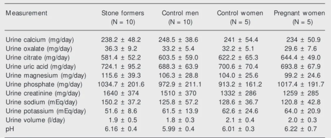
The chemical composition of urine from patients was similar to that of matched con-trols, with no significant difference detected between groups, as shown in Table 1.
After applying SDS-PAGE and silver
ni-Table 1. Chemical data for urine of nephrolithiasis in patients and control subjects.
M easurement Stone formers Control men Control w omen Pregnant w omen
(N = 10) (N = 10) (N = 5) (N = 5)
Urine calcium (mg/day) 238.2 ± 48.2 248.5 ± 38.6 241 ± 54.4 234 ± 50.9 Urine oxalate (mg/day) 36.3 ± 9.2 33.2 ± 5.4 32.2 ± 5.1 29.6 ± 7.6 Urine citrate (mg/day) 581.4 ± 52.2 603.5 ± 59.0 622.2 ± 65.3 644.4 ± 49.0 Urine uric acid (mg/day) 724.1 ± 95.2 688.3 ± 63.9 700.6 ± 70.4 693.8 ± 67.9 Urine magnesium (mg/day) 115.6 ± 39.3 106.3 ± 28.8 104.0 ± 25.6 99.2 ± 24.6 Urine phosphate (mg/day) 1034.7 ± 201.6 972.9 ± 211.1 913.2 ± 161.2 1017.4 ± 191.7 Urine creatinine (mg/day) 1640 ± 374 1510 ± 370 1332 ± 286 1259 ± 285 Urine sodium (mEq/day) 150.2 ± 37.2 125.8 ± 57.2 128.6 ± 36.7 120.8 ± 42.8 Urine potassium (mEq/day) 51.6 ± 8.6 61.5 ± 13.9 62.6 ± 24.6 64.0 ± 20.9
Urine volume (l/day) 1.9 ± 0.5 1.8 ± 0.3 2.1 ± 0.4 2.0 ± 0.3
pH 6.16 ± 0.4 5.99 ± 0.4 6.01 ± 0.3 6.22 ± 0.7
trate staining, THP and UM were detected as single bands of ~80 kDa. Both proteins re-acted against THP antibody raised in rabbits. The apparent content of hexoses in UM was at least 1.3 times higher than that of THP. The concanavalin A-Sepharose column recognized only UM, probably because of exposure of mannose residues due to cleavage of terminal carbohydrate chains such as the Neu/GalNAc/ GluNAc and GalNAc/GluNAc moieties. The urinary excretion rate of THP and UM was similar among stone formers and controls (49 ± 1.1 mg/day for THP and 49.3 ± 1.2 mg/day for UM from stone formers vs 51 ± 0.5 and 49.3 ± 2.2 mg/day for THP and UM from normal men, respectively; 50.2 ± 1.9 and 50.8 ± 1.9 mg/day for THP and UM from normal women, respectively; 52 ± 0.7 and 51.3 ± 2.7 mg/day for THP and UM from pregnant women, respectively; P = NS for all compari-sons). The THP/UM ratio obtained for each group was also similar: 1.04 ± 0.7 for non-stone-forming males, 1 ± 0.56 for patients with nephrolithiasis, 0.91 ± 0.37 for normal fe-males, and 0.97 ± 0.41 for pregnant women (P = NS).
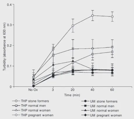
The data in Figure 1 report the time course of the measurement of absorbance at 630 nm. After the induction with sodium oxalate there was an initial steady increase of ab-sorbance in all groups in the first 20 min. These curves reached their plateau around 40 min, and some of them slightly decreased after this period of time due to crystal
aggre-T
u
rb
id
it
y
(
a
b
s
o
rb
a
n
c
e
a
t
6
3
0
n
m
)
0.4
0.3
0.2
0.1
0
No Ox 3 20 40 60
Time (min)
THP stone formers THP normal men THP normal w omen THP pregnant w omen
UM stone formers UM normal men UM normal w omen UM pregnant w omen
Figure 1. Time course of increased turbidity. Data are reported as means ± SD of absorbance at 630 nm. No Ox indicates oxalic acid w as not added. THP = Tamm-Horsfall protein; UM = uromodulin.
Table 2. Effects of Tamm-Horsfall protein (THP) and uromodulin (UM ) on calcium oxalate crystallization.
M easurement Stone formers Control men Control w omen Pregnant w omen
(N = 10) (N = 10) (N = 5) (N = 5)
THP UM THP UM THP UM THP UM
Crystal 53.53 ± 10.33* 8.76 ± 1.24 11.05 ± 2.10 6.19 ± 0.77 20.02 ± 5.81* 13.14 ± 3.72 6.10 ± 1.21 4.43 ± 0.42
number (x 103)
Total crystal 1.68 ± 0.15 2.23 ± 0.21* 1.22 ± 0.15 1.26 ± 0.10 1.51 ± 0.17 1.63 ± 0.12 1.24 ± 0.19 1.21 ± 0.12 volume (x 106)
Data w ere collected at the end of the experiment (60 min) and are reported as means ± SD. * P<0.002 compared w ith other groups (one-w ay ANOVA).
gation. The stone formers THP curve was significantly different from all other groups (P<0.0001 for all comparisons) at 40 and 60 min and also at 3 and 20 min (P<0.01), except for normal women.
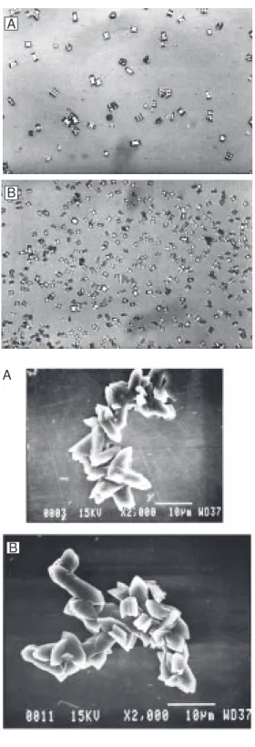
deFigure 2. Ef f ect s of Tam m -Horsf all prot ein f rom norm al men (A) and stone formers (B) on calcium oxalate crystallization (light microscopy, 500X).
Figure 3. Ef f ect s of Tam m -Horsfall protein from norm al men (A) and stone formers (B) on calcium oxalate crystallization (scanning electron microscopy).
creased the particle diameter of the stone formers (P<0.002), when analyzed by the mode of the volume distribution curve (5.64 ± 0.55 compared to 11.41 ± 0.48 µm of UM; 15.94 ± 3.93 and 12.45 ± 0.97 µm of normal men THP and UM, respectively; 8.17 ± 1.57
and 9.82 ± 0.95 µm of normal women THP and UM, respectively; 12.17 ± 1.41 and 12.99 ± 0.51 µm of pregnant women THP and UM, respectively). UM produced fewer particles than THP in all groups. Nonethe-less, the total volume of the crystals pro-duced by UM tended to be larger than THP in controls and the total volume of stone former UM was slightly but significantly different from the UM of THP stone formers and from all other groups (Table 2).
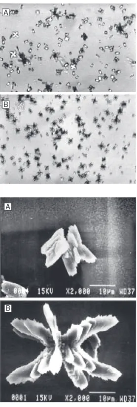
Figures 2-5 show representative light and scanning electron microscopy photographs of crystals formed in the presence of THP and UM from normal men and stone form-ers, respectively. The THP of the nephroli-thiasis group formed a large number of small crystals and preserved the typical CaOx monohydrate and dihydrate morphology. Uromodulin allowed formation of few crys-tals of large volume and a wide variety of shapes such as hexagonal plates, mulberry formations, and aggregates.
D iscussio n
Urinary tract stones form because of the tendency for urine to be supersaturated with stone salts. However, stone formers have slightly higher levels of supersaturation than non-stone formers, and the distribution of supersaturation within the two populations shows a substantial overlap (19). Urinary inhibitors of crystal growth and aggregation have long been assumed to play a protective role in nephrolithiasis. Urine from patients with recurrent stones contains larger crystals than urine from normal individuals and in-hibits crystallization less than normal urine does. These facts support the idea that defec-tive inhibitors could contribute to stone for-mation (11).
We studied a highly selected population of patients with nephrolithiasis who did not have any of the most common urinary bio-chemical abnormalities related to kidney stone formation. Using this approach, we A
A
B
tried to target stone formers with a high probability of harboring defects in macro-molecules that inhibit several steps of crys-tallization.
THP is one of these macromolecules and has been extensively studied, sometimes with paradoxical results concerning CaOx crys-tallization reported by different investiga-tors, depending on the conditions and meth-ods employed. Most authors agree that THP is a weak inhibitor of nucleation and growth, but in solutions with high pH, low ionic strength, and low concentrations of divalent ions, the glycoprotein acts as a powerful inhibitor of CaOx crystal aggregation (20).
Our results agree with several reports that have shown no significant difference in the daily urinary excretion of THP between normal subjects and patients with nephroli-thiasis. This finding led to the hypothesis of structural differences in THP obtained from these groups. In fact, Hess et al. (21) showed that THP isolated from the urine of stone formers contained less carbohydrate (mainly sialic acid) than the THP from control sub-jects. Although THP and UM have the same amino acid sequence, UM isolated by the concanavalin A method possesses more high-mannose oligosaccharides. This protein was considerably more potent in inhibiting anti-gen-induced T cell proliferation than THP isolated from non-pregnant urine by salt pre-cipitation (22). The sugar moiety was as-sumed to be responsible for the observed immunosuppressive differences.
Garcia et al. (23) studied the adhesion of CaOx monohydrate crystals to renal cells in the presence of THP or UM. The adhesion of crystals coated with THP to monolayer cul-tures of nontransformed monkey renal epi-thelial cells (BSC-1 line) was decreased by 30%. However, adhesion of CaOx crystals coated with UM did not differ from that of uncoated crystals. Sialic acid residues may be important for this action of THP because neuraminidase treatment abolished the ef-fect (23).
Figure 4. Effects of uromodulin from normal men (A) and stone formers (B) on calcium oxalate crystallization (light microscopy, 500X).
Figure 5. Effects of uromodulin from normal men (A) and stone formers (B) on calcium oxalate crystallization (scanning electron microscopy).
In the present study, we used absorbance at 630 nm that represents an exact measure of particle concentration per unit volume (24), and a Coulter Counter to assess nucleation and aggregation of CaOx crystals. The latter
tech-A
B
nique was devised by Robertson and Peacock and modified by Ryall et al. (18). The results showed a different effect of THP and UM on CaOx crystallization. As demonstrated in Table 2, UM produced fewer particles than THP in all groups and the total volume of crystals produced by UM was larger than that pro-duced by THP. In addition, THP decreased the diameter of the particles formed, mainly in the stone former group.
All experiments were confirmed by light and scanning electron microscopy (Figures 2-5). THP maintained the preferred structure in this calcium-rich system, forming octahe-dral crystals typical of CaOx dihydrate.
In-terestingly, UM caused the crystal structure to exhibit marked polymorphism. Our data agree with those reported by Wesson et al. (19), who stated that the formation of CaOx dihydrate crystals offers a potential biologi-cal advantage by decreasing the affinity for the inner medullary collecting duct cell mem-brane surface, the region where symptomatic stones probably form.
These data support the view that the dual role of THP and UM suggests different func-tions. Under the conditions of this assay sys-tem, THP affects nucleation and inhibits crys-tal aggregation while UM may promote aggre-gation of highly polymorphic CaOx crystals.
Re fe re nce s
1. Coe FL & Parks JH (1997). New insights into the pathophysiology and treatment of nephrolithiasis: new research venues.
Journal of Bone and M ineral Research, 12: 522-533.
2. Hess B & Kok DJ (1996). Nucleation, grow th, and aggregation of crystals. In: Coe FL, Favus M J, Pak CY, Parks JH & Preminger GM (Editors), Kidney Stones. M edical and Surgical M anagement. Lip-pincott-Raven, Philadelphia, PA, USA. 3. Robertson WG & Peacock M (1972).
Cal-cium oxalate crystalluria and inhibitors of crystallization in recurrent renal stone formers. Clinical Science, 43: 499-506. 4. Rose M B (1975). The inhibitory effect of
urine on calcium oxalate precipitation. In-vestigative Urology, 12: 428-433. 5. Nakagaw a Y, Abram V, Parks JH, Lau S-H,
Kaw ooya JK & Coe FL (1985). Urine gly-coprotein crystal grow th inhibitors. Evi-dence for a molecular abnormality in cal-cium oxalate nephrolithiasis. Journal of Clinical Investigation, 76: 1455-1462. 6. Worcester E (1996). Inhibitors of stone
formation. Seminars in Nephrology, 16: 474-486.
7. Hoyer JR & Seiler M W (1979). Pathophysi-ology of Tamm-Horsfall protein. Kidney International, 16: 279-289.
8. Kumar S & M uchmore A (1990). Tamm-Horsfall protein-uromodulin (1950-1990).
Kidney International, 37: 1395-1401. 9. Worcester EM, Nakagawa Y, Wabner CL,
Kumar S & Coe FL (1988). Crystal adsorption and growth slowing by nephrocalcin, albu-min and Tamm-Horsfall protein. American Journal of Physiology, 255: F1197-F1205.
10. Hess B (1994). Tamm-Horsfall glycopro-tein and calcium nephrolithiasis. M ineral and Electrolyte M etabolism, 20: 393-398. 11. Hess B, Nakagaw a Y, Parks JH & Coe FL (1991). M olecular abnormality of Tamm-Horsfall glycoprotein in calcium oxalate nephrolithiasis. American Journal of Phys-iology, 260: F569-F578.
12. M uchmore AV & Decker JM (1985). Uro-modulin: A unique 85 kD immunosuppres-sive glycoprotein isolated from urine of pregnant w omen. Science, 229: 479-481. 13. Pennica D, Kohr WJ, Kuang WJ, Glaister D, Aggarw al BB, Chen EY & Goeddel DV (1987). Identification of human uromodu-lin as the Tamm-Horsfall urinary glycopro-tein. Science, 236: 83-88.
14. Lieske JC & Coe FL (1996). Urinary inhibi-tors and renal formation. In: Coe FL, Fa-vus M J, Pak CY, Parks JH & Preminger GM (Editors), Kidney Stones. M edical and Surgical M anagement. Lippincott-Raven, Philadelphia, PA, USA.
15. Porile JL, Asplin JR, Parks JH, Nakagaw a Y & Coe FL (1996). Normal calcium oxa-late crystal grow th inhibition in severe cal-cium oxalate nephrolithiasis. Journal of the American Society of Nephrology, 7: 602-607.
16. Tamm I & Horsfall FL (1950). Character-ization and separation of an inhibitor of viral hemagglutination present in urine.
Proceedings of the Society for Experimen-tal Biology and M edicine, 74: 108-114. 17. Achilles W (1997). In vitro crystallization
sys-tems for the study of urinary stone forma-tion. World Journal of Urology, 15: 244-251. 18. Ryall RL, Bagley CJ & M arshall VR (1981).
Independent assessment of the grow th and aggregation of calcium oxalate crys-tals using the Coulter Counter. Investiga-tive Urology, 18: 401-405.
19. Wesson JA, Worcester EM , Wiessner JH, M andel NS & Kleinman JG (1998). Control of calcium oxalate crystal structure and cell adherence by urinary m acrom ol-ecules. Kidney International, 53: 952-957. 20. Dussol B & Berland Y (1996). Urinary kid-ney stone inhibitors. Where are w e? Ne-phrology, Dialysis, Transplantation, 11: 1222-1224.
21. Hess B, Jaggi M , Zipperle L & Jaeger P (1995). Reduced carbohydrate content of Tamm-Horsfall glycoprotein (THP) from severe recurrent renal calcium stone formers (RCSF). Journal of the American Society of Nephrology, 6: 949 (Abstract). 22. Hession C, Decker JM , Sherblom AP,
Kumar S, Yue CC, M attaliano RJ, Tizard R, Kaw ashima E, Schmeissner U & Heletky S (1987). Uromodulin (Tamm Horsfall gly-coprotein): A renal ligand for lymphokines.
Science, 237: 1479-1484.
23. Garcia WE, Lieske JC, Nakagaw a Y & Toback FG (1998). Tamm-Horsfall protein, but not uromodulin, inhibits adhesion of calcium oxalate monohydrate (COM ) crys-tals to renal cells. Journal of the American Society of Nephrology, 9: 554 (Abstract). 24. Hess B, M einhardt U, Zipperle L,