A m o de rate de cre ase in te m pe rature
inhibits the calcium signaling
m e chanism (s) o f the re gulato ry
vo lum e de cre ase in chick e m bryo
cardio m yo cyte s
Department of Cell Biology, Duke University Medical Center, Durham, NC, USA M.M. Souza
and R.T. Boyle
Abstract
Chick cardiomyocytes, when submitted to hyposmotic swelling, ex-hibit a partial regulatory volume decrease (RVD). A Ca2+
influx by stretch-activated channels signals a taurine efflux and the RVD at 37oC. We evaluated the cells performance at room temperature. Cardiomyocytes isolated and cultured from 11-day-old chick embryos were submitted to a hyposmotic solution (180 mOsm/kg H2O) at 37
o C and at room temperature (26o
C). Under these conditions we measured the changes in cell volume as well as the intracellular free Ca2+ (using fura-2). During hyposmotic swelling, cells at 37oC displayed a peak relative volume of 1.61 ± 0.03 and recovery to 1.22 ± 0.04 (N = 14), while cells at 26o
C presented a peak swell relative volume of 1.74 ± 0.06 and did not recover (1.59 ± 0.09, N = 9). Transient increases in intracellular Ca2+, which are characteristic of the normal RVD, were observed at both temperatures (29.1 ± 4.5% (N = 8) and 115.2 ± 42.8% (N = 5) increase at 37o
and 26o
C (P<0.05), respectively). A delay in the Ca2+ transient increase was also observed when the cells were at 26oC (109 ± 34 s compared to 38 ± 9 s at 37oC, P<0.05). At room temperature the RVD does not occur because the calcium transient increase, which is an early event in the signaling of the RVD, is delayed. Also, free calcium is not cleared as in the 37oC RVD. In the normal RVD the free calcium returns to baseline levels. The very high and persistent free calcium levels seen at room temperature can lead to unregulated enzyme activities and may promote irreversible injury and cell death.
Co rre spo nde nce
M.M. Souza
Departamento de Ciências Fisiológicas, CCB, UEL Caixa Postal 6001 86051-990 Londrina, PR Brasil
Fax: + 55-43-371-4207 E-mail: mmsouza@ uel.br
Presented at the XV Annual Meeting of the Federação de Sociedades de Biologia Experimental, Caxambu, MG, Brazil, August 23-26, 2000.
Received April 13, 2000 Accepted September 13, 2000
Ke y wo rds
·Cell volume
·RVD
·Room temperature
·High free calcium
Volume regulatory mechanisms play a critical role in the maintenance of structural integrity, and thus in the proper function of living cells. These regulatory processes in-volve signal transduction pathways and cel-lular transport systems.
Cultured chick cardiomyocytes, when
submitted to hyposmotic swelling (from 290 to 180 mOsm/kg H2O) exhibit a partial regu-latory volume decrease (RVD) mediated by a loss of amino acids (mostly taurine). There are no significant changes in inorganic ion contents in this condition (1). This RVD is
dependent on the influx of Ca2+
stretch-activated channels which in turn signals a
taurine efflux and the RVD at 37oC (2-5).
Studies with chick cardiomyocytes under isosmotic conditions at low temperatures
reveal a dissipation of the Na+ gradient and
low levels of Na+
/K+
ATPase activity, with
the Na+
/Ca2+
exchanger functioning only minimally (6). On the basis of these consid-erations, we evaluated the RVD under hy-posmotic swelling at low temperature.
Cardiomyocytes isolated and cultured from 11-day-old chick embryos (7) were transferred from isosmotic (290 mOsm/kg H2O) to a hyposmotic solution (180 mOsm/
kg H2O) at 37o
C and at room temperature
(26o
C). The cells were perfused at a rate of ~4 ml/min with HEPES-buffered saline so-lution (142.2 mM NaCl, 5.4 mM KCl, 1.0 mM NaH2PO4, 10 mM HEPES, 5.6 mM dextrose, 0.8 mM MgSO4, and 1 mM CaCl2; the hyposmotic solution contained 50% of isosmotic NaCl) on the heated (or not) stage
of an inverted microscope (Nikon Diaphot, TMD, Tokyo, Japan). Measurements of cell area from video images of spherical cells (model KP MIL camera, Hitashi Denshi, Ltd.; model PVM-137 monitor, SONY, 1000X resolution) were made using JAVA system software (Jandel Scientific, San Rafael, CA, USA) and were then converted into cell volume. Values were normalized to the last measurement made in the isosmotic solution before volume challenge.
Cells microinjected with fura-2 (1 mM in 140 mM KCl, 10 mM MOPS-3-[N-morpho-lino] propane-sulfonic acid and passed through a chelex-100 column, pH 7.4) were
used for intracellular free Ca2+
measurements by the ratiometric method. The excitation and emission wavelengths were 350/380 and 505 nm, respectively. The coverslips were affixed to a heated (or not) stage on a Zeiss IM35 inverted epifluorescence microscope coupled to a Spex model CM3 dual-wave-length excitation spectrofluorometer (Spex Industries, Edison, NJ, USA).
The mean and standard errors were cal-culated from relative values and plotted on graphs. Data for control and experimental
groups were compared by the Student t-test.
Single cells, when submitted to a change in osmolality (from 290 to 180 mOsm/kg
H2O) at 37o
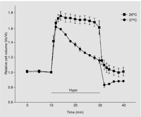
C, exhibited a peak swelling of 1.61 ± 0.03 (N = 14) within 2 min, followed by a partial RVD with an end point at 1.22 ± 0.04 (N = 14) (Figure 1). However, when the cells were exposed to the same hyposmotic
condition, but at 26o
C, the peak of swelling
was higher than at 37o
C (1.74 ± 0.06, N = 9; P<0.05) and the relative volume was not recovered (1.59 ± 0.09, N = 9; P>0.05) (Fig-ure 1). Also, upon return to isosmotic saline at 30 min, the cells at room temperature returned to pre-swollen levels (relative vol-ume of 1.0), thus further substantiating that no net loss of osmolytes and no RVD results
from hyposmotic swelling at 26o
C.
When chick cardiac myocytes are sub-mitted to hyposmotic swelling they exhibit a
R
e
la
ti
v
e
c
e
ll
v
o
lu
m
e
(
V
o
/V
i)
1.8
26oC 37oC
1.6
1.4
1.2
1.0
0.8
0.6
Time (min)
0 10 20 30 40
Hypo
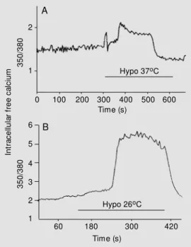
transient increase in intracellular free Ca2+ (2,3), which was also demonstrated in our
experiments. The intracellular Ca2+
transient increase (as measured by 350/380) was ob-served at both temperatures (29.1 ± 4.5%, N
= 8, with a delay of 38 ± 9 s at 37o
C) (Figure
2). While at 26o
C the cells exhibited a 350/ 380 fluorescence ratio increase of 115.2 ± 42.8% (N = 5) and a delay of 109 ± 34 s (P<0.05, when both parameters were com-pared with the control temperature) (Figure 2). Post-experiment calculations were per-formed to calibrate and transform ratio
val-ues to Ca2+
concentrations. Values for the different conditions were approximately:
con-trol (0.16 µM), hyposmotic solution at 37oC
(0.5 µM) and at 26o
C (1.3 µM).
In order for cells to maintain a constant volume when subjected to anisosmotic envi-ronments, they must have the ability to mo-bilize osmolytes. The RVD response to hyposmotic swelling is usually
character-ized by a loss of K+
, Cl
and some organic osmolytes (8). In chick cardiac myocytes exposed to a hyposmotic solution, there is a
concurrent influx of Ca2+
(2,3) by stretch-activated channels (5), and it has been pro-posed that protein kinase C, activated by free
Ca2+
, mediates the amino acid efflux (tau-rine) that leads to RVD (9). Moreover, it has been shown that cytoskeletal components are involved in the volume regulation of cardiac myocytes. Electrophysiological stud-ies and immunolabeling assays of cytoskel-etal proteins reveal suppression of a swelling current and disruption of subplasmalemmal F-actin dynamics induced by cytochalasin B and phalloidin, and the capacity of volume regulation is significantly affected (10,11).
Tissues of mammals exposed to low tem-peratures show alterations in intracellular ion contents (6,12). For homeotherms, hy-pothermia can attenuate enzymatic reactions leading to impairment of cell integrity. Con-sidering the differences in temperature de-pendence for the active and passive trans-port components of the cells pump-leak
system, which maintains a stable intracellu-lar ion concentration, a change in tempera-ture represents a significant challenge to cel-lular functions.
Embryonic chick cardiomyocytes in isos-motic solution at low temperatures exhibit
an increase in intracellular Na+
concentra-tion. It has been suggested that Na+ influx
from Na+
/H+
occurs and may not be
cor-rected by Na+
/K+
-ATPase, which in this situ-ation shows only 15% of its original activity (6). No change was observed in intracellular
K+
concentration, and total intracellular Ca2+
presented a small increase at low
tempera-ture (6), while intracellular free Ca2+
demon-strated a substantial increase (13).
Few reports regarding cell volume regu-lation at low temperature are available. Mal-phigian tubules of a New Zealand mountain insect exposed to hyperosmotic conditions
at low temperature (0o
C) showed a greater change in cell volume as compared to higher
temperatures (20o
C) and no regulatory vol-ume increase was observed (14). Some data about chick cardiac myocytes at low
tem-peratures are available (10o
C), but only un-der isosmotic conditions, and in this situa-tion the cells shrink (6). The authors believe
3
5
0
/3
8
0
2
1
0 100 200 300 400 500 600
Time (s) Hypo 37oC
3
5
0
/3
8
0
6
4
1
60 180 300 420
Time (s) Hypo 26oC 5
3
2
A
B
Figure 2 - Intracellular free Ca2+ m easurem ents expressed as fluorescence ratio. Representa-tive experiments show ing Ca2+ transient increases in hypos-motic solutions (hypo) at 37oC (A) and 26oC (B).
In
tr
a
c
e
llu
la
r
fr
e
e
c
a
lc
iu
that the shrinkage is a consequence of
inhibi-tion of the inwardly directed Na+/K+/2Cl
-cotransporter.
Our findings reveal that embryonic chick cardiac myocytes during hyposmotic
chal-lenge at 26o
C (room temperature) swell more
than at 37o
C, and that at room temperature no RVD is observed. Also, in this situation
the intracellular free Ca2+
exhibits a huge increase, much more than a transient
in-crease during hyposmotic swelling at 37oC.
As mentioned previously, during a
hy-posmotic challenge there is a Ca2+
influx for signaling the RVD which occurs via
stretch-activated channels, and the rapid free Ca2+
transient increase at 37o
C is a result of Ca2+
influx activating Ca2+ release from
intracel-lular stores (15). At room temperature the transient is also present, but is delayed. Our
findings suggest that the Ca2+ release
medi-ated by receptor activities is inefficient at the lower temperature and the same lack of re-sponsiveness may exist with calmodulin, which normally is instrumental in clearing high cytoplasmic free calcium levels through its stimulatory effect on plasma membrane
Ca2+
ATPase (16). It is unclear at this point if low temperatures prevent the conformational changes in calmodulin which are necessary
to activate Ca2+
ATPase, or if the activated ATPase is less efficient in hydrolyzing ATP
at the decreased temperatures. However, the latter is most likely the case and may explain
why Ca2+
ATPases in the sarcoplasmic reticu-lum, which are normally highly efficient in transporting calcium out of the cytoplasm,
appear to be inactive at 26o
C. In addition,
mitochondrial uptake of Ca2+
through its
low-affinity/high-capacity Ca2+
pump ap-pears to be temperature sensitive in our mo-del as this mechanism is known to operate when intracellular free calcium becomes per-ilously high (17). Also, even with abundant
free Ca2+, signal transduction pathways that
lead to the activation of protein kinase C (and thus to the RVD) are also interrupted.
Taken together, the evidence obtained in this study as well as in other investigations on the chick embryo cardiomyocyte sug-gests that even moderate hypothermia is a potent inhibitor of active transport. This is manifest in the cardiomyocyte inability to mobilize osmolytes for the RVD response and to control and reduce cytoplasmic cal-cium concentrations to normal levels. It is
well known that if the intracellular free Ca2+
concentration rises to high levels, it levies toxic effects on the cells. The very high and persistent free calcium seen at room temper-ature can lead to unregulated enzyme activi-ties and promote irreversible cell injury and perhaps cell death.
Re fe re nce s
1. Rasmusson RL, Davis DG & Lieberman M (1993). Amino acid loss during volume regulatory decrease in cultured chick heart cells. American Journal of Physiolo-gy, 264(Cell Physiology, 33):C136-C145. 2. Smith TW, Rasmusson RL, Freudenrich CC & Lieberman M (1992). Role of Ca2+ in myocardial volume regulation. Circula-tion, 86: I-480 (Abstract).
3. Aloi L, Smith JJ, M oore ES, Sw aminathan M & Lieberman M (1995). Cardiac cell sw elling activates a calcium-dependent signal transduction mechanism. FASEB Journal,9: A355 (Abstract).
4. Hall SK, Zhang J & Lieberman M (1997).
An early transient current is associated w ith hyposmotic sw elling and volume regulation in cardiac cells. Experimental Physiology, 82: 43-54.
5. Souza M M , Boyle RT & Lieberman M (2000). Different physiological mechan-isms control isovolumetric regulation and regulatory volume decrease in chick em-bryo cardiomyocytes. Cell Biology Inter-national, 24:(in press).
6. Knerr SM M & Lieberman M (1993). Ion transport during hypothermia in cultured heart cells: Implications for protection of the immature myocardium. Journal of M o-lecular and Cellular Cardiology, 25:
277-288.
7. Jacob R, Lieberman M & Liu S (1987). Effects of sodium-potassium pump inhibi-tion and low sodium on membrane poten-tial in cultured embryonic chick heart cells.
Journal of Physiology, 387: 549-566. 8. Hoffmann EK & Dunham PB (1995). M
em-brane mechanisms and intracellular sig-naling in cell volume regulation. Interna-tional Review of Cytology, 161: 172-262. 9. Smith JJ, Spizz G, Aloi L, M oore ES,
FASEB Journal, 10: A314 (Abstract). 10. Zhang J, Larsen TH & Lieberm an M
(1997). F-actin modulates sw elling-acti-vated chloride current in cultured chick cardiac myocytes. American Journal of Physiology, 273: C1215-C1224. 11. Larsen TH, Dalen H, Boyle RT, Souza M M
& Lieberman M (2000). Cytoskeletal in-volvement during hyposmotic sw elling and volume regulation in cultured chick cardiac myocytes. Histochemistry and Cell Biology, 113: 479-488.
12. Sudo J & M orel J (1984). Na+ and K+ cell
concentration in collagenase treated rat kidney tubules incubated at various tem-peratures. American Journal of Physiolo-gy, 246: C407-C414.
13. Liu B, Wang LCH & Belke D (1991). Effect of low temperature on the cytosolic free calcium in rat ventricule myocytes. Cell Calcium, 12: 11-18.
14. Neufeld DS & Leader JP (1998). Cold inhi-bition of cell volume regulation during the freezing of insect M alphigian tubules.
Journal of Experimental Biology, 201: 2195-2204.
15. Souza M M , Boyle RT & Lieberman M (2000). Comparisons of different stages of embryonic development by the physi-ological regulatory response to hypos-motic challenge. Comparative Biochemis-try and Physiology, 125A: 451-458. 16. Head JF (1992). A better grip on
calmodu-lin. Current Biology, 2: 609-611. 17. Carafoli E (1987). Intracellular calcium