http://dx.doi.org/10.1590/1519-6984.26712
Characterization of the microbial community in a lotic environment
to assess the effect of pollution on nitrifying and
potentially pathogenic bacteria
Medeiros, JD.
a*
, Araújo, LX.
a, Silva, VL. da
b, Diniz, CG.
b, Cesar, DE.
a,
Del’Duca, A.
cand Coelho, CM.
aaDepartament of Biology, Institute of Biological Science, Federal University of Juiz de Fora – UFJF,
José Lourenço Kelmer, Martelos, CEP 36036-900, Juiz de Fora, MG, Brazil
bDepartament of Parasitology, Microbiology and Immunology, Institute of Biological Science, Federal University of Juiz
de Fora – UFJF, José Lourenço Kelmer, Martelos, CEP 36036-900, Juiz de Fora, MG, Brazil
cFederal Institute of Southeastern of Minas Gerais, CEP 36080-001, Juiz de Fora, MG, Brazil
*e-mail: [email protected]
Received: December 12, 2012 – Accepted: May 20, 2013 – Distributed: August 31, 2014 (With 4 Figures)
Abstract
This study aimed to investigate microbes involved in the nitrogen cycle and potentially pathogenic bacteria from urban and rural sites of the São Pedro stream. Water samples were collected from two sites. A seasonal survey of bacterial abundance was conducted. The dissolved nutrient content was analysed. PCR and FISH analysis were performed to identify and quantify microbes involved in the nitrogen cycle and potentially pathogenic bacteria. The seasonal survey revealed that the bacterial abundance was similar along the year on the rural area but varied on the urban site. Higher concentration of dissolved nutrients in the urban area indicated a eutrophic system. Considering the nitrifying microbes, the genus Nitrobacter was found, especially in the urban area, and may act as the principal bacteria in converting nitrite into nitrate at this site. The molecular markers napA, amoA, and nfrA were more accumulated at the urban site, justifying the higher content of nutrients metabolised by these enzymes. Finally, high intensity of amplicons from
Enterococcus, Streptococcus, Bacteroides/Prevotella/Porphyromonas, Salmonella, S. aureus, P. aeruginosa and the diarrheagenic lineages of E. coli were observed at the urban site. These results indicate a change in the structure of the microbial community imposed by anthrophic actions. The incidence of pathogenic bacteria in aquatic environments is of particular importance to public health, emphasising the need for sewage treatment to minimise the environmental impacts associated with urbanisation.
Keywords: lotic environment, urbanisation, pollution, nitrifying microbes, pathogenic bacteria.
Caracterização da comunidade microbiana em um ambiente
lótico para acessar o efeito da poluição em bactérias
nitrificantes e potencialmente patogênicas
Resumo
Este estudo objetivou investigar os micro-organismos envolvidos no ciclo do nitrogênio e bactérias potencialmente patogênicas das áreas urbanas e rurais do Córrego São Pedro. Amostras de água foram coletadas dos dois locais. Um levantamento sazonal da densidade bacteriana foi realizado. O teor de nutriente dissolvido foi avaliado. As técnicas de PCR e FISH foram realizadas para identificar e quantificar os micro-organismos envolvidos no ciclo do nitrogênio e bactérias potencialmente patogênicas. O levantamento sazonal revelou que a abundância bacteriana foi semelhante ao longo do ano na área rural, porém variou na região urbana. Altas concentrações de nutrientes dissolvidos na área urbana indicaram este como um sistema eutrófico. Considerando os micro-organismos nitrificantes, o gênero Nitrobacter foi encontrado, especialmente na região urbana, e pode estar atuando como a principal bactéria convertendo nitrito em nitrato nessa área. Os marcadores moleculares napA, amoA, e nfrA foram mais acumulados na área urbana, justificando o
alto teor dos nutrientes metabolizados por essas enzimas. Finalmente, alta intensidade de amplicons para Enterococcus, Streptococcus, Bacteroides/Prevotella/Porphyromonas, Salmonella, S. aureus, P. aeruginosa e linhagens diarreagênicas de E. coli foram observados na área urbana. Estes resultados indicaram uma mudança na estrutura da comunidade microbiana imposta pelas ações antropogênicas. A incidência de bactérias patogênicas em ambientes aquáticos é de particular importância para a saúde pública, enfatizando a necessidade de tratamentos de esgoto para minimizar os impactos ambientais associados com a urbanização.
Palavras-chave: ambiente lótico, urbanização, poluição, micro-organismos nitrificantes, bactéria patogênica.
1. Introduction
Rivers and streams are important reserves of water available for human consumption, animal life, agriculture, and industry (Kenzaka et al., 2001). Therefore, there is need to conserve this resource. However, in the modern world, extensive and growing urbanisation is a threat to the ecosystem of streams, through factors associated with the discharge of sewage. This phenomenon has been called “urban stream syndrome” (Paul and Meyer, 2001). Some paper explores mechanisms driving the syndrome, and identifies appropriate goals and methods for ecological restoration of urban streams (Komínková, 2012).
Water systems must meet certain criteria to be considered healthy. In the past, these criteria included only physicochemical parameters (Murray et al., 2001). Indeed, several studies have shown that urbanisation increases the concentration of some nutrients, such as nitrogen, ammonia, nitrate, and phosphorus, decrease the concentration of oxygen and is responsible for pH changes in rivers and streams (Hoare, 1984; Meybeck, 1998; Wernick et al., 1998; Martinelli et al., 2010; Cumar and Nagaraja, 2011; Padmalal et al., 2012). However, the analysis of the chemical and physical characteristics of an ecosystem becomes limited when the objective is the understanding of its complexity as the biological components should also be taken into account. In this regard, many studies have demonstrated that knowledge of the structure and dynamics of the microbial community in rivers and streams is essential, mainly due to their role in several biogeochemical cycles (Brümmer et al., 2000; Araya et al., 2003; Kostanjšek et al., 2005). This is especially important for environments modified by anthropogenic action (Böckelmanna et al., 2000; Kenzaka et al., 2001; Tiquia, 2010).
Since it has been shown that urbanisation is related to the incresase in nitrogen, ammonium and nitrate levels (Hoare, 1984; Meybeck, 1998; Wernick et al., 1998; Martinelli et al., 2010; Cumar and Nagaraja, 2011; Padmalal et al., 2012), one can hypothesise that it could influence the density and diversity of nitrifying and denitrifying microbes in impacted ecosystems. Considering the global nitrogen cycle, nitrification and denitrification are important steps. Microorganisms are essentially the group that mediates these processes; bacteria are the major players, followed by fungi and archaea. The nitrifying bacteria include a number of genera. Nitrosomonas and Nitrosospira are generally known as ammonia oxidising bacteria (AOB), while Nitrobacter and Nitrospira are nitrite oxidising bacteria (NOB) (Head et al., 1993; Teske et al., 1994; Herbert, 1999).
In addition, urbanisation can be a source of pathogenic bacteria through the discharge of human sewage into water environments (Girones et al., 2010). Through the monitoring of microbes in water, it is possible to identify potential pathogens. Species such as Pseudomonas aeruginosa, Escherichia coli (including diarrheagenic
E. coli), Aeromonas hydrophila, Staphylococcus aureus,
Salmonella sp., Enterococcus sp., Streptococcus sp., and
Bacteroides sp. have been found in urban systems, and the vast majority of these are associated with the fecal material of humans and other animals (Savichtcheva et al., 2007; Gonzalez et al., 2010; Willems et al., 2011). Therefore, it is acknowledged that the presence of these groups in water environments constitutes a potential health hazard.
In spite of the great impact that urbanisation imposes on the microbial community in freshwater, the determination of microbial composition has been a great challenge to microbial ecologists. Conventional methods based on microbiological culture cannot provide a representative composition of the microbial community (Böckelmanna et al., 2000). Alternatively, molecular biology techniques can be used. Although several studies have used molecular tools to study the microbial community in polluted freshwater, to the best of our knowledge, few of those targeted group-specific bacteria (He and Jiang, 2005; Muniesa et al., 2006; Savichtcheva et al., 2007; Gonzalez et al., 2010). Because urbanisation has been related to the increase of nitrogen compounds in the aquatic systems and also a source of pathogenic bacteria through the discharge of sewage, we decided to target the microbes related to those specific aspects posed by the urbanisation phenomenon. Thus, the aim of this study was to investigate microbes from the nitrogen cycle and potentially pathogenic bacteria at urban and rural sites along a stream that receive discharge of domestic sewage and does not have appropriated wastewater management. We hypothesized that there would be a change on the structure of microbial community between the urban and rural region and our finds contribute to the understanding of the anthropogenic impacts on aquatic ecosystems.
2. Material and Methods
2.1. Study area
The São Pedro stream is located in the city of Juiz de Fora, Brazil, and is responsible for supplying water to approximately 10% of the population of this city. A part of the stream that passes through the city is polluted, especially due to the discharge of domestic sewage (Latuf, 2004). A previous study in this area that analysed chemical and biological parameters indicated that the São Pedro stream can be divided in two distinct sites and that this difference may be caused by anthropic actions through the urbanisation process (G. Alfenas et al. manuscript in preparation). Site 1 (661799E/7591070N) is an urban area with homes nearby. At this point, the water has an unpleasant odour and very dark colour. The site 2 (668307E/7591772N) is located in a rural area in a farming region with clean and clear water.
2.2. Seasonal survey
cells were concentrated from the water sample (1.0 mL) on polycarbonate filters (25 mm in diameter, pore size 0.2 mm, Whatman). Filters were labelled with 100 µL of DAPI at a final concentration of 2 mg/mL for 3 minutes at room temperature. Bacterial cells on the filter sections were observed using a BX60 microscope (Olympus, Japan). The microbes were analysed in 10 random fields from each sample. From all the fields analysed, the mean abundance and standard deviations were calculated.
2.3. Point sampling
Approximately 1 L and 10 L of water samples from the subsurface at the urban and rural sites, respectively, of the São Pedro Stream, were collected in April 2010. The water samples were kept separately in a 15 L bottle previously rinsed three times with a sample from each site.
2.4. Analysis of physicochemical parameters
Water temperature and pH were measured in situ with Handheld meter pH 330i (WTW, Germany). An aliquot of water from each site was used to analyse the dissolved nutrients. The concentration of nitrite, nitrate, ammonium nitrogen, total organic nitrogen, and total phosphorus were measured following the methodology described by Wetzel and Likens (1991). The total nitrogen content was calculated as a sum of the concentration of nitrite, nitrate, ammonium nitrogen, and total organic nitrogen.
2.5. Detection of nitrifying bacteria
In order to identify nitrifying microbes and molecular markers of the different steps of the nitrogen cycle, PCR analyses were performed. The collected water samples were sonicated on ice using a Vibra Cell VCX130PB (Sonics & Materials, U.S.A) three times for 60 seconds, at an amplitude of 90%. The samples were filtered using a 3M filter in order to eliminate insects and small leaves, followed by GF/F filter to eliminate zooplankton and phytoplankton. The filtered water was centrifuged at 8000 rpm for 15 minutes in 500 mL bottles.
DNA was extracted by chemical digestion with phenol-chloroform, according to Smith and Callihan (1992). Detailed, the pellet was resuspended in 500 µL of lysis solution (20% sucrose, 10 mM EDTA, 40 mM Tris-HCl and 1.3 mg/µL lysozyme) plus RNase (Promega Corporation, Madison, WI, U.S.A). The solution was incubated for 20 minutes at room temperature, followed by 10 minutes at 37 °C in a water bath. SDS was added to a final concentration of 0.2% and incubated 30 minutes at room temperature. The resulting lysate was extracted once with equal volumes of phenol:chloroform:isoamylalcohol (25:24:1; Sigma, St Louis, MO) and twice with chloroform:isoamylalcohol (24:1). Finally, nucleic acids were precipitated with an equal volume of cold isopropanol at –20 °C for 30 minutes. The pellet was washed with cold 70% ethanol, air dried and resuspended in 1 µL of RNase and DNase-free water. DNA integrity was checked by agarose gel electrophoresis and quantified spectrophotometrically in a NanoDrop ND 1000 instrument (Thermo Scientific, DE, USA). The DNA was stored at -20 °C until use.
Table 1 shows the primer sequences used to amplify specific DNA fragments from microbes related to the nitrogen cycle (Nitrosomonadaceae, Nitrosospira, Nitrospira,
Nitrobacter, amoA - ammonia monooxygenase, napA - nitrate reductase A, nfrA - nitrate ammonification). PCR
amplifications were carried out as previously described. All the primers used in this study were fully characterised in previous studies, in which they were used as primers or fluorescent
in situ hybridisation (FISH) probes (Amann et al., 1990; Mobarry et al., 1996; Hovanec et al., 1998; Watanabe et al., 2001; Mohan et al., 2004; Geets et al., 2007; Smith et al., 2007). The PCR products were separated by electrophoresis on a 1% agarose gel and visualised by staining with 0.5 mg of ethidium bromide per mL of gel.
The PCR assays were performed at least in duplicate. The data were analysed through a comparison of the patterns of band obtained on the urban and rural areas. The more intense the band, the more abundant is the amplified product since the initial concentration of DNA template were the same (20 ng) of all PCR reactions. To verify if the quantity and quality of the DNA extracted from both sites were comparable, PCR analysis was performed using primers for the domain Bacteria (positive control). The negative control was the PCR reaction containing all components needed to perform this analysis, except for DNA.
After the identification of microbes involved in the nitrogen cycle, FISH analysis was carried out in order to quantify some of the microbes in the environment. For better characterise our studied areas, we also investigated the density of the domains Bacteria and Archaea through FISH analysis. As the first step, 20% (w/v) paraformaldehyde in phosphate buffered saline (PBS) was added to an aliquot of the collected sample to a final concentration of 2%. Then, the cells were concentrated from the water sample (1.0 mL) on polycarbonate filters (25 mm in diameter, pore size 0.2 mm, Whatman). The filters were placed on glass slides and covered with 40 µL of hybridisation solution containing the probes at a final concentration of 2.5 ng/
µL (0.9 M NaCl; 20 mM Tris-HCl pH 7.2; 5 mM EDTA; 0.01% SDS; a variable concentration of formamide). The probe sequences and hybridisation conditions are given in Table 2. The filters were incubated in a hybridisation chamber at 42 °C overnight. Then, they were transferred to a 96-well plate with 1 mL of pre-warmed washing solution (20 mM Tris-HCl pH 7.2; 10 mM EDTA; 0.01% SDS; a variable concentration of NaCl) and incubated at 48 °C for 15 minutes. Filters were labelled with 100
Table 1. Sequence of primers used for PCR analysis in this study.
Target organism/gene Primer Sequence (5’-3’) Amplicon Reference
Bacteria EUB338f ACTCCTACGGGAGGCAGC 550bp Amann et al.
1990
926Rr CCCGTCAATTCMTTTGAGTTT Watanabe et al.
2001
Nitrosomonadaceae EUB338f ACTCCTACGGGAGGCAGC 950bp Amann et al.
1990
Nso1225r CGCCATTGTATTACGTGTGA Mobarry et al.
1996
Nitrosospira Nsv443f CGGAACGAAACGGTCACG 500bp Mobarry et al.
1996
926Rr CCCGTCAATTCMTTTGAGTTT Watanabe et al.
2001
Nitrospira EUB338f ACTCCTACGGGAGGCAGC 350bp Amann et al.
1990
Ntspa0685r GGGAATTCCGCGCTCCT Hovanec et al.
1998
Nitrobacter EUB338f ACTCCTACGGGAGGCAGC 750bp Amann et al.
1990
NIT3r CCTGTGCTCCATGCTCCG Mobarry et al.
1996
amoA amoAf GGGGTTTCTACTGGTGGT 491bp Geets et al.
2007
amoAr CCCCTCKGSAAAGCCTTCTTC
napA napA V66 TAYTTYYTNHSNAARATHATGTAYGG 415bp Smith et al.
2007
napA V67 NGGRTGCATYTCNGCCATRTT
nrfA nrfAf GCNTGYTGGWSNTGYAA 500bp Mohan et al.
2004
nrfAr TWNGGCATRTGRCARTC
Streptococcus EUB338f ACTCCTACGGGAGGCAGC 320bp Amann et al.
1990
STRr GTGCAGAAGGGGAGAGTGG Trebesius et al.
2000
Enterococcus spp. ENRf CCCTTATTGTTAGTTGCCATCATT 144bp Rinttilä et al.
2004
ENTr ACTCGTTGTACTTCCCATTGT
Bacteroides/
PrevotellaPorphyromonas
BPPf GGTGTCGGCTTAAGTGCCAT 140bp Rinttilä et al.
2004
BPPr CGGA(C/T)GTAAGGGCCGTGC
Salmonella sp. invA139 GTGAAATTATCGCCACGTTCGGGCAA 284bp Fukushima et al.
2003
invA141 TCATCGCACCGTCAAAGGAACC
Aeromonashydrophila AHCf GAGAAGGTGACCACCAAGAACA 232bp Fukushima et al.
2003
AHCr AACTGACATCGGCCTTGAACTC
Staphylococcus aureus SA-1 GCGATTGATGGTGATACGGTT 276bp Fukushima et al.
2003
SA-2 CAAGCCTTGACGAACTAAAGC
Pseudomonas aeruginosa OPR-1 GCTCTGGCTCTGGCTGCT 230bp Qin et al. 2003
OPR-2 AGGGCACGCTCGTTAGCC
Escherichia coli ECOL1 GCTTGACACTGAACATTGAG 660bp Chotár et al.
2006
ECOL2 GCACTTATCTCTTCCGCATT
E.coli EPEC (eae) eae1 CTGAACGGCGATTACGCGAA 917bp Aranda et al. 2004
eae2 CCAGACGATACGATCCAG
E.coli EPEC (bfpA) BFP1 AATGGTGCTTGCGCTTGCTGC 326bp Aranda et al. 2004
BFP2 GCCGCTTTATCCAACCTGGTA
E.coli ETEC (elt) LTf GGCGACAGATTATACCGTGC 450bp Aranda et al. 2004
LTr CGGTCTCTATATTCCCTGTT
E.coli ETEC (est) STf ATTTTTMTTTCTGTATTRTCTT 190bp Aranda et al. 2004
were calculated. All counts were corrected by subtracting the counts obtained with the negative control probe. The experiments were performed in duplicate.
2.6. Detection of potentially pathogenic bacteria
In order to identify bacteria of human health interest in the water of the studied environments, PCR analysis were performed using primers for potentially pathogenic bacteria:
Streptococcus, Enterococcus, Bacteroides/Prevotella/ Porphyromonas, Salmonella, A. hydrophila, S. aureus,
P. aeruginosa and diarrheagenic lineages of E. coli (see Table 1). The DNA used was the same as that used for the nitrifying bacteria analyses, and PCR amplifications were carried out as previously described by (Amann et al., 1990; Trebesius et al., 2000; Watanabe et al., 2001; Fukushima et al., 2003; Qin et al., 2003; Aranda et al., 2004; Rinttilä et al., 2004; Chotár et al., 2006). The negative control was the PCR reaction containing all the components needed to perform this analysis, except DNA. As the positive control, PCR amplifications were carried out on DNA extracted from a reference culture of the bacterial group targeted by the primer. For the lineage ETEC of E. coli,
PCR amplification specific for the gene elt was carried out and its amplicon was purified using the QIAquick Gel Extraction Kit (Qiagen, Inc.). The DNA fragment obtained was sequenced and blasted against the nucleotide database of the National Center for Biotechnology Information (NCBI) to confirm its specificity.
2.7. Statistical analysis
The data were tested for normality and homogeneity of variance. The single criterion variance analysis (ANOVA - one way) and an a posteriori Tukey’s test were used for normal data (seasonal survey) and the Mann-Whitney
test was used for non-normal data (FISH analysis) using the program SigmaPlot 11.0. There was an attempt to transform the non-normal data to a normal distribution but to no avail. In both cases, values of P < 0.05 were considered significant.
3. Results
3.1. Seasonal survey
The annual mean of bacterial abundance were 17.4×10⁶
mL–1 (±6.97) on the urban area and 4.85×10⁶ mL–1 (±1.83)
on the rural. The bacterial abundance obtained in all months analysed was significantly higher in the urban site compared to the rural. Considering the rural area, the bacterial abundance was similar throughout the year, except for the months October and May. However, the urban area presented a seasonal variation. April was the month that had bacterial abundance closer to the mean annual abundance (17.97×10⁶ mL–1). Furthermore, the
bacterial abundance obtained in April was statistically similar to the abundance detected in most of the months analysed, thus justifying the choice of the month April for the posterior analysis.
3.2. Physicochemical parameters
The water temperature was 22 °C in both studied areas. The water pH at the urban site was 6.5 and at the rural site it was 6.8. The concentration of dissolved nutrients is shown in Table 3. The concentration of nitrite, nitrate, ammonium nitrogen, total organic nitrogen, and total phosphorus were higher in the urban area. The total nitrogen obtained in the urban area was 4.107 mg/L; this concentration was 0.482 mg/L in the rural area.
Target organism/gene Primer Sequence (5’-3’) Amplicon Reference
E.coli EHEC (stx1) Stx1f ATAAATCGCCATTCGTTGACTAC 180bp Aranda et al. 2004
Stx1r AGAACGCCCACTGAGATCATC
E.coli EHEC (stx2) Stx2f GGCACTGTCTGAAACTGCTCC 255bp Aranda et al. 2004
Stx2r TCGCCAGTTATCTGACATTCTG
E.coli EIEC (ipaH) IpaH1 GTTCCTTGACCGCCTTTCCGATACCGTC 600bp Aranda et al. 2004
IpaH2 GCCGGTCAGCCACCCTCTGAGAGTAC
Table 1. Continued...
Table 2. Sequence of Oligonucleotide probes used for Fluorescence in situhybridisation in this study.
Target organism Probe Sequence (5’-3’) Form
(%)†
NaCl
(mM)‡ Reference
Bacteria EUB338 GCTGCCTCCCGTAGGAGT 30 102 Amann et al. 1990
EUB338II GCAGCCACCCGTAGGTGT 30 102 Daims et al. 2001
EUB338III GCTGCCACCCGTAGGTGT 30 102 Daims et al. 2001
Archaea Arc915 GTGCTCCCCCGCCAATTCCT 20 225 Stahl and Amann. 1991
Nitrospiraceae* Ntspa712 CGCCTTCGCCACCGGCCTTCC 50 28 Daims et al. 2001
Nitrosomonadaceae Nso 1225 CGCCATTGTATTACGTGTGA 35 80 Mobarry et al. 1996
Nitrobacter NIT3 CCTGTGCTCCATGCTCC 40 56 Mobarry et al. 1996
Negative Control NON ACTCCTACGGGAGGCAGC 30 102 Wallner et al. 1993
3.2. Nitrifying bacteria
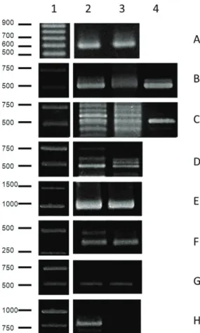
Through PCR analysis shown in Figure 1, the groups Nitrosomonadaceae and Nitrosospira that function as AOB and Nitrospira (one of the NOB groups) were detected in both areas of study. Bacteria of the genus
Nitrobacter, which convert nitrite into nitrate, were present at urban area and in the rural area were less than the detection limit of our assay. The genes amoA, napA, and nfrA, important molecular markers of the nitrogen cycle, were present in both areas. However, the higher intensity of the bands from the urban area compared to the rural one suggests that the amplicons for amoA, napA
and nfrA were more abundant in this site (see Figure 1). Through FISH analysis, it was observed that the groups Nitrosomonadaceae, Nitrospiraceae, and Nitrobacter
had significantly higher abundance in the urban area (see Figure 2). Considering the domains Bacteria and
Archaea, the abundance of Bacteria was significantly
higher in the urban area (9.48×10⁶ mL–1) compared to
the rural area (0.80×10⁶ mL–1). For the domain Archaea,
the abundance was 2.76×10⁶ mL–1 in the urban area and
0.35×10⁶ mL–1 in the rural area.
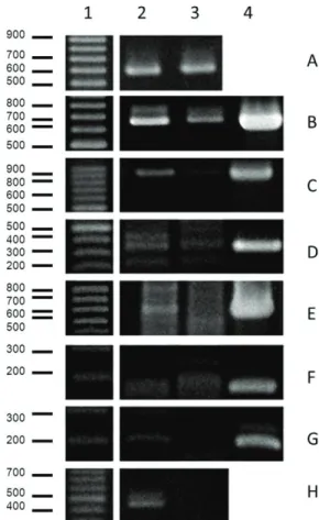
3.3. Potentially pathogenic bactéria
The Figure 3 and 4 show the results of PCR using primers specific for genera and species of already known human pathogens. The genera Enterococcus sp. was present in the urban area and was not detected in the rural area. The amplicons for the genera Salmonella sp., Streptococcus sp., and Bacteroides/Prevotella/Porphyromonas had a higher intensity, suggesting that they were more accumulated in the urban area. The same results were observed for the species
P. aeruginosa and S. aureus. In contrast, the amplicons for
A. hydrophila were present at the same intensity at both sites (as shown in Figure 3).
For E. coli, the amplicons were more abundant in the urban area. Enteropathogenic E. coli (EPEC) was identified
according to the genotype eae+/bfp+ in both studied areas. Enterohemorrhagic E. coli (EHEC) was identified
according to the genotype stx1+/stx2+ only in the urban area and stx1+/stx2- in the rural area. Enteroinvasive
E. coli (EIEC) was classified as ipaH+ in the two areas and the strain ETEC (enterotoxigenic E. coli) was identified
according to the genotype elt+/est- in the urban area and
elt-/est- in the rural area. It is important to mention that the amplicons obtained for all the diarrheagenic E. coli
were more intense at the urban site when compared to the rural site (see Figure 4).
4. Discussion
Increasing urbanisation has serious impacts on freshwater ecosystems. It is known that the pollution and discharge of sewage alters the microbial community of freshwater environments (Paul and Meyer, 2001; Walsh et al., 2005; Girones et al., 2010). However, there is still a need to establish biological standards that can evaluate the water quality. The study presented here attempted to identify the specific differences in the microbial community at two sites along the São Pedro stream that is influenced by urbanisation.
Table 3. Concentration of Nutrients (mg/L) dissolved in the studied areas.
Nitrite (mg/L) Nitrate (mg/L) Ammonium
nitrogen (mg/L)
Total Org. NIT.
(mg/L) Total P (mg/L)
Rural ND* 0.121 0.081 0.28 0.021
Urban 0.029 0.558 1.811 1.708 0.420
*Not detected.
The abundance of bacteria (1×10⁷) obtained in the urban
area of study was in agreement with Kenzaka et al. (2001) and within the range reported for other polluted rivers that were considered eutrophic systems by Yamaguchi et al. (1997). The concentration of dissolved nutrients obtained in this study in the urban area also corroborated with classifying this area as an eutrophic system since this classification is within the range of nutrients found at this site. This high abundance can be justified by the influx of organic material in this environment, resulting from domestic sewage. The high abundance of Archaea in the urban area can be explained since these microbes can easily survive in environments with a higher content of nutrients and could be active in the recycling of nitrogen (Dridi et al., 2011).
PCR analysis showed that the group Nitrobacter, was less than the detection limit of our assay in the rural area. This result corroborates with FISH analysis, through which we observed a higher abundance of Nitrobacter
Figure 2. Density of bacteria from groups Nitrosomonadaceae (Nso), Nitrospiraceae (Ntspa), Nitrobacter (NIT) in Site 1 (urban) and Site 2 (rural) by FISH analysis.
Figure 3. Identification of pathogenic bacteria by PCR analysis. The images are representative of the experiments. A- Bacteria, B- Streptococcus sp., C- Enterococcus sp., D- Salmonella sp., E- Bacteroides/Prevotella/Porphyromonas, F- Pseudomonas aeruginosa, G- Aeromonashydrophila, H- Staphylococcus aureus. 1- Molecular Mass Marker; 2- Site 1; 3- Site 2; 4- Positive Control.
in the urban area compared to the rural site (as shown in Figure 1 and 2). The high density of the Nitrobacter group is in agreement with the data of dissolved nutrients; these results showed,(at the urban site) a low level of nitrite which was below the detection level, and a high quantity of nitrate (see Table 3). For the group Nitrospiraceae, which includes the genus Nitrospira, no difference was observed between the two habitats by PCR analysis (see Figure 1). These results suggest that, in these habitats, the genus responsible for converting nitrite into nitrate is Nitrobacter. The high amount of nitrate compared to nitrite in both studied areas, especially the urban one, could explain the presence of the napA gene in these environments since the enzyme codified by this gene needs it substrate (nitrate) to perform its function. The majority of the Nitrosomonadaceae group was observed in the urban area, according to FISH analysis (as shown in Figure 2). This result is in agreement with and explains the extremely high amount of ammonium nitrogen found in this area, since Nitrosomonadaceae, including the genus Nitrosomonas, is one of the groups responsible for oxidising ammonia to nitrite. The presence of the amoA and nrfA genes could also be explained by the high amount of ammonium nitrogen in the urban area since the enzyme ammonium monooxygenase needs its substrate (ammonium nitrogen) to perform the nitrification process; the gene nfrA is responsible for supplying this nutrient to the environment. It is important to mention that the analysis of the nutrient content and the presence of the group or enzyme that metabolises it must be considered within the timescale of nutrient metabolism.
In addition to the nitrifying bacteria, our study also investigated bacteria with important clinical interests that are potential human pathogens, since a high number of these microbes are found in urban sewage and may be considered environmental contaminants. Most of those pathogens can be removed from the water by a sewage treatment but, when no treatment is performed, these pathogens can be discharged into effluents and can be a threat to public health (Girones et al., 2010). In Brazil, non-treated sewage is a major cause of water pollution. An important genus of this family Enterobacteriaceae is the genus Salmonella. It is composed of two species, Salmonella enterica and Salmonella bongori, with many subgroups and serovars, all of which are capable of causing human illness. Contamination with
Salmonella could occur through drinking contaminated water, swimming in contaminated water, or eating food washed with contaminated water (Boyd et al., 1996; Hsu et al., 2011). The presence of Salmonella sp. in more abundance in the urban area of this study (Figure 3) is in agreement with the findings of Gonzalez et al. (2010), indicating a high prevalence of Salmonella sp. in the Brazilian Lagoon that is an urban ecosystem undergoing accelerated degradation especially due to pollution. Savichtcheva et al. (2007) also found that Salmonella sp. was the most frequently detected enteropathogen in samples of rivers with different levels of pollution, as well as in samples of sludge and wastewater treatment.
Finally, we considered the most studied organisms in the Enterobacteriaceae family, E. coli. Several virulence
properties are well-characterised among E. coli populations, differentiating them from commensal and pathogenic strains. Considering the pathogenic E. coli, these virulence determinants are genetically encoded either by chromosomal, plasmid, or bacteriophage DNA and are represented by selected genes such as eae (attaching and effacing lesions), bfpA (localized adherence), ipaH (enteroinvasive mechanism), the genes encoding heat-labile toxin (elt), and heat-stable toxin (est), and the genes encoding the Shiga toxins, stx1 and stx2 (Aranda et al., 2004). Several studies have aimed at investigating the incidence of diarrheagenic
E. coli in different water environments, and the majority of them have demonstrated that these lineages and the related virulence genes are present in urban and polluted environments. Our data (see Figure 4) confirm these findings. The presence of EHEC and ETEC in sludge and rivers with different degrees of pollution was detected by Savichtcheva et al. (2007). The stx genes were found in different streams with high urbanisation and also in preserved forest (García-Aljaro et al., 2005; Higgins et al., 2005; Muniesa et al., 2006). Our results show that EHEC was genotyped as stx1+/stx2+ at the urban site; however
the stx2 gene was less than the detection limit in the rural area of the study, which is relevant and worrisome since the stx2 gene is believed to be commonly associated with more severe illnesses (Oliveira et al., 2008). An interesting observation is that the same pathotypes were also found in regional studies with urban pigeons and isolates from human diarrheic feces, indicating the circulation of the same genes in different samples from the city of Juiz de Fora (Silva et al., 2009; Garcia et al., 2011).
The genera Enterococcus, Streptococcus, and
Staphylococcus are low-GC Gram-positive bacteria belonging to the phylum Firmicutes. All these genera have species that are classified as human commensals, and as such are part of the normal human microbial community. However, studies have shown that under adverse conditions (uncolonised areas or imbalanced homeostasis) these species can behave as opportunistic pathogens and became virulent and resistant to multiple antibiotics (Willems et al., 2011). Our results (see Figure 3) indicate the presence of
Enterococcus sp., Streptococcus sp., and S. aureus in a water environment. He and Jiang (2005) also detected, using molecular techniques, the presence of Enteroccocus
in coastal waters and sewage. S. aureus is not associated with the fecal material of humans, but the presence of this species in water suggests that this environment is a potential source of community-acquired S. aureus infections (Goodwin and Pobuda, 2009).
and are more sensitive than coliforms, since they can be found in greater quantities in humans. This bacteria is an obligate anaerobe and the high level of Bacteroides in water indicates recent contamination (Fiksdal et al., 1985).
The genera Aeromonas is involved in several diseases in humans and other animals. A hydrophila was found in both study areas (see Figure 3). This result was expected because these microbes are primarily aquatic organisms and, as reported by Scoaris et al. (2008), A. hydrophila can be found in treated and non-treated water. P. aeruginosa, a free-living bacteria ubiquitous in the environment, is another potential human pathogen. This species can be found either in oligotrophic environments or in high nutrient environments (Mena and Gerba, 2009), which is in agreement with our data. Our results show that P. aeruginosa was present in the rural area with poor nutrients, but it was more abundant in the urban area considered a eutrophic system. Garcia-Armisen et al. (2011) have recently shown the incidence of P. aeruginosa resistant to multiple antibiotics in rivers contaminated with sewage.
In conclusion, our study presents a comparison of the community composition of a polluted and preserved stream. The results show a major incidence of nitrifying and potentially pathogens at the polluted site, which is influenced by urbanisation, especially by the discharge of sewage. These findings, specially the high incidence of bacteria from gastrointestinal tract of human and others animals indicate a change in the structure of the microbial community imposed by human occupation. The survival and persistence of pathogenic bacteria in natural environments is of particular importance to public health. This study confirms the need for sewage treatment and policies to minimise the environmental impacts associated with urbanisation. Our results show the scenario of a local stream but reflect the situation of many rivers and streams in Brazil. This is alarming, since these resources are responsible for supplying water for part of the population. Our results suggest a range of genes and microbes that can be used as marker of water deteriorated by human action through urbanisation. In the future, these markers can be included in the biological parameters evaluated for urban streams and can be used to better classify the water quality.
Acknowledgements
The research described in this manuscript was supported by a grant from the Fundação de Amparo a Pesquisa do Estado de Minas Gerais (FAPEMIG), Brazil. Special thanks go to members of the Laboratório de Ecologia Aquática at the Universidade Federal de Juiz de Fora for crucial support with the nutrient content analyses.
References
AMANN, RI., BINDER, BJ., OLSON, RJ., CHISHOLM, SW., DEVEREUX, R. and STAHL, DA., 1990. Combination of 16S rRNA-targeted oligonucleotide probes with flow cytometry for analyzing mixed microbial populations. Applied and Environmental Microbiology, vol. 56, no. 6, p. 1919-1925. PMid:2200342.
ARANDA, KRS., FAGUNDES-NETO, U. and SCALETSKY, ICA., 2004. Evaluation of multiplex PCRs for diagnosis of infection with diarrheagenic Escherichia coli and Shigella spp. Journal of Clinical Microbiology, vol. 42, no. 12, p. 5849-5853. http:// dx.doi.org/10.1128/JCM.42.12.5849-5853.2004. PMid:15583323 ARAYA, R., TANI, K., TAKAGI, N. and NASU, M., 2003. Bacterial activity and community composition in stream water and biofilm from an urban river determined by fluorescent in situ hybridization and DGGE analysis. FEMS Microbiology Ecology, vol. 43, no. 1, p. 111-119. Available from: <http://www.ncbi.nlm. nih.gov/pubmed/19719701>.
BERNHARD, AE. and FIELD, KG., 2000. Identification of nonpoint sources of fecal pollution in coastal waters by using host-specific 16S ribosomal DNA genetic markers from fecal anaerobes. Applied and Environmental Microbiology, vol. 66, no. 4, p. 1587-1594. http://dx.doi.org/10.1128/AEM.66.4.1587-1594.2000. PMid:10742246
BöCKELMANNA, U., MANzA, W., NEUB, TR. and SzEWzYKA, U., 2000. Characterization of the microbial community of lotic organic aggregates (‘river snow’) in the Elbe River of Germany by cultivation and molecular methods. FEMS Microbiology Ecology, vol. 33, no. 2, p. 157-170. PMid:10967215.
BOEHM, AB., FUHRMAN, JA., MRSE, RD. and GRANT, SB., 2003. Tiered approach for identification of a human fecal pollution source at a recreational beach: case study at Avalon Bay, Catalina Island, California. Environmental Science & Technology, vol. 37, no. 4, p. 673-680. http://dx.doi.org/10.1021/es025934x. PMid:12636264
BOYD, EF., WANG, FS., WHITTAM, TS. and SELANDER, RK., 1996. Molecular genetic relationships of the salmonellae. Applied and Environmental Microbiology, vol. 62, no. 3, p. 804-808. PMid:8975610.
BRÜMMER, IHM., FEHR, W. and WAGNER-DöBLER, I., 2000. Biofilm community structure in polluted rivers: abundance of dominant phylogenetic groups over a complete annual cycle. Applied and Environmental Microbiology, vol. 66, no. 7, p. 3078-3082. Available from: <http://www.ncbi.nlm.nih.gov/ pubmed/10877809>.
CHOTáR, M., VIDOVá, B. and GODáNY, A., 2006. Development of specific and rapid detection of bacterial pathogens in dairy products by PCR. Folia Microbiologica, vol. 51, no. 6, p. 639-646. http://dx.doi.org/10.1007/BF02931632. PMid:17455804 CUMAR, SKM. and NAGARAJA, B., 2011. Environmental impact of leachate characteristics on water quality. Environmental Monitoring and Assessment, vol. 178, no. 1-4, p. 499-505. http:// dx.doi.org/10.1007/s10661-010-1708-9. PMid:20859680 DAIMS, H., NIELSEN, JL., NIELSEN, PH., SCHLEIFER, KH. and WAGNER, M., 2001. In situ characterization of Nitrospira-like nitrite-oxidizing bacteria active in wastewater treatment plants. Applied and Environmental Microbiology, vol. 67, no. 11, p. 5273-5284. http://dx.doi.org/10.1128/AEM.67.11.5273-5284.2001. PMid:11679356
DRIDI, B., RAOULT, D. and DRANCOURT, M., 2011. Archaea as emerging organisms in complex human microbiomes. Anaerobe, vol. 17, no. 2, p. 56-63. http://dx.doi.org/10.1016/j.anaerobe.2011.03.001. PMid:21420503
FUKUSHIMA, H., TSUNOMORI, Y. and SEKI, R., 2003. Duplex real-time SYBR green PCR assays for detection of 17 species of food- or waterborne pathogens in stools. Journal of Clinical Microbiology, vol. 41, no. 11, p. 5134-5146. http:// dx.doi.org/10.1128/JCM.41.11.5134-5146.2003. PMid:14605150 GARCíA-ALJARO, C., MUNIESA, M., BLANCO, JE., BLANCO, M., BLANCO, J., JOFRE, J. and BLANCH, AR., 2005. Characterization of Shiga toxin-producing Escherichia coli isolated from aquatic environments. FEMS Microbiology Letters, vol. 246, no. 1, p. 55-65. http://dx.doi.org/10.1016/j. femsle.2005.03.038. PMid:15869962
GARCIA-ARMISEN, T., VERCAMMEN, K., PASSERAT, J., TRIEST, D., SERVAIS, P. and CORNELIS, P., 2011. Antimicrobial resistance of heterotrophic bacteria in sewage-contaminated rivers. Water Research, vol. 45, no. 2, p. 788-796. http://dx.doi. org/10.1016/j.watres.2010.09.003. PMid:20870262
GARCIA, PG., SILVA, VL. and DINIz, CG., 2011. Occurrence and antimicrobial drug susceptibility patterns of commensal and diarrheagenic Escherichia coli in fecal microbiota from children with and without acute diarrhea. Journal of Microbiology, vol. 49, no. 1, p. 46-52. http://dx.doi.org/10.1007/s12275-011-0172-8. PMid:21369978
GEETS, J., COOMAN, M., WITTEBOLLE, L., HEYLEN, K., VANPARYS, B., DE VOS, P., VERSTRAETE, W. and BOON, N., 2007. Real-time PCR assay for the simultaneous quantification of nitrifying and denitrifying bacteria in activated sludge. Applied Microbiology and Biotechnology, vol. 75, no. 1, p. 211-221. http://dx.doi.org/10.1007/s00253-006-0805-8. PMid:17256118 GIRONES, R., FERRúS, MA., ALONSO, JL., RODRIGUEz-MANzANO, J., CALGUA, B., CORRêA, AA., HUNDESA, A., CARRATALA, A. and BOFILL-MAS, S., 2010. Molecular detection of pathogens in water—the pros and cons of molecular techniques. Water Research, vol. 44, no. 15, p. 4325-4339. http:// dx.doi.org/10.1016/j.watres.2010.06.030. PMid:20619868 GONzALEz, AM., PARANHOS, R. and LUTTERBACH, MS., 2010. Relationships between fecal indicators and pathogenic microorganisms in a tropical lagoon in Rio de Janeiro, Brazil. Environmental Monitoring and Assessment, vol. 164, no. 1-4, p. 207-219. http://dx.doi.org/10.1007/s10661-009-0886-9. PMid:19365609
GOODWIN, KD. and POBUDA, M., 2009. Performance of CHROMagar Staph aureus and CHROMagar MRSA for detection of Staphylococcus aureus in seawater and beach sand: comparison of culture, agglutination, and molecular analyses. Water Research, vol. 43, no. 19, p. 4802-4811. http://dx.doi. org/10.1016/j.watres.2009.06.025. PMid:19577788
HE, JW. and JIANG, S., 2005. Quantification of enterococci and human adenoviruses in environmental samples by real-time PCR. Applied and Environmental Microbiology, vol. 71, no. 5, p. 2250-2255. http://dx.doi.org/10.1128/AEM.71.5.2250-2255.2005. PMid:15870308
HEAD, IM., HIORNS, WD., EMBLEY, TM., MCCARTHY, AJ. and SAUNDERS, JR., 1993. The phylogeny of autotrophic ammonia-oxidizing bacteria as determined by analysis of 16S ribosomal RNA gene sequences. Journal of General Microbiology, vol. 139, Pt. 6, p. 1147-1153. http://dx.doi.org/10.1099/00221287-139-6-1147. PMid:7689633
HERBERT, RA., 1999. Nitrogen cycling in coastal marine ecosystems. FEMS Microbiology Reviews, vol. 23, no. 5, p. 563-590. http://dx.doi.org/10.1111/j.1574-6976.1999.tb00414.x. PMid:10525167
HIGGINS, JA., BELT, KT., KARNS, JS., RUSSELL-ANELLI, J. and SHELTON, DR., 2005. tir- and stx-positive Escherichia coli in stream waters in a metropolitan area. Applied and Environmental Microbiology, vol. 71, no. 5, p. 2511-2519. http://dx.doi.org/10.1128/ AEM.71.5.2511-2519.2005. PMid:15870341
HOARE, RA., 1984. Nitrogen and phosphorus in Rotorua urban streams. New Zealand Journal of Marine and Freshwater Research, vol. 18, no. 4, p. 451-454. http://dx.doi.org/10.1080/ 00288330.1984.9516066.
HOVANEC, TA., TAYLOR, LT., BLAKIS, A. and DELONG, EF., 1998. Nitrospira-like bacteria associated with nitrite oxidation in freshwater aquaria. Applied and Environmental Microbiology, vol. 64, no. 1, p. 258-264. PMid:16349486.
HSU, BM., HUANG, KH., HUANG, SW., TSENG, KC., SU, MJ., LIN, WC., JI, DD., SHIH, FC., CHEN, JL. and KAO, PM., 2011. Evaluation of different analysis and identification methods for Salmonella detection in surface drinking water sources. The Science of the Total Environment, vol. 409, no. 20, p. 4435-4441. http://dx.doi.org/10.1016/j.scitotenv.2011.05.052. PMid:21782212 KOMíNKOVá, D., 2012. The Urban Stream Syndrome: a mini-review. The Open Environmental & Biological Monitoring Journal, vol. 5, no. 1, p. 24-29. http://dx.doi.org/10.2174/1875 040001205010024.
KENzAKA, T., YAMAGUCHI, N., PRAPAGDEE, B., MIKAMI, E. and NASU, M., 2001. Bacterial community composition and activity in urban rivers in Thailand and Malaysia. Journal of Health Science, vol. 47, no. 4, p. 353-361. http://dx.doi. org/10.1248/jhs.47.353.
KOSTANJŠEK, R., LAPANJE, A., DROBNE, D., PEROVIĆ, S., PEROVIĆ, A., zIDAR, P., ŠTRUS, J., HOLLERT, H. and KARAMAN, G., 2005. Bacterial Community structure analyses to assess pollution of water and sediments in the lake Shkodra/ Skadar, Balkan Peninsula. Environmental Science & Pollution Research, vol. 12, no. 6, p. 361-368.
LATUF, MO., 2004. Diagnostic study of the São Pedro’s creek waters, Juiz de Fora town, Minas Gerais state, Brazil. Geografia, vol. 13, no. 1, p. 21-55.
MARTINELLI, LA., COLETTA, LD., RAVAGNANI, EC., CAMARGO, PB., OMETTO, JPHB., FILOSO, S. and VICTORIA, RL., 2010. Dissolved nitrogen in rivers: comparing pristine and impacted regions of Brazil. Revista Brasileira de Biologia = Brazilian Journal of Biology, vol. 70, no. 3, Suppl., p. 709-722. http://dx.doi.org/10.1590/S1519-69842010000400003. PMid:21085777
MENA, KD. and GERBA, CP., 2009. Risk assessment of Pseudomonas aeruginosa in water. Reviews of Environmental Contamination and Toxicology, vol. 201, p. 71-115. http://dx.doi. org/10.1007/978-1-4419-0032-6_3. PMid:19484589
MEYBECK, M., 1998. Man and river interface: multiple impacts on water and particulates chemistry illustrated in the Seine river basin. Hydrobiologia, vol. 373-374, p. 1-20. http://dx.doi. org/10.1023/A:1017067506832.
MOBARRY, BK., WAGNER, M., URBAIN, V., RITTMANN, BE. and STAHL, DA., 1996. Phylogenetic probes for analyzing abundance and spatial organization of nitrifying bacteria. Applied and Environmental Microbiology, vol. 62, no. 6, p. 2156-2162. PMid:8787412.
Ecology, vol. 49, no. 3, p. 433-443. http://dx.doi.org/10.1016/j. femsec.2004.04.012. PMid:19712292
MUNIESA, M., JOFRE, J., GARCíA-ALJARO, C. and BLANCH, AR., 2006. Occurrence of Escherichia coli O157:H7 and other enterohemorrhagic Escherichia coli in the environment. Environmental Science & Technology, vol. 40, no. 23, p. 7141-7149. http://dx.doi.org/10.1021/es060927k. PMid:17180960 MURRAY, KS., FISHER, LE., THERRIEN, J., GEORGE, B. and GILLESPIE, J., 2001. Assessment and use of indicator bacteria to determine sources of pollution to an urban river. Journal of Great Lakes Research, vol. 27, no. 2, p. 220-229. http://dx.doi. org/10.1016/S0380-1330(01)70635-1.
OLIVEIRA, MG., BRITO, JRF., GOMES, TAT., GUTH, BEC., VIEIRA, MAM., NAVES, zVF., VAz, TMI. and IRINO, K., 2008. Diversity of virulence profiles of Shiga toxin-producing Escherichia coli serotypes in food-producing animals in Brazil. International Journal of Food Microbiology, vol. 127, no. 1-2, p. 139-146. http://dx.doi.org/10.1016/j.ijfoodmicro.2008.06.023. PMid:18678426
PADMALAL, D., REMYA, SI., JYOTHI, SJ., BAIJULAL, B., BABU, KN. and BAIJU, RS., 2012. Water quality and dissolved inorganic fluxes of N, P, SO4, and K of a small catchment river in the Southwestern Coast of India. Environmental Monitoring and Assessment, vol. 184, no. 3, p. 1541-1557. http://dx.doi. org/10.1007/s10661-011-2059-x. PMid:21544504
PAUL, MJ. and MEYER, JL., 2001. Streams in the urba,n landscape. Annual Review of Ecology and Systematics, vol. 32, no. 1, p. 333-365. http://dx.doi.org/10.1146/annurev.ecolsys.32.081501.114040. QIN, X., EMERSON, J., STAPP, J., STAPP, L., ABE, P. and BURNS, JL., 2003. Use of real-time PCR with multiple targets to identify Pseudomonas aeruginosa and other nonfermenting gram-negative bacilli from patients with cystic fibrosis. Journal of Clinical Microbiology, vol. 41, no. 9, p. 4312-4317. http:// dx.doi.org/10.1128/JCM.41.9.4312-4317.2003. PMid:12958262 RINTTILä, T., KASSINEN, A., MALINEN, E., KROGIUS, L. and PALVA, A., 2004. Development of an extensive set of 16S rDNA-targeted primers for quantification of pathogenic and indigenous bacteria in faecal samples by real-time PCR. Journal of Applied Microbiology, vol. 97, no. 6, p. 1166-1177. http:// dx.doi.org/10.1111/j.1365-2672.2004.02409.x. PMid:15546407 SAVICHTCHEVA, O., OKAYAMA, N. and OKABE, S., 2007. Relationships between Bacteroides 16S rRNA genetic markers and presence of bacterial enteric pathogens and conventional fecal indicators. Water Research, vol. 41, no. 16, p. 3615-3628. http://dx.doi.org/10.1016/j.watres.2007.03.028. PMid:17507075 SCOARIS, DO., COLACITE, J., NAKAMURA, CV., UEDA-NAKAMURA, T., ABREU FILHO, BA. and DIAS FILHO, BP., 2008. Virulence and antibiotic susceptibility of Aeromonas spp. isolated from drinking water. Antonie van Leeuwenhoek, vol. 93, no. 1-2, p. 111-122. http://dx.doi.org/10.1007/s10482-007-9185-z. PMid:17636377
SILVA, VL., NICOLI, JR., NASCIMENTO, TC. and DINIz, CG., 2009. Diarrheagenic Escherichia coli strains recovered from urban pigeons (Columba livia) in Brazil and their antimicrobial susceptibility patterns. Current Microbiology, vol. 59, no. 3, p. 302-308. http://dx.doi.org/10.1007/s00284-009-9434-7. PMid:19504156
SMITH, CJ. and CALLIHAN, DR., 1992. Analysis of rRNA restriction fragment length polymorphisms from Bacteroides spp. and Bacteroides fragilis isolates associated with diarrhea in humans and animals. Journal of Clinical Microbiology, vol. 30, no. 4, p. 806-812. PMid:1374078.
SMITH, CJ., NEDWELL, DB., DONG, LF. and OSBORN, AM., 2007. Diversity and abundance of nitrate reductase genes (narG and napA), nitrite reductase genes (nirS and nrfA), and their transcripts in estuarine sediments. Applied and Environmental Microbiology, vol. 73, no. 11, p. 3612-3622. http://dx.doi. org/10.1128/AEM.02894-06. PMid:17400770
STAHL, DA. and AMANN, R., 1991. Development and application of nucleic acid probes. In GOODFELLOW, M. (Ed.). Nucleic acid techniques in bacterial systematics. Chichester: John Wiley & Sons. TESKE, A., ALM, E., REGAN, JM., TOzE, S., RITTMANN, BE. and STAHL, DA., 1994. Evolutionary relationships among ammonia- and nitrite-oxidizing bacteria. Journal of Bacteriology, vol. 176, no. 21, p. 6623-6630. PMid:7961414.
TIQUIA, SM., 2010. Metabolic diversity of the heterotrophic microorganisms and potential link to pollution of the Rouge River. Environmental Pollution, vol. 158, no. 5, p. 1435-1443. http://dx.doi.org/10.1016/j.envpol.2009.12.035. PMid:20106574 TREBESIUS, K., LEITRITz, L., ADLER, K., SCHUBERT, S., AUTENRIETH, IB. and HEESEMANN, J., 2000. Culture independent and rapid identification of bacterial pathogens in necrotising fasciitis and streptococcal toxic shock syndrome by fluorescence in situ hybridisation. Medical Microbiology and Immunology, vol. 188, no. 4, p. 169-175. http://dx.doi.org/10.1007/ s004300000035. PMid:10917153
WALLNER, G., AMANN, R. and BEISKER, W., 1993. Optimizing fluorescent in situ hybridization with rRNA-targeted oligonucleotide probes for flow cytometric identification of microorganisms. Cytometry, vol. 14, no. 2, p. 136-143. http://dx.doi.org/10.1002/ cyto.990140205. PMid:7679962
WALSH, CJ., ROY, AH., FEMINELLA, JW., COTTINGHAM, PD., GROFFMAN, PM. and MORGAN, IRP., 2005. The urban stream syndrome: current knowledge and the search for a cure. Journal of the North American Benthological Society, vol. 24, no. 3, p. 706-723. http://dx.doi.org/10.1899/04-028.1. WATANABE, K., KODAMA, Y. and HARAYAMA, S., 2001. Design and evaluation of PCR primers to amplify bacterial 16S ribosomal DNA fragments used for community fingerprinting. Journal of Microbiological Methods, vol. 44, no. 3, p. 253-262. http://dx.doi.org/10.1016/S0167-7012(01)00220-2. PMid:11240048 WERNICK, BG., COOK, KE. and SCHREIER, H., 1998. Land use and streamwater nitrate-n dynamics in an urban-rural fringe watershed. Journal of the American Water Resources Association, vol. 34, no. 3, p. 639-650. http://dx.doi.org/10.1111/j.1752-1688.1998. tb00961.x.
WETzEL, RG. and LIKENS, GE., 1991. Limnological analyses. New York: Springer-Verlag. http://dx.doi.org/10.1007/978-1-4757-4098-1.