Ana Margarida Teixeira Saramago
Dissertation presented to obtain the Ph.D degree in Biology
Instituto de Tecnologia Química e Biológica
Universidade Nova de Lisboa
Oeiras, March, 2014
Financial Support from Fundação para a Ciência e Tecnologia (FCT) – Ph.D: grant - SFRH/BD/65607/2009
Work performed at:
Control of Gene Expression Laboratory
Instituto de Tecnologia Química e Biológica
Av. da República (EAN)
2781-901 Oeiras – Portugal Tel: +351-21-4469548
Supervisor :
Professora Doutora Cecília Maria Pais de Faria de Andrade Arraiano – Investigadora Coordenadora, Instituto de Tecnologia Química e Biológica, Universidade Nova de Lisboa. (Head of the Laboratory of Control of Gene Expression, where the work of this Dissertation was performed)
Co-supervisor :
Doutora Susana Margarida Lopes Domingues – Investigadora Pós-Doutorada, Instituto de Tecnologia Química e Biológica, Universidade Nova de Lisboa.
(Post-doc Fellow in the Laboratory of Control of Gene Expression, where the work of this Dissertation was performed)
President of the Jury :
Examiners:
Professora Doutora Mónica Amblar Esteban – Investigadora Principal do Instituto de Salud Carlos III, Madrid (Principal Examiner).
Professor Doutor Arsénio do Carmo Sales Mendes Fialho – Professor Associado com Agregação, Instituto Superior Técnico, Universidade Técnica de Lisboa (Principal Examiner).
Professor Doutor Miguel Agostinho Sousa Pinto Torres Fevereiro – Investigador Principal do Instituto Nacional de Investigação Agrária e Veterinária - INIAV
“A journey of a thousand miles begins with a single step.”
I want express my gratitude to all that supported me in the realization of my Ph.D. and dedicate to them several words:
- To Instituto de Tecnologia Química e Biológica (ITQB) for hosting me, for providing good work conditions, and to give me the excellent opportunity to develop exceptional scientific projects, even with a surrounding environment of economic crisis. Once more, we proved that we (Portuguese people) are plastic and creative people who do not let themselves go down easily. I also would like to thank to the past and present ITQB directors, namely Professor Doutor José Artur Martinho Simões, Professor Doutor Luís Paulo Rebelo, and currently Professor Doutor Cláudio Soares.
- To Fundação para a Ciência e Tecnologia (FCT) for financial support that made my Ph.D. possible.for
- I also would like to thank to Sandra Viegas,my friend and officemate, for all the good advices, support and good friendship. She has been precious on both academic and at personal level, for which I am extremely grateful!!
- To Inês Silva and Andreia Aires,also my very good and special friends! I want to thank you for being wonderful persons, for sharing so many pleasant moments (never forgetting the professional side), and for being such good friends.
- To Rute Matos for the friendship and for giving me the pleasure to work with her! I hope that we continue to work together!
- To may labmate Igor Ruiz de los Mozos for being an exceptional friend and hard worker, and for all the fun and crazy times during my last year of Ph.D… and to Nabila Haddad for being an extremely nice and helpful person!! I will never forget you both!!
- To the members of the CMA group that have contributed greatly to my personal and professional time. The group has been a source of friendships as well as good advices, fun and team spirit. My greatest thanks to Inês Silva, Sandra Viegas,
Susana Domingues, Andreia Aires, Cátia Bárria, Susana Barahona, Patrícia Apura, Ricardo Moreira, Rute Matos, Teresa Pinto (thank you for helping me to rescue my polyacrylamide gels), Ricardo Santos, Raquel Santos, Lisete Galego,
Clémentine Dressaire, Michal Malecki, Filipa Reis, Vânia Pobre, Zé Andrade,
Patrick Freire and Ana Barbas!
- To Iñigo Lasa, Alejandro Toledo-Arana and Igor Ruiz de los Mozos from Instituto de Agrobiotecnología, Pamplona, Spain, and to Nabila Haddad and
Hervé Prévost from LUNAM Université, Nantes, France for their collaboration and precious help in this Doctoral work. I hope that we can continue our collaboration.
- I thank to my good friends and Ph.D. colleagues for all the fun, friendship, and for all the memorable moments altogether!
- I also want to express my greatest gratitude to my biggest friends who are always by my side (they know who they are) for their unconditional friendship, support and encouragement throughout all the important moments of my life. - I also want to express my gratitude for my two lovely feline friends, Pantufa
and Lucky for their unconditional love.
- Lastly, I would like to thank to my mom who raised me with an unconditional love, and supported me in all my pursuits. Without her continuous support and encouragement I never would have been able to achieve my goals! To my dad for all the motivation and also for being always by my side! And to my family for all the love and encouragement.
ABSTRACT
... XVII
RESUMO
... XXIII
LIST OF PUBLICATIONS
... XXXI
DISSERTATION OUTLINE
... XXXVII
ABBREVIATIONS
... XLI
CHAPTER 1
... 1
CHAPTER 2
... 43
CHAPTER 3
... 81
CHAPTER 4
... 119
CHAPTER 5
... 191
CHAPTER 6
... 231
Ribonucleases (RNases) are key factors in the control of all biological
processes, since they modulate the stability of RNA transcripts, allowing rapid
changes in gene expression. Some RNases are up-regulated under stress
situations and are involved in virulence processes in pathogenic microorganisms.
RNases also control the levels of regulatory RNAs, which play very important
roles in cell physiology.
In general, RNA degradation starts with an internal cleavage performed
by an endoribonuclease. This is followed by degradation from one extremity by
one or more exoribonucleases. RNase E has been considered the main enzyme
that catalyses the initial cleavage event in negative bacteria. In
Gram-positives, this degradation step has been attributed to its functional equivalents:
RNase Y and RNases J1/2. However, the ubiquitous RNase III-like enzymes,
which cleave the RNA molecule internally within double-stranded segments, are
also emerging as important players in the control of bacterial mRNA stability.
The first objective of this doctoral work was to perform a functional and
biochemical analysis of RNase III from Campylobacter jejuni. This microorganism
is a foodborne pathogen for which there is little information regarding
ribonucleases. However, there are hints that they can be instrumental in the
virulence mechanisms of this bacterium. Namely, Polynucleotide phosphorylase
(PNPase) was shown to allow survival of C. jejuni in cold conditions, and to have
a role in bacterial swimming, cell adhesion, colonization and invasion. Since
PNPase synthesis is auto-controlled in an RNase III-dependent mechanism, it was
also relevant to study the role of RNase III in C. jejuni. Hence, we have cloned,
overexpressed, purified and studied the activity of C. jejuni RNase III (Cj-RNase
III) over a range of different conditions. Throughout this study, we showed that
Plasticity of Cj-RNase III suggests that this enzyme may be important for all the
environmental demands that C. jejuni must face during its lifestyle. The results
also demonstrated that Mn2+ seems to be the preferred co-factor of the enzyme,
contrarily to what was described for other RNase III orthologues. Therefore,
Cj-RNase III may have an important role under a Mn2+-rich environment, namely
inside macrophages. We have also demonstrated that Cj-RNase III is able to
complement an RNase III- Escherichia coli strain in the maturation of 30S rRNA
and in the regulation of PNPase production. Mutational analysis of residues
involved in the catalytic activity of Cj-RNase III confirmed a conservation of the
catalytic mechanism characteristic of this family. Altogether these results
reinforce the notion that members of the RNase III family of enzymes from
different species retain some conserved functions. This study brought relevant
information regarding functionality and biochemical properties of Cj-RNase III.
Despite the functional conservation, the peculiarities found in Cj-RNase III
activity lead us to speculate that this enzyme might be also a main player in the
survival of this pathogen to environmental demands.
The second goal of this dissertation was to examine the role of RNase III
and other Salmonella Typhimurium ribonucleases in the control of antibiotic
susceptibility and biofilm formation, two important factors in bacterial survival.
Salmonella infections are a serious medical and veterinary problem worldwide
and there is an increasing need for new strategies for prevention and control. This
study showed that in particular, endoribonucleases E and III affect the
susceptibility of Salmonella to ribosome-targeting antibiotics, and affect biofilm
formation. We also studied in vitro the role of these two endoribonucleases in the
degradation of the small non-coding RNA (sRNA) MicA, which has a role in
biofilm development and in the control of outer membrane porins in
silencing” mechanism resembles eukaryotic RNA interference process. By
contrast, RNase E is only able to efficiently degrade free MicA sRNA. Our results
allowed us to propose a model explaining the cooperation of both enzymes in the
cell in order to achieve the fine-tuned control of the posttranscriptional regulator
MicA.
In the third part of this work, we have studied the role of RNase III in
Staphylococcus aureus, particularly in biofilm production. Biofilm development is a
highly complex process, and constitutes one of the major medical concerns within
S. aureus pathogenesis. S. aureus icaR encodes a key transcription regulator that
represses the ica operon, which has a key role in biofilm production. It was
demonstrated that the 3’untraslated region (3’-UTR) of icaR mRNA affects the
expression level of the IcaR protein by modulating the stability of its own
transcript. The modulation of icaR is performed through RNA base-pairing
between the 3’-UTR and the 5’-UTR regions, which triggers the degradation by
RNase III, leading to the destruction of the icaR transcript. So far the relevance of
3’-UTRs in the regulation of bacterial mRNA functionality has been disregarded.
Our data illustrate that bacterial 3’-UTRs can provide strategies for fine-tuning
control of gene expression.
From the previous work some questions remained unanswered, namely
the influence of environmental signals in the modulation of IcaR levels. In the last
stage of this doctoral work, we have collected evidences that a temperature shift
may dictate the formation/destabilization of the 5’-3’-UTR duplex, adjusting IcaR
levels according to the temperature sensed in the surrounding environment. We
have also seen that a S. aureus strain lacking PNPase is not able to form biofilm,
mechanism provides a rapid and efficient activation/inactivation of IcaR
production in response to a sudden change in environmental conditions, and a
fine-tuned adjustment of biofilm production in S. aureus.
In this work it was shown that RNase III seems to be important to the
survival of Campylobacter to different environments, with consequences for its
pathogenicity. In Salmonella, RNase III contributes to antibiotic resistance, biofilm
development and is involved in the control in the coupled-degradation of MicA
sRNA and its targets. RNase III is also a major player in the control of biofilm
production in S. aureus, and 3´ untranslated regions of prokaryotic mRNAs can
regulate gene expression. Overall, the work developed during this Dissertation
provided important results for a better understanding of the versatile
contribution of RNase III to several relevant cellular mechanisms in different
pathogenic microorganisms. This reinforces the importance of this
As Ribonucleases (RNases) são factores chave no controlo de todos os
processos biológicos, pois são responsáveis pela modulação da estabilidade dos
transcriptos de RNA, permitindo assim rápidas mudanças na expressão génica.
Algumas RNases são altamente expressas em situações de stress e estão
envolvidas em processos de virulência em microrganismos patogénicos. As
RNases são também responsáveis pelo controlo dos níveis de RNAs regulatórios
que, por sua vez, também têm um papel igualmente importante para a fisiologia
da célula.
Em geral, a degradação de RNA é iniciada por uma clivagem interna
efectuada por uma endoribonuclease. Segue-se depois a degradação a partir de
uma das extremidades levada a cabo por uma ou várias exoribonucleases. A
RNase E tem sido considerada como a principal enzima que catalisa o evento de
clivagem inicial, em bactérias Gram-negativas. Em bactérias Gram-positivas, este
passo de degradação tem sido atribuído a outras RNases funcionalmente
equivalentes: RNase Y e RNase J1/2. No entanto, a RNase III é uma enzima
ubíqua, cuja família está a emergir como importantes intervenientes no controlo
da estabilidade de RNA em bactérias. Estas enzimas têm a particularidade de
serem específicas para o corte interno de segmentos de RNA em cadeia dupla.
O primeiro objectivo deste trabalho doutoral consistiu numa análise
funcional e bioquímica da RNase III de Campylobacter jejuni. Este é um
microrganismo responsável por provocar intoxicações alimentares, para o qual
existe muito pouca informação relativamente a RNases. No entanto, existem
pistas para o facto estas enzimas poderem ser instrumentais para os mecanismos
de virulência nesta bactéria. Nomeadamente, a enzima polinucleotídeo
fosforilase (PNPase) demonstrou ser necessária à sobrevivência de C. jejuni em
colonização e invasão. Tendo em conta que a síntese da PNPase é auto-controlada
num mecanismo dependente da RNase III, pode-se inferir que esta última possa
também ter um papel importante para Campylobacter. Como tal, procedeu-se à
clonagem, sobreexpressão, purificação e estudo da actividade da RNase III de C.
jejuni (Cj-RNase III) numa gama de diferentes condições. Com este estudo, foi
possível demonstrar que esta enzima é activa numa larga gama de pHs e
temperaturas. A plasticidade da Cj-RNase III é sugestiva de que esta enzima
poderá ter um papel importante nos vários desafios ambientais que C. jejuni tem
de enfrentar ao longo do seu ciclo de vida. Os resultados sugeriram que o ião
divalente manganês (Mn2+) é o co-factor preferencial desta enzima,
contrariamente ao que foi previamente descrito para os outros ortólogos da
RNase III. Estes dados levantam a hipótese de que a Cj-RNase III possa ter uma
função relevante em ambientes ricos em manganês, nomeadamente dentro dos
macrófagos. Neste trabalho também se demonstrou que Cj-RNase III é capaz de
complementar uma estirpe de Escherichia coli RNase III- na maturação do rRNA
30S e na regulação da síntese da PNPase, reforçando a ideia de que os membros
desta família de enzimas provenientes de diferentes espécies possuem funções
conservadas. Finalmente, uma análise mutacional dos resíduos envolvidos na
actividade catalítica da Cj-RNase III confirmou a conservação do mecanismo
catalítico que é característico dos membros já reportados desta família de
enzimas. Em geral, este estudo revelou informação relevante relativamente à
funcionalidade e propriedades bioquímicas da Cj-RNase III. Apesar da
conservação funcional, as peculiaridades encontradas na actividade desta enzima
levantam a hipótese de que este pode ser um interveniente com um papel
importante na sobrevivência deste microrganismo patogénico aos diferentes
bem como de outras ribonucleases, no controlo da susceptibilidade a antibióticos
e na formação de biofilme em Salmonella Typhimurium, dois factores importantes
para a sobrevivência das bactérias. As infecções por Salmonella reflectem-se a
nível mundial e têm graves repercussões na medicina humana e veterinária.
Como tal, há uma necessidade crescente de novas estratégias de prevenção e
controlo. Com este estudo demonstrou-se que, em particular, as
endoribonucleases E e III afectam a susceptibilidade aos antibióticos que actuam
ao nível do ribossoma em Salmonella, bem como a capacidade de formação de
biofilmes. Também se estudou in vitro, o papel destas duas endoribonucleases na
degradação de um pequeno RNA não codificante designado de MicA, que por
sua vez está implicado na formação de biofilmes e que adicionalmente tem como
função o controlo de porinas da membrana externa em Enterobacteriaceae. Os
nossos resultados revelaram que a RNase III apenas é capaz de degradar o
pequeno RNA MicA quando este se encontra hibridado com o seu RNA
mensageiro alvo. Desta forma o alvo é também destruído. Este mecanismo de
silenciamente génico assemelha-se, de certa forma, ao fenómeno de RNA de
interferência que existe em células eucariótas. Contrariamente a este cenário, a
RNase E é capaz de degradar o pequeno RNA quando este se encontra livre, ou
seja, sem estar hibridado com nenhum dos seus alvos. Desta forma, pode-se
concluir que a RNase III apenas cliva o pequeno RNA num mecanismo
dependente do alvo, enquanto que a RNase E degrada o MicA quando este está
desemparelhado. Os resultados obtidos permitiram propor um modelo que
explica a cooperação de ambas as enzimas na obtenção de um controlo
pós-transcricional preciso e minucioso na célula.
A terceira parte deste trabalho teve como objectivo estudar a função da
de biofilmes é um processo altamente complexo e, no âmbito da patogénese de S.
aureus, constitui uma das principais preocupações a nível médico. O gene icaR de
S. aureus codifica para um regulador transcricional que reprime o operão ica, que
por sua vez tem um papel na produção de biofilmes. Neste trabalho,
demonstrou-se que a região não traduzida a 3’ (3’-UTR) do mRNA do icaR afecta o nível de
expressão da proteína IcaR através da modulação da estabilidade deste próprio
transcrito. A modulação do icaR é efectuada através de um emparelhamento entre
as regiões 3’-UTR e 5’-UTR e, consequentemente, o duplex de RNA formado é
clivado pela RNase III, levando à destruição do transcrito icaR. Até há bem pouco
tempo a relevância de regiões 3’-UTR na regulação de RNAs mensageiros
bacterianos foi negligenciada. Estes dados são ilustrativos de que as regiões 3’
-UTR em bactérias também possuem a capacidade de controlar a expressão génica.
Este trabalho originou algumas questões, nomeadamente quais os sinais
ambientais eventualmente responsáveis pela modulação da interacção 5’-3’-UTR
do transcrito icaR. Na última fase deste trabalho doutoral, obtiveram-se
evidências de que alterações de temperatura ditam a formação/destabilização do
duplex 5’-3’-UTR, ajustando desta forma os níveis de IcaR de acordo com a
temperatura sentida no ambiente circundante. Adicionalmente, o mutante da
PNPase não é apto na formação de biofilmes. Os dados obtidos sugerem um
provável papel desta enzima no controlo dos níveis de IcaR na célula. Este
complexo mecanismo regulatório permite uma rápida e eficiente
activação/inactivação da produção de IcaR em reposta a uma mudança ambiental
repentina e assim um ajustamento fino na produção de biofilmes em S. aureus.
Nesta dissertação demonstrou-se que a enzima RNase III parece ser
importante para a sobrevivência de Campylobacter a diferentes ambientes com
está implicada no controlo de um pequeno RNA, em particular quando este está
hibridado com os RNAs mensageiros-alvo. Neste trabalho, a RNase III revelou ser
um interveniente global na produção de biofilmes em Staphylococcus e
desmistificou-se a ideia de que as extremidades a 3’ dos RNAs mensageiros
procarióticos não têm um papel regulatório. O trabalho desenvolvido ao longo
deste doutoramento permitiu a obtenção de dados importantes para uma melhor
compreensão da contribuição da versátil RNase III para vários mecanismos
celulares relevantes em diferentes microrganismos patogénicos. Todos estes
SARAMAGO M, Bárria C, Santos R, Silva IJ, Pobre V, Domingues S, Andrade J,
Viegas SC and Arraiano CM (2013). Ribonucleases and the regulation of small
non-coding RNAs. Accepted in Curr Opin Microbiol.
SARAMAGO M, Domingues S, Viegas SC and Arraiano CM (2013). Biofilm
formation and antibiotic resistance in Salmonella Typhimurium are affected by
different ribonucleases. J Microbiol Biotechnol
Haddad N*, SARAMAGO M*, Matos RG, Prévost H and Arraiano CM (2013).
Characterization of the biochemical properties of Campylobacter jejuni RNase III.
Accepted in Bioscience Reports. *These authors contributed equally to this work
de los Mozos IR, Vergara-Irigaray M, Segura V, Villanueva M, Bitarte N,
SARAMAGO M, Domingues S, Arraiano CM, Fechter P, Romby P, Valle J,
Solano C, Lasa I, Toledo-Arana A (2013).Base pairing interaction between 5’- and
3’UTRs controls icaR mRNA translation in Staphylococcus aureus. Accepted in
PLoS Genetics
Santos R*, Barahona S*, Bárria C, Pinto T, Apura P, SARAMAGO M, Pobre V,
Andrade JM, Moreira R, Domingues S, Matos RG, Silva IJ, Viegas SC, Arraiano
CM. (2013). RNA Degradation Pathways. Invited Review in Cellular and
Molecular Life Sciences -CMLS Springer *These authors contributed equally to
this work
Silva IJ, SARAMAGO M, Dressaire C, Domingues S, Viegas SC, Arraiano CM
(2011). Importance and key events of prokaryotic RNA decay: the ultimate fate of
Viegas SC*, Silva IJ*, SARAMAGO M, Domingues S, Arraiano CM (2011).
Regulation of the small regulatory RNA MicA by ribonuclease III: a
target-dependent pathway. Nucleic Acids Research; 39(7):2918-30 *These authors
contributed equally to this work
Arraiano CM, Andrade JM, Domingues S, Guinote IB, Malecki M, Matos RG,
Moreira RN, Pobre V, Reis FP, SARAMAGO M, Silva IJ, Viegas SC. (2010). The
Critical Role of RNA Processing and Degradation in the Control of Gene
This Dissertation is divided in six chapters.
Chapter one consists of a general introduction on the RNA degradation
mechanisms, highlighting the main differences between the RNA degradation
models for the two main Gram-negative and Gram-positive bacteria - Escherichia
coli and Bacillus subtilis. This chapter describes the importance of ribonucleases
and other factors such as regulatory RNAs during the decay process. Part of this
section was published in the Journals FEMS Microbiology Reviews, WIREs RNA
and Current Opinion in Microbiology. The author of this thesis is co-author of these
publications, being second author in WIREs RNA and first author in Current
Opinion in Microbiology.
Chapter two focuses on the functional and biochemical analysis of RNase III from
Campylobacter jejuni. From this work resulted a publication in Bioscience Reports in
which the author of this dissertation played a major contribution and is the first
author.
Chapter three describes the investigation of the contribution of the
endoribonucleases III and E in antibiotic resistance, biofilm formation and on the
degradation of MicA sRNA, in Salmonella Typhimurium. This work resulted in
two articles, one published in Nucleic Acids Research, and the other in Journal of
Microbiology and Biotechnology. In both articles the author of this dissertation
played a major contribution, as a second and first author, respectively.
Chapter four consists in the study of RNase III in Staphylococcus aureus, namely in
the regulation of IcaR protein levels, a transcriptional regulator which is involved
mechanism of gene regulation by long 3’ untranslated regions in prokaryotes. The
results reported in this chapter were published on PlosGenetics in which the
author of this Dissertation is a co-author.
In chapter five, we have further addressed some questions regarding the
existence of other players in the regulation of IcaR levels, and how the 5’-3’UTR
interaction can be modulated by environmental signals. The results described in
this chapter gave additional and important information that will be instrumental
for future publications.
To finalize, chapter six consists of an integrated discussion of the global results
Amp ampicillin
A. aeolicus Aquifex aeolicus
ATP adenosine triphosphate
BHI brain heart infusion
bp base pair
B. subtillis Bacillus subtillis
°C degree Celsius
C cytosine
Ca2+ calcium
cDNA complementary DNA
CFU colony-forming unit
C. jejuni Campylobacter jejuni
Cm chloramphenicol
cpm counts per minute
CRISPR clustered regularly interspaced short palindromic repeats
deletion
DEPC diethyl pyrocarbonate
DG Gibbs free energy
DSS disuccinimidyl suberate
DTT dithiothreitol
DNA deoxyribonucleic acid
DNase deoxyribonuclease
dsRBD double-stranded RNA binding domain
dsRNA double stranded RNA
E. coli Escherichia coli
EDTA ethylenediaminetetraacetic acid
Em erythromycin
G guanine
g relative centrifugal force
g grams
GMP deoxyguanosine monophosphate
h hour
His histidine
IPTG isoPropyl--D-thiogalactopyranoside
Kb kilobase
Kcal kilocalories
kDa kilodalton
L. lactis Lactococcus lactis
L. monocytogenes Listeria monocytogenes
LB luria-bertani broth
M molar/ molarity (mol/L)
Mg2+ magnesium mg milligram
g microgram
l microliter
ml milliliter
min minute
mM milliMolar
Mn2+ manganese mRNA messenger RNA
ng nanogram
nM nanomolar
nt nucleotide
OD optical density
Oligo oligonucleotide
OMP outer membrane protein
32P phosphorus 32 radionucleotide P p-value
PAA polyacrylamide
PAGE polyacrylamide gel electrophoresis
PAP I poly(A) polymerase I
PBS phosphate-buffered saline buffer
PCR polymerase chain reaction
PDB protein data bank
PIA-PNAGPoly-N-Acetyl-β-(1,6)-lucosamine
pmol picomol
PNPase polynucleotide phosphorylase
Poly(A) polyadenylate
qRT-PCR quantitative real-time polymerase chain reaction
psi pressure unit
RACE rapid amplification of cDNA ends
RBS ribosome binding site
RNA ribonucleic acid
RNAi RNA interference
RNase ribonuclease
rpm rotations per minute
rRNA ribosomal RNA
RT-PCR reverse transcriptase polymerase chain reaction
SD Shine-Dalgarno
SDS sodium dodecyl sulphate
sec second
siRNA small interfering RNA
sRNA small RNA
SSC sodium chloride/sodium citrate
S. aureus Staphylococcus aureus
T thymine
TAE tris/acetic acid/EDTA
TAP tobacco acid pyrophosphatase TBE tris/borate/EDTA
tmRNA transfer messenger RNA
Tris trishydroxymethylaminomethane
(2-Amino-2-(hydroxymethyl)propane-1,3-diol)
tRNA transfer RNA
TSB-gluc Trypticase soy broth with glucose
TSS transcriptional start site
TTS transcriptional termination site
U uracil
U units
UTP uracil triphosphate
UTR untranslated region
UV ultraviolet radiation
V volt
v/v volume/volume
wt wild-type
w/v weight/volume
Chapter 1
I
NTRODUCTION
Part of this chapter was based on:
Saramago M, Bárria C, Santos R, Silva IJ, Pobre V, Domingues S, Andrade J, Viegas SC and
Arraiano CM (2013). Ribonucleases and the regulation of small non-coding RNAs. Accepted
in Curr Opin Microbiol
Silva IJ, Saramago M, Dressaire C, Domingues S, Viegas SC, Arraiano CM. 2011. Importance
and key events of prokaryotic RNA decay: the ultimate fate of an RNA molecule. WIREs
RNA. 2(6):818-36
Arraiano CM, Andrade JM, Domingues S, Guinote IB, Malecki M, Matos RG, Moreira RN,
Pobre V, Reis FP, Saramago M, Silva IJ, and Viegas SC. 2010. The critical role of RNA
Introduction ... 5
Bacterial RNA degradation mechanisms ... 7
General model for RNA decay ... 7 RNA decay involving different endonucleases ... 13
Functional and Structural determinants of RNA degrading
enzymes ... 16
Ribonuclease E... 16 Ribonuclease III ... 19
Regulatory RNAs ... 23
RNase E, RNase III and small RNAs ... 24 RNases, Riboswitches and RNA thermometers ... 27
Aim of this Dissertation ... 29
INTRODUCTION
Many cellular mechanisms cannot be fully understood without a profound knowledge of the RNA metabolism. Protein production depends not only on the levels of mRNAs but also on other RNA species. The translation of mRNAs is mediated by tRNAs and rRNAs and functional RNAs also intervene in the regulation of gene expression. Regulation of bacterial mRNAs allows microorganisms to rapidly adapt to changing environments. Even though transcription is important to determine steady-state levels, the role of post-transcriptional control is also critical in the regulation of gene expression. Analyzing RNA degradation in prokaryotes has been particularly difficult due to the coupling of transcription, translation and mRNA degradation, and to a rapid exponential decay with an average of 1.3 min at 37°C. rRNAs and tRNAs are usually more stable, but in order to be functionally active, they have to be processed to the mature form. It has been shown that the levels of small non-coding RNAs (sRNAs) are also highly dependent on post-transcriptional events. The collected knowledge makes it clear how far our understanding of RNA degradation has come in the last few years and how much remains to be discovered about this important genetic regulatory process.
contributing to the recycling of ribonucleotides, and also carry out surveillance, destroying aberrant RNAs detrimental to the cell. Individual RNA species differ widely with respect to their stability. The rate of turnover has no relation to the length of the gene. The segments that decay more rapidly can be anywhere in the mRNA and the stability of the gene transcripts seems to be regulated by determinants localized in specific mRNA segments. Secondary structure features can also influence the degradation by RNases. Several factors can intervene in the decay mechanisms: the sequence/structure of RNAs can act as stabilizer or destabilizer elements to specific RNases; the presence of ribosomes during active translation can hide some RNA loci that are vulnerable to RNases; poly(A) stretches are the preferred substrate for several RNases – therefore, the addition of poly(A) tails can modulate the stability of full-length transcripts and degradation intermediates, and accelerate the decay of normally stable RNAs;
trans-acting factors such as regulatory small non-coding RNAs (sRNAs) can bind
to the RNAs and expose or hide RNA sites that are preferential targets for RNases; the host factor Hfq is known to bind sRNAs and affect their turnover; and other factors such as helicases can act in trans unwinding RNA structures and changing their accessibility to RNases.
BACTERIAL RNA DEGRADATION MECHANISMS
General model of RNA decay
Turnover of RNA molecules involves cleavage reactions that are carried out by RNases, a diverse collection of cellular enzymes, whose functions and properties have been mainly elucidated through the study of mutants (Arraiano
et al., 2010; Arraiano et al., 1988). Although E. coli possesses a plethora of RNases only a few are devoted to the RNA degradation. The conventional model for RNA decay in this bacterium usually begins with an endonucleolytic cleavage at one or more internal sites on the RNA molecule (Figure 1A). Two endonucleases have been associated with the initial cleavage event: RNase III and RNase E. However, RNase E is believed to be the main endonuclease involved in the RNA turnover in E. coli (Arraiano et al., 2010). In fact, in the absence of RNase E 60% of the annotated coding sequences were either increased or decreased in their steady-state levels (Stead et al., 2011). In contrast, only 12% of the coding sequences were affected by the absence of RNase III (Stead et al., 2011).
RNase E is an essential single-stranded endonuclease that exhibits a preference for A/U-rich regions in close proximity to stem-loops (Mackie, 1998; McDowall et al., 1994). This characteristic is also shown by its paralogue, RNase G. This endonuclease, which has a strong resemblance with the amino-terminal portion of RNase E (McDowall et al., 1993), is also involved in the degradation and processing of RNA (Carpousis et al., 2009). Both enzymes display higher activity over substrates bearing a monophosphorylated than over substrates with
E through a different pathway, called ‘bypass’ or ‘internal entry’ (Baker and Mackie, 2003; Kime et al., 2010).
RppH is an RNA pyrophosphohydrolase that removes the
pyrophosphate from the 5’-termini and preferentially acts on single-stranded RNA. The discovery of this enzyme presented an alternative pathway in which the initial event is non-nucleolytic (Deana et al., 2008). Conversion of 5’
-triphosphate to 5’-monophosphate by RppH provides the ideal substrate for RNase E, and the preference of RppH for single-stranded RNA explains why 5’ -stem-loops are mediators of stability. Ribosome loading is also known to mediate RNA stability. A poor ribosome binding site, possibly by increasing the distance between the actively translating ribosomes, exposes putative internal cleavage sites and may increase message instability.
assures thecoordination of the endo- and exonucleolyticdegradation of an RNA molecule. After the initial endonucleolytic cleavage step the upstream fragment,
lacking the 3’-terminal hairpin, can be readily digested by 3’-exonucleases. The activity of these enzymes is impaired by a 3’-stem-loop, which protects the majority of the primary transcripts (Andrade et al., 2009). The downstream fragment generated after the initial endonucleolytic cleavage is usually more prone to degradation. It bears a monophosphorylated 5’-end and therefore may be the ideal substrate for an additional cleavage by RNase E. The turnover of
malEF transcript illustrates how the endo- andexonucleolytic enzymes can act in
a concerted way. PNPase degradation of malEF is only accomplished in the presence of RNase E and RhlB, indicating that thedegradosome participates in its degradation (Stickney et al., 2005).Even though RNase E has been considered the main enzyme in E. coli that catalyses the initial cleavage event, the RNase III family of enzymes has emerged as an important group of endonucleases in the control of RNA stability (Jaskiewicz and Filipowicz, 2008). RNase III deletion in E. coli causes a slow growth phenotype (Babitzke et al., 1993). Its homologue in B. subtilis was first considered essential for viability (Herskovitz and Bechhofer, 2000). However, Durand et al. (Durand et al., 2012b) showed that RNase III is not essential, but it is required for the protection of the cell against toxic molecules. A second B. subtilis RNase III-like enzyme (called Mini-III) was also described (Redko et al., 2008). Both enzymes seem to act mostly in bacteriophage mRNA and rRNA processing (Bechhofer, 2009; Durand et al., 2012b).
Arraiano, 2008; Viegas et al., 2011). This phenomenon closely resembles siRNA– direct RNA cleavage in eukaryotes, a process that also involves enzymes of the RNase III family.
Three exonucleases are mainly involved in RNAdecay in E. coli: PNPase, RNase R and RNase II (Figure 1A). All of these enzymes degrade RNA processively and non-specifically from the 3’-end. While PNPase is a phosphorolytic exonuclease yielding nucleoside diphosphates as reaction products, both RNase R and RNase II catalyse the hydrolysis of the RNA substrates, producing nucleoside monophosphates.Among the three, only RNase R is able todigest structured RNA by itself (Andrade et al., 2009). The degrading activity of PNPase or RNase II is stalled by the presence of secondary structures (Spickler and Mackie, 2000). However, PNPase can also proceed through extensive folded RNA when acting in association with other proteins. Its associationwith the helicase RhlB or integration into the degradosome allows the unwinding of the RNA stem-loops (Liou et al., 2002). Surprisingly, the PNPase homologue of Thermus termophilus, whose optimal temperature is 65°C, has been reported to completely degrade RNAs with stable intramolecular secondary structures without the aid of a helicase (Falaleeva et al., 2008). Nonetheless, both
PNPase and RNase R require a minimal 3’-overhang of 7–10 unpaired nucleotides in order to be able to bind and initiate digestion of an RNA molecule (Vincent and Deutscher, 2006). By providing a single-stranded platform for the initiation of the
exonucleolytic attack, the degradation of RNA molecules containing 3’
None of the three 3’-exonucleases seems to be indispensable for E. coli
growth at optimal temperature. However the combined absence of both PNPase and RNase II or PNPase and RNase R is lethal for the cell, indicating some overlapping role between these exonucleases (Cheng and Deutscher, 2003). For instance, both RNase R and PNPase are involved in the degradation of rRNA fragments, whose accumulation was proposed to lead to cell death (Cheng and Deutscher, 2003). A transcriptome analysis revealed that, although RNase II accounts for 90% of exonuclease activity in the cell, PNPase probably plays a greater role in mRNA degradation than previously thought (Deutscher and Reuven, 1991; Mohanty and Kushner, 2003). RNase II is the major exonuclease involved in E. coli RNA decay and other enterobacteriaceae but, this enzyme is absent in several other bacterial species,such as B. subtilis, Legionella pneumophila
and Streptococcus pneumoniae, in which RNase R is the only hydrolytic 3’–5’
exonuclease (Charpentier et al., 2008; Domingues et al., 2009). In B. subtilis the RNA decay is primarily phosphorolytic and this major activity is attributed to PNPase (Arraiano et al., 2010). Regarding the main exonucleases, PNPase is the only one found in Streptomyces, thus constituting an essential protein in these organisms (Bralley et al., 2006). Conversely, the uniqueexonuclease in Mycoplasma genitalium is RNase R,and therefore it is essential (Hutchison et al., 1999).
Figure 1 - Mechanisms of RNA decay in the Gram-negative and Gram-positive bacterial models.(A) In E. coli the decay of the majority of transcripts starts with an endonucleolytic cleavage by RNase E. The enzyme has a preference for 5’-monophosphorylated substrates. A possible pathway for RNase E cleavage involves a primary cleavage by the RNA pyrophosphohydrolase RppH, which converts the 5’-triphosphorylated terminus of primary transcripts to monophosphate. However, some substrates are cleaved by RNase E regardless of the 5’-phosphorylation status, through an alternative pathway called ‘bypass’
or ‘internal entry’, which involves the direct entry of RNase E at single-stranded sites. RNase III is double-stranded specific and can also initiate the decay of structured RNAs. After endonucleolytic cleavage, breakdown products are ready for exonucleolytic digestion by any of the three main exonucleases in this bacterium. Unlike RNase R, both RNase II and PNPase are sensitive to secondary structures. Exonucleolytic activity is promoted by the 3’-polyadenylation of substrates. The activity of PAP I, the main polyadenylating enzyme in E. coli, is modulated by the RNA-chaperone Hfq. PNPase can synthesize heteropolymeric tails that also facilitate degradation. Cycles of polyadenylation and exonucleolytic degradation have been proposed as one way to overcome secondary structures. A minor alternative pathway in the cell is the direct exonucleolytic degradation of full length transcripts (represented by a dashed arrow). Exonucleolytic degradation releases short fragments which are subsequently degraded to mononucleotides by oligoribonuclease. (B) In B. subtilis, transcripts can be degraded from the 5’-end through the 5’–3’exonuclease activity of RNase J1, or they can be first endonucleolytically cleaved. Since the 5’–3’exonuclease activity of RNase J1 is blocked by 5’-PPP, before this cleavage the recently discovered BsRppH removes the pyrophosphate. The endonucleolytic cleavage can be either performed by RNase J1/RNase J2 or RNase Y. The breakdown products can be then further degraded by the 3’–5’exonucleases, PNPase and RNase R (unprotected 3’-ends), or by the 5’–3’ exonuclease activity of RNase J1 (newly generated monophosphorylated 5’-ends). RNase J1 is able to fully degrade its RNA substrates to mononucleotides. The final products released by RNase R and PNPase are further degraded by the oligoribonuclease homologues in B. subtilis. Here we represent ribonucleases acting independently. However some of these enzymes can act together in degradation complexes. For instance in E. coli the degradosome (RNase E, PNPase, RhlB and enolase) and in B. subtilis the putative complex formed by RNase J1/J2, RNase Y, PNPase, the RNA helicase CshA and two glycolytic enzymes {Silva, 2011 #8}.
RNA decay involving different endonucleases
role of RNase E was not found, leaving no clue for the process of B. subtilis RNA degradation. One answer came from the discovery of two different ribonucleases, the paralogous RNase J1 and J2, which are present in almost all bacteria lacking RNase E (Even et al., 2005). Curiously, both RNase E and RNase J1 orthologs have been found in Sinorhizobium meliloti (Madhugiri and Evguenieva-Hackenberg, 2009). Although there is no sequence homology, the RNase J enzymes share a similar architecture with RNase E (Li de la Sierra-Gallay et al., 2008) and exhibit equivalent endonucleolytic activity. RNase J1, which is essential for cell viability, is involved in RNA turnover (Mader et al., 2008) (Figure 1B). Surprisingly, this enzyme is also able to catalyse the exonucleolytic degradation of RNA in the 5’-3’ direction (Mathy et al., 2007). To date this is the only 5’-exonuclease known in prokaryotes and its discovery has had important implications in the RNA decay
model. The exonucleolytic decay from the 5’-end may explain the stabilizing
effect conferred by 5’-stem-loops, 5’-protein binding, 5’-ribosome stalling and the
presence of a 5’-triphosphate in B. subtilis (Bechhofer, 2009). It has been suggested that this dual-function enzyme (alone or in complex with RNase J2) catalyses not only the endonucleolytic cleavage of an RNA substrate but also continues the
degradation of the generated 5’-end by switching to the 5’-exonucleolytic mode (Li de la Sierra-Gallay et al., 2008). In fact, global analysis of RNase J-depleted B.
subtilis strains showed an altered abundance for a large number of mRNA
transcripts, indicating that this ribonuclease affects gene expression on a global scale (Durand et al., 2012a; Mader et al., 2008). Interestingly, only the
exonucleolytic activity of RNase J1 is dependent on the 5’-end phosphorylation status, as it is blocked by triphosphorylated RNA (Li de la Sierra-Gallay et al., 2008). The complex formed by RNase J1 and J2 changes their individual cleavage activities and specificities (Mathy et al., 2010).
(Figure 1B), that shares extensive functional homologies with RNase E. In fact, several reports suggest that the predominant activity of RNase J1 is the 5’-3’ exonucleolytic activity (Condon, 2010; Durand et al., 2012a; Lehnik-Habrink et al., 2012; Newman et al., 2011), while RNase Y has an effect on the global mRNA half-life in B. subtilis comparable to that of RNase E in E. coli. There are several studies reporting an increase of the bulk mRNA half-life in B. subtilis upon RNase Y deletion (Durand et al., 2012a; Lehnik-Habrink et al., 2011; Shahbabian et al., 2009).
Like RNase E, RNase Y is sensitive to the phosphorylation state of the 5’-end, exhibiting a marked preference for monophosphorylated RNA. Hence, two essential enzymes in B. subtilis RNA decay are dependent on a
monophosphorylated 5’-end. Indeed, it was recently discovered the existence of an RNA pyrophosphohydrolase in B. subtilis (BsRppH) (Richards et al., 2011). BsRppH was shown to remove the γ and β phosphates from the 5’-end of the RNA as orthophosphate (Richards et al., 2011), whereas the corresponding from
E. coli releases them mainly as a pyrophosphate (Deana et al., 2008). Interestingly,
the results obtained with rppH deletion mutant suggested the existence of other(s) RNA pyrophosphohydrolase(s) in B. subtilis (Richards et al., 2011).
Based on the amino acid sequence, RNase Y seems to comprise at least four domains: a short N-terminal trans-membrane domain; a coiled-coil domain seemingly important for oligomerization (Lehnik-Habrink et al., 2011); a KH domain required for RNA binding; and an HD domain that contains the catalytic site (Condon, 2003). The presence of an N-terminal trans-membrane domain in RNase Y suggests membrane localization further extending the analogy to RNase E (Shahbabian et al., 2009) (Figure 1B). In fact, the proper membrane localization of RNase Y is essential for B. subtilis (Lehnik-Habrink et al., 2011).
phosphofructokinase, has been reported (Commichau et al., 2009). This complex brings together some of the degrading activities necessary to achieve full degradation of an RNA molecule. The RNA fragments released by the RNase Y endonucleolytic cleavage could be good substrates for the 3’-exonucleolytic
activity of PNPase and for the 5’-exonucleolytic degradation by RNase J1/J2 (Bechhofer, 2009). This putative degradosome-like complex indicates that the presence of such an RNA degradative machine may be a common feature in prokaryotes, even those that lack an RNase E homologue. Nonetheless, the existence of this complex in B. subtilis is still controversial. The presence of a degradosome-like complex in this organism has been challenged by the failure of isolating it as a complex in its native state and by the absence of detection of degradosome interactions in yeast two- and three-hybrid screens (Mathy et al., 2010). However, these screens failed to detect also the established self-interactions of the RNase J1 and J2 (Commichau et al., 2009; Li de la Sierra-Gallay et al., 2008; Newman et al., 2011). Further studies are still required to definitely establish the presence or absence of an RNA degradosome in this bacterium.
FUNCTIONAL AND
STRUCTURAL DETERMINANTS OF
RNA
DEGRADING ENZYMES
Ribonuclease E
RNase E was first identified by temperature-sensitive mutations on rne
bulk mRNA (Apirion and Lassar, 1978; Arraiano et al., 1988; Babitzke and Kushner, 1991; Melefors and von Gabain, 1991; Mudd et al., 1990; Ono and Kuwano, 1979; Taraseviciene et al., 1991). It has been reported that RNase E plays a central role in the processing of precursors of the 5S rRNA (Apirion and Lassar, 1978; Misra and Apirion, 1979), the 16S rRNA gene (Li et al., 1999), tRNAs (Ow and Kushner, 2002), transfer-messenger RNA (tmRNA) (Lin-Chao et al., 1999) and the M1 RNA component of the RNase P ribozyme (Ko et al., 2008; Lundberg and Altman, 1995). Homologues of RNase E have been identified in 450 bacteria, archaea and plants (Lee and Cohen, 2003).
E. coli RNase E is a 1061-residue enzyme composed of two distinct
functional regions. The amino terminal half forms the catalytic domain (residues 1-529), which is organized as a dimer of dimers in a final homotetramer quaternary structure (Callaghan et al., 2005) (Figure 2). Each protomer contains the following structural domains: S1, RNase H, DNase I and a small domain that is responsible for dimer–dimer interaction. The arrangement of the domains within each dimer resembles the blades and handles of an open pair of scissors. The crystal structure explains some features of the enzyme and suggests a mechanism for RNA recognition and cleavage (Koslover et al., 2008). The
influence of 5’-phosphorylation is a consequence of the pocket formed between
the S1 and the RNase H subdomains, which binds 5’-monophosphorylated RNA
and promotes downstream degradation. A 5’-monophosphate in substrate RNAs serves as an allosteric activator of RNase E activity (Jiang and Belasco, 2004; Mackie, 1998). After binding, a conformational change induced by the movement of the RNA-binding domains clamps the substrate down and organizes the active site (Koslover et al., 2008). The catalytic site contains conserved residues of the DNase I domain and a single metal-binding site that coordinates an Mg2+ ion
may be related to the deformation required to accommodate structured RNA for processing by internal entry. An amphipathic segment (called segment A) at the C-terminal region of RNase E (residues 530-1061) directs the enzyme to the inner membrane (Khemici et al., 2008). The existence of a second RNase E-membrane binding involving the catalytic domain of the enzyme was also reported (Murashko et al., 2012). The N-terminal RNase E membrane binding alters its enzymatic activity by increasing the substrate affinity, and affects the secondary structure of the catalytic domain stabilizing the folding state of the protein (Murashko et al., 2012). Membrane localization of this enzyme could be determinant for spatial discrimination of the RNA substrates (Khemici et al., 2008; Liou et al., 2001; Taghbalout and Rothfield, 2008). The efficiency of RNase E cleavage also depends on the structure of the substrates and the accessibility of putative cleavage sites.
Figure 2 - Structure of RNase E in complex with RNA substrate. Crystal structure of the catalytic domain of E. coli RNase E, PDB ID 2C4R. RNA substrate in complex with the enzyme is coloured in orange and the metal ions that assist catalysis are shown as red spheres. Purple spheres denote the Zn2+ ions
important for maintenance of the principal dimers in the RNase E quaternary structure. Structure was drawn using PyMOL (http://pymol.sourceforge.net).
RNase activity A and B, respectively) that interact with RNase E and inhibit RNase E endonucleolytic cleavages of a selective group of transcripts (Gao et al., 2006; Lee and Cohen, 2003).
The cellular level and activity of RNase E are subject to complex regulation. The enzyme concentration in the cell is regulated by a feedback loop in which RNase E modulates the decay of its own mRNA, maintaining the level of the enzyme within a narrow range (Diwa et al., 2000; Jain and Belasco, 1995; Mudd and Higgins, 1993; Ow et al., 2002; Sousa et al., 2001).
Ribonuclease III
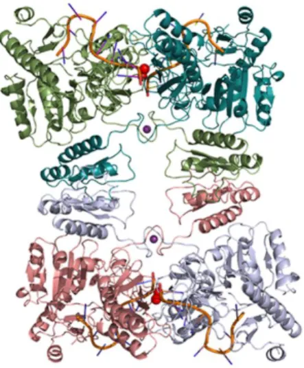
Argonaute Zwille (PAZ) domain and a C-terminal configuration like Drosha, consisting of two NucD and one dsRBD (Drider and Condon, 2004; MacRae and Doudna, 2007). Finally, the class IV is only represented, to date, by the Mini-RNase III of B. subtilis, which is constituted by a single NucD domain (Redko et al., 2008). The class I members of the RNase III family are ubiquitously found in bacteria, bacteriophages and some fungi (MacRae and Doudna, 2007). Escherichia coli RNase III has served as the prototypical member of the family. In this model microorganism, RNase III is encoded by the rnc gene, and is active as a 52 kDa homodimer (Li and Nicholson, 1996) (Figure 3). Each monomer contains a C-terminal dsRBD, located in the last 74 amino acids, which is responsible for substrate recognition and adopts a tertiary fold with the characteristic 1-β1-β
2-β3-2 structure that is conserved throughout the RNase III family (Blaszczyk et al., 2001). Additionally, each monomer contains an N-terminal NucD. When the two monomers are combined (RNase III homodimer), they form a single processing center in the subunit interface, in which each monomer contributes to the hydrolysis of one RNA strand of the duplex substrate. Ji and co-workers (Blaszczyk et al., 2004; Gan et al., 2006) resolved the structure of the hyperthermophilic bacteria Aquifex aeolicus RNase III and the data have revealed two functional forms of dsRNA binding by RNase III: a catalytic form, functioning as a dsRNA-processing enzyme, cleaving both natural and synthetic dsRNA, and a noncatalytic form, in which RNase III plays the role of a dsRNA-binding protein (without cleaving). The latter activity is in agreement with previous studies in which this enzyme binds certain substrates in order to influence gene expression (Calin-Jageman and Nicholson, 2003; Dasgupta et al., 1998; Oppenheim et al., 1993). Magnesium (Mg2+) is the preferred cofactor. It was
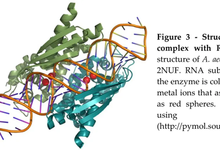
Figure 3 - Structures of RNase III in complex with RNA substrate. Crystal structure of A. aeolicus RNase III, PDB ID 2NUF. RNA substrate in complex with the enzyme is coloured in orange and the metal ions that assist catalysis are shown as red spheres. Structures were drawn
using PyMOL
(http://pymol.sourceforge.net).
The RNase III substrate selection consists of a combination of structural determinants and sequence elements referred to as reactivity epitopes, such as the helix length, the strength of base-pairing or the occurrence of specific nucleotide pairs (termed proximal and distal boxes) located at defined positions related to the cleavage site. In addition, there are two classes of double-helical elements that can function as negative determinants, which can either inhibit the recognition of this endoribonuclease or suppress the cleavage (without affecting recognition) (Pertzev and Nicholson, 2006; Zhang and Nicholson, 1997).
Regarding the maturation of rRNA, RNase III is involved in the processing of 16S and 23S rRNAs (Babitzke et al., 1993). In Salmonella and other members of Alphaproteobacteria, the enzyme is also responsible for the cleavage of the intervening sequences (IVS) found in their 23S rRNA gene (Evguenieva-Hackenberg and Klug, 2000), and in the decay of several mRNA species. For example, in E. coli, RNase III participates in the first step of the decay of pnp
endoribonuclease also has the ability to regulate its own synthesis with a specific cleavage near the 5’end of its own mRNA that removes a stem loop, which acts as a degradation barrier (Bardwell et al., 1989; Matsunaga et al., 1996). A genome-wide study in E. coli revealed a broad function of RNase III in gene regulation (Stead et al., 2011). Indeed, some studies have reported the involvement of RNase III in the control of steady-state levels of genes involved in cellular adaptation to stress (Freire et al., 2006; Santos et al., 1997; Sim et al., 2010). Additionally, this double-stranded enzyme is implicated in the decay of sRNA/mRNA complexes upon translational silencing {Viegas, 2008 #79}.
Bs-RNase III is a homologue of E. coli RNase III in B. subtilis. It is a 28-kDa protein (Mitra and Bechhofer, 1994), encoded by the rncS gene (Herskovitz and Bechhofer, 2000; Mitra and Bechhofer, 1994). Although the local environment of the site of Bs-RNase III cleavage appears to be very similar to that of E. coli RNase III, there are important differences in their substrate specificity (Mitra and Bechhofer, 1994; Wang and Bechhofer, 1997). More recently, the B. subtilis Mini-III was reported to be involved in 23S rRNA gene maturation (Redko et al., 2008). Interestingly, like Bs-RNaseIII, Mini-III does not seem to have endogenous mRNA substrates (Bechhofer, 2009). In Lactococcus lactis, RNase III plays a determinant role in the control of the stability of citQRP mRNA, which encodes for an operon involved in citrate metabolism (Drider et al., 1999; Drider et al., 1998). Complementation assays performed in E. coli showed that L. lactis RNase III can process E. coli rRNAs and regulate the levels of PNPase mRNA, substituting the endogenous RNase III (Amblar et al., 2004). The role of RNase III was even more strengthened in a recent study that showed that this enzyme in S. aureus is involved in the processing of hybridized sense/antisense RNA (Lasa et al., 2011).
considered the major RNase that catalyzes the initial rate determining cleavage of several transcripts, the RNase III family of enzymes has emerged as one of the most important groups of endoribonucleases in the control of RNA stability (Jaskiewicz and Filipowicz, 2008).
REGULATORY RNAS
The universality of regulatory RNAs playing a role in gene regulation in bacteria, primarily at the posttranscriptional level, is now well established. These elements are responsible to influence the translation and/or stability of mRNAs using several modes of action. There are many RNA regulatory elements (Waters and Storz, 2009). sRNAs mostly exert their regulatory functions by interacting with proteins or by pairing with mRNA targets. Besides sRNAs, a variety of large mRNA leaders sense environmental cues or intracellular concentrations of small metabolites to adopt structures that prevent/activate their extended transcription or translation. These RNA elements are the so-called riboswitches and thermosensors. Finally, the CRISPR RNAs are central to the defense against foreign DNA in many bacteria and archaea.
In order to understand the action of the regulatory RNAs, it is fundamental to study the processing and turnover of these molecules. Here we will give some details about the contribution of RNase E and RNase III, which are the main focus of this dissertation, into the regulatory role of some of these RNA elements.
RNase E, RNase III and small RNAs
All sRNAs participate in regulatory circuits involved in metabolism, stress adaptation and virulence. Due to bioinformatic approaches and genome-wide identification, the number of sRNAs identified in bacteria has considerably increased. The mechanisms by which sRNAs modulate gene expression are diverse, but two general modes of action have been established. sRNAs can act by interaction with a protein to modify its activity, but the most common are the antisense sRNAs that act by base-pairing with one or more targets. These antisense RNAs can be located in the complementary strand to the mRNA they regulate (cis-encoded sRNAs), containing perfect complementarity, and/or can be localised in a different genomic region apart from the target (trans-encoded sRNAs), thus they exhibit limited base-pairing. Such trans-encoded sRNAs typically require the bacterial RNA chaperone, Hfq, both for target interaction and for intracellular stability. It is generally assumed that Hfq binds both the regulator and the target RNA, favoring their interaction (Waters and Storz, 2009).
Most commonly, base-pairing between sRNA and the mRNA targets can cause the inhibition or activation of mRNA translation, mRNA stabilization or mRNA degradation (Lalaouna et al., 2013; Storz et al., 2005; Waters and Storz, 2009).
The majority of sRNAs reported in the literature are known to block translation by hybridization with the ribosome binding site (RBS) in the