Soma in Addition to the Germline
Caroline Kumsta, Malene Hansen*
Program of Development and Aging, Sanford-Burnham Medical Research Institute, Del E. Webb Neuroscience, Aging and Stem Cell Research Center, La Jolla, California, United States of America
Abstract
Gene inactivation through RNA interference (RNAi) has proven to be a valuable tool for studying gene function inC. elegans. When combined with tissue-specific gene inactivation methods, RNAi has the potential to shed light on the function of a gene in distinct tissues. In this study we characterizedC. elegans rrf-1mutants to determine their ability to process RNAi in various tissues. These mutants have been widely used in RNAi studies to assess the function of genes specifically in theC. elegans germline. Upon closer analysis, we found that two rrf-1mutants carrying different loss-of-function alleles were capable of processing RNAi targeting several somatically expressed genes. Specifically, we observed that the intestine was able to process RNAi triggers efficiently, whereas cells in the hypodermis showed partial susceptibility to RNAi in rrf-1 mutants. Other somatic tissues inrrf-1mutants, such as the muscles and the somatic gonad, appeared resistant to RNAi. In addition to these observations, we found that therrf-1(pk1417) mutation induced the expression of several transgenic arrays, including the FOXO transcription factor DAF-16. Unexpectedly,rrf-1(pk1417)mutants showed increased endogenous expression of the DAF-16 target genesod-3; however, the lifespan and thermo-tolerance ofrrf-1(pk1417) mutants were similar to those of wild-type animals. In sum, these data show thatrrf-1mutants display several phenotypes not previously appreciated, including broader tissue-specific RNAi-processing capabilities, and our results underscore the need for careful characterization of tissue-specific RNAi tools.
Citation:Kumsta C, Hansen M (2012)C. elegans rrf-1Mutations Maintain RNAi Efficiency in the Soma in Addition to the Germline. PLoS ONE 7(5): e35428. doi:10.1371/journal.pone.0035428
Editor:Ellen A.A. Nollen, University Medical Center Groningen, The Netherlands
ReceivedNovember 9, 2011;AcceptedMarch 16, 2012;PublishedMay 4, 2012
Copyright:ß2012 Kumsta, Hansen. This is an open-access article distributed under the terms of the Creative Commons Attribution License, which permits unrestricted use, distribution, and reproduction in any medium, provided the original author and source are credited.
Funding:CK is funded by an American Association for Cancer Research -Astellas Pharma Global Development Inc. Fellowship in Basic Cancer Research Grant (Grant Number: 11-40-28-KUMS - http://www.aacr.org/home/scientists/aacr-research-funding.aspx). MH is funded by National Insititutes of Health/National Insititute on Aging (Grant numbers: 1 R01 AG038664-01A1 and 1 R01 AG039756). MH is also an Ellison Medical Foundation New Scholar in Aging (Grant number: AG-NS-0481-08). The funders had no role in study design, data collection and analysis, decision to publish, or preparation of the manuscript.
Competing Interests:CK was funded by the American Association for Cancer Research. This does not alter the authors’ adherence to all the PLoS ONE policies on sharing data and materials.
* E-mail: [email protected]
Introduction
Double-stranded (ds) RNA-induced gene silencing, also called RNA interference (RNAi), was initially discovered in the nematode Caenorhabditis elegans as a defense mechanism against viruses and transposable elements. Since then, RNAi has proven to be a powerful experimental tool to gain insight into gene function in multiple organisms [1]. During RNAi in C. elegans, dsRNA serves as a trigger for sequence-specific degradation of mRNA as a mechanism of gene silencing [2,3]. The dsRNA is first cleaved into short-interfering (si) RNAs by a complex containing Dicer, an RNase III family endoribonuclease. These siRNAs are then loaded into the RNA-induced silencing complex (RISC) where they guide recognition of the target-complementary mRNA, which is subsequently cleaved and degraded. Intriguingly, only a few molecules of dsRNA are sufficient to direct degradation of a much larger population of mRNAs [4,5]. This is due to an amplification process mediated by RNA-dependent RNA polymerases (RdRPs). The RdRPs use siRNAs derived from Dicer-complex processing as primers to produce more dsRNA, with the mature endogenous mRNA as a template. These newly synthesized dsRNA molecules may also feed back into the Dicer complex to be cleaved into additional siRNA (for review, see [4,5]).
against genes expressed in the germline, but are completely resistant to muscle-specific RNAi triggers [7]. Based on these RNAi experiments, therrf-1mutant strain has become the most widely used tool in theC. elegansfield for determining the site of action of genes specifically in the germline. As an illustration, close to 50 publications to date have used the rrf-1(pk1417) allele to conclude that a gene of interest functions in the germline and not in the soma. The initial analysis of the rrf-1 strain examined muscle-expressed genes as examples of somatic gene expression, and different genes predominantly expressed in the germline [7]. Here, we describe an analysis of somatic tissues besides muscles in whichrrf-1could function.
Key advantages of using C. elegans as a model organism for genetic studies is its experimental tractability while displaying physiological complexity with multiple specialized tissues. The study of tissue-specific functions of genes using RNAi techniques requires robust tissue-selective tools. To address whether therrf-1 mutant is an efficient tool for probing the function of genes specifically in the germline, we have characterized the effect of rrf-1mutations on processing RNAi in various specialized tissues in C. elegans. We confirmed that rrf-1 mutants are able to process RNAi efficiently in the germline, and are resistant to RNAi in the muscle, as reported previously [7]. However, we found thatrrf-1 mutants are capable of processing RNAi in the intestinal soma against several endogenous genes as well as GFP reporters expressed in the intestine. We also used transgenic reporters to assess the RNAi-processing capabilities of other somatic tissues in rrf-1 mutants, and found that transgene reduction occurred in a subset of hypodermal cells, the seam cells, whereas the somatic gonad was resistant togfpRNAi. In contrast, the hypodermis was resistant to RNAi against at least two endogenous targets, suggesting that this somatic tissue may be able to process RNAi in some, but not all, cells. Taken together, these results show that therrf-1 mutants are sensitive to at least some RNAi triggers in somatic tissues, most notably in the intestine. In addition to imparting an ability to process somatic RNAi, we found that the rrf-1(pk1417) mutation also increased the expression of some transgenes in somatic tissues. Lastly, we discovered that therrf-1 mutant displayed increased endogenous expression of the FOXO transcription factor DAF-16 target gene sod-3, a superoxide dismutase, suggesting that rrf-1 mutants may have increased DAF-16 activity. While DAF-16 is a key modulator ofC. elegans longevity, the possible increase in its activity inrrf-1mutants does not translate into an extended lifespan or increased stress resistance. Our characterization of the rrf-1 mutant highlights the need for researchers to exercise caution in using this mutant as a tissue-specific RNAi tool, including in the study of longevity pathways with relevance to the FOXO transcription factor DAF-16.
Results
RNAi is Processed in the Intestine of Two Differentrrf-1
Mutants
Because we were interested in using theC. elegans rrf-1strain as a tool for tissue-specific RNAi experiments, we sought to charac-terize the potency of RNAi in various tissues in this mutant. To this end, we employed two different strategies. In the first, we selected several RNAi clones reported to be exclusively or primarily expressed in specific tissues and monitored their corresponding tissue-specific phenotypes in C. elegans. We tested these RNAi clones for effects in two differentrrf-1loss-of-function alleles; rrf-1(pk1417) and rrf-1(ok589) [7]. The pk1417 allele contains a large deletion that removes 400 amino acids in the
RRF-1 protein, including most of the conserved RdRP-specific residues [7]. Theok589allele similarly contains a 303-amino acid deletion in RRF-1, which is also presumed to be a null mutation. As a complementary approach, we crossed several transgenic strains with different tissue-specific GFP expression with the rrf-1(pk1417) loss-of-function mutant, and initiated RNAi by feeding these new strains bacteria expressinggfp dsRNA. Using these two approaches, we initially reexamined the two tissues that previously had been investigated in rrf-1 mutants, namely the germline and body-wall muscle [7]. As expected, we found that bothrrf-1mutants were capable of processing endogenous andgfp RNAi triggers in the germline (Figure S1) but not in the muscles (Figure S2), as shown in [7].
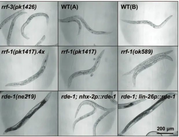
We next tested the ability of therrf-1strain to process RNAi in the intestine. The intestine is the tissue that takes up dsRNA when delivered by ingestion of bacteria, and might therefore be especially sensitive to RNAi by feeding. To test this, we searched for genes that were expressed only in the intestine, and would induce an easily detectable intestinal phenotype upon RNAi-mediated silencing. The GATA-type transcription factor ELT-2 fit these criteria as it is expressed exclusively in the intestine, where it is required for intestinal differentiation [9]. Accordingly, theelt-2mutation is known to cause lethality at the L1 stage, with animals displaying a blocked intestinal lumen [9,10]. Consistent with a role for elt-2in the intestine, we found that wild-typeC. elegansandrrf-3(pk1426)mutants (which display enhanced somatic RNAi sensitivity due to a loss-of-function mutation in the RdRP RRF-3 [8]), both experienced constricted and occluded intestines and a clear appearance when subjected toelt-2RNAi (Figure 1). Moreover, we found thatrde-1(ne219)mutants (which are resistant to RNAi due to a loss-of-function mutation in the Argonaut protein RDE-1, a key component of the RISC complex [11]) expressing RDE-1 under the intestinal nhx-2 promoter [12], responded to elt-2RNAi similarly to the wild-type animals (Figure 1). As expected, a strain expressing RDE-1 in the hypodermis (rde-1(ne219); lin-26p::rde-1) was unaffected by elt-2 RNAi [13] (Figure 1). Notably, when we subjected either rrf-1(pk1417)orrrf-1(ok589) mutants toelt-2RNAi, we observed comparable phenotypes to those observed in wild-type animals grown on bacteria expressingelt-2dsRNA (Figure 1), implying thatrrf-1mutants are capable of processingelt-2RNAi.
RNAi is Processed in the Intestine ofrrf-1(pk1417)
Mutants Expressing Different GFP Reporters
To further investigate the finding thatrrf-1mutants can process RNAi in the intestine, we employed two different transgenic strains with strong GFP expression in the intestine. The first strain overexpresses a GFP-tagged version of the protein LGG-1, a homolog of the mammalian protein LC3, with prominent expression observed in the intestine, pharynx, hypodermis, and somatic gonad [16]. When we fed bacteria expressinggfpdsRNA to anrrf-1(pk1417)strain carrying thelgg-1reporter, we observed a strong reduction in intestinal GFP that was readily apparent by day 1–2 of adulthood (Figure 2A). To exclude the possibility that GFP silencing depended on thelgg-1transgene, we used a second reporter strain with strong intestinal GFP fluorescence. This strain expresses the FOXO transcription factor DAF-16 with an N-terminal GFP tag, under the control of thedaf-16promoter [17]. When we subjected the rrf-1(pk1417) strain carrying the daf-16 reporter construct to gfp RNAi, this treatment resulted in a noticeable albeit partial loss of intestinal GFP expression compared to wild-type animals (Figure 2B). Taken together, our results demonstrate that C. elegans rrf-1 mutants are at least partially capable of processing RNAi in the intestinal soma.
The Hypodermis ofrrf-1Mutants shows Mixed Susceptibility to RNAi
To determine if other somatic tissues were able to process RNAi in rrf-1 mutants, we next investigated the hypodermis, a tissue comprised of multiple cell types that establish the basic body form and which secretes the cuticle of the animal. We tested RNAi processing in the hypodermis by inhibiting different genes that are reported to be exclusively or primarily expressed in the hypodermis. The dual oxidase BLI-3 generates hydrogen peroxide required for the crosslinking of collagen dityrosines during cuticle formation [18]. RNAi-induced reduction of the bli-3gene, reported to be expressed exclusively in the hypodermis, led to an extreme molting defect in the rrf-3(pk1426) strain, in wild-type animals as well as in hypodermally rescued rde-1 mutants (Figure 3A). Interestingly, the intestinally rescued rde-1mutant was similarly affected bybli-3dsRNA, suggesting thatbli-3may be more broadly expressed than reported. Importantly, we observed that the rrf-1(pk1417) and rrf-1(ok589) strains were largely unaffected by bli-3 RNAi (Figure 3A and Figure S4A), even when treated for multiple generations (data not shown). Therrf-1 strains were similarly resistant to RNAi against another gene reported to be expressed exclusively in the hypodermis, the membrane protein tetraspanin encoded bytsp-15[19]. We found thatrrf-1mutants subjected totsp-15RNAi were resistant to blister formation and defects in body morphology, whereastsp-15 RNAi-treated wild-type animals were severely affected (data not shown). Figure 1. Intestinalelt-2RNAi induces prominent phenotypes inrrf-1mutants.The indicatedC. elegansstrains were raised on bacteria expressingelt-2dsRNA and imaged by DIC microscopy on day 3 of adulthood. The rde-1strain and therde-1strain expressing RDE-1 in the hypodermis (lin-26p::rde-1) were unaffected by RNAi, whereas all other strains tested show growth defects, a clear appearance, and an abnormal intestine. This experiment was repeated four times with,100 worms per strain with similar results. WT: wild-type N2 (A – Hansen lab, B – Tuck lab), rrf-1(pk1417).4x: rrf-1(pk1417)outcrossed 4 times to WT(A).
In contrast to our analysis ofbli-3and tsp-15, RNAi against two other genes that are largely expressed in the hypodermis, bli-4 (subtilisin-like serine endoprotease) anddie-1(zinc finger protein), induced comparable phenotypes in rrf-1 mutants and wild-type animals (Figure S4A, B). However, the interpretation of these results was compromised by the fact thatbli-4 anddie-1 are not exclusively expressed in the hypodermis, but are also present in other somatic tissues including the intestine. Consistent with this notion, we found that both bli-4 and die-1 RNAi induced phenotypes in both hypodermally and intestinally rescued rde-1
strains (Figure S4B and data not shown). Taken together, our analysis of endogenous genes does not support an RNAi-processing ability of the hypodermis.
To extend these findings, we analyzed RNAi processing in seam cells, a specialized hypodermal cell type important for the formation of cuticular structures called alae. For these studies, we introduced a seam cell-specific GFP reporter into the rrf-1(pk1417) mutant and compared the GFP signal in these mutants and wild-type animals after they had been fed from hatching with bacteria expressing gfp dsRNA. Interestingly, we Figure 2. GFP reporters in the intestine ofrrf-1mutants are repressed bygfpRNAi.The indicatedC. elegansstrains were raised on bacteria expressinggfpdsRNA, and imaged by DIC and fluorescence microscopy (overlays shown here) on day 1 of adulthood.rrf-1mutants carried the
pk1417allele. (A) Thelgg-1p::gfp::lgg-1reporter is expressed in the intestine, pharynx, somatic gonad, and hypodermis. GFP expression is dramatically reduced upon treatment withgfpRNAi in both wild-type andrrf-1animals. The exposure time for the GFP channel under control conditions was 15 ms and forgfpRNAi 25 ms. In thegfpRNAi micrographs, note that the pharynx appears largely unaffected by RNAi in both wild-type and inrrf-1
Figure 3.rrf-1mutants are resistant to RNAi against the hypodermal genebli-3, but are able to processgfpRNAi in seam cells.The indicatedC. elegansstrains were raised on bacteria expressingbli-3 dsRNA (A) andgfp dsRNA (B) and imaged by bright-field or fluorescence microscopy, respectively, on day 1 of adulthood. (A) Therrf-1mutants, as well as the RNAi-resistantrde-1strain are unaffected bybli-3RNAi, whereas all other strains tested show severe molting defects and varying degrees of blister formation. See Figure S4A for additional information. This experiment was repeated three times with 100–200 worms per strain with similar results, also for several generations. WT: wild-type N2 (A – Hansen lab, B – Tuck lab),rrf-1(pk1417).4x: rrf-1(pk1417)outcrossed 4 times to WT(A). (B) TheSCMp::gfpreporter is exclusively expressed in hypodermal seam cells, which are denoted with arrows. GFP expression is abolished in the wild-type background and reduced in therrf-1(pk1417)background. Therrf-1
mutant shows visibly higher GFP expression levels compared to the wild-type strain. The exposure time for the GFP channel was 100 ms. See Figure S4C for quantification of transgene intensity. These experiments were repeated 3 times with 30–50 worms per strain and imaging of ,10 per experiment, with similar results.
observed that, similar to wild-type animals,rrf-1(pk1417)mutants had a significantly reduced GFP signal in their seam cells in response togfpRNAi(Figure 3B and Figure S4C). Thus, in our transgenic analysis we observed that hypodermal seam cells were at least partially capable of processing gfp RNAi, which was in contrast to the results we obtained when analyzing endogenous genes exclusively expressed in the hypodermis. Taken together, these experiments highlight the possibility that a restricted subset of cells in the hypodermis is able to process RNAi.
The Somatic Gonad is Resistant to RNAi in rrf-1(pk1417)
Mutants
Because therrf-1(pk1417)mutant has been used in many studies to determine whether a gonadal gene functions in the germline or in the somatic gonad, we were specifically interested in determin-ing if the somatic gonad ofrrf-1mutants was able to process RNAi. To do this, we examined the persistence of GFP expression after feedinggfpRNAi to therrf-1(pk1417) strain carrying alag-2p::gfp transcriptional reporter strain that displays GFP fluorescence in the distal tip cell (DTC) [20]. In the rrf-1background, the GFP signal in the DTC persisted upon treatment withgfpRNAi, which is consistent with previous reports that somatic tissues of rrf-1 mutants are resistant to RNAi (Figure S5A). Moreover, GFP fluorescence was maintained in the spermatheca and vulva when rrf-1(pk1417)animals expressing thelgg-1p::gfp::lgg-1reporter were treated withgfpRNAi (Figure S5B). Taken together, these results indicate that the somatic gonad may be resistant to RNAi inrrf-1 mutants.
Somatic RNAi Effects inrrf-1(pk1417)Mutants are not Due to the Negative RNAi Regulator ERI-6/7
It is possible that the ability ofrrf-1mutants to process RNAi in at least some somatic tissues is not an intrinsic feature of therrf-1 mutation, but instead could be due to mutations in negative regulators of the RNAi pathway in that strain [4]. For example, disruption of the adjacent genes eri-6 and eri-7 can lead to an enhanced RNAi response [4]. To exclude the possibility that knowneri-6/eri-7background mutations have accumulated in the rrf-1strains used in this study, we amplified and sequenced the eri-6/eri-7 locus. Neither of the two rrf-1 alleles carried the mutation mg441 (data not shown), a mutation that leads to an enhanced RNAi response due to erroneous splicing and genomic rearrangements in theeri-6/7locus (Figure S6) [4]. In addition, we designed primers flanking three other reported deletion mutations (tm1887, tm1716, tm1917), and found no evidence that therrf-1strains used in this study harbored any of these reported eri-6/7 mutations (Figure S6). While this analysis does not exclude the possibility that unidentified RNAi-enhancing muta-tions exist in therrf-1mutant, our results are consistent with the rrf-1 strain possessing an intrinsic ability to process dsRNA in somatic tissues, in addition to the germline.
rrf-1(pk1417)Mutants Display Elevated Expression of Transgenic Arrays
During the course of our experiments with thedaf-16p::gfp::daf-16 reporter strain, we noticed a significantly higher expression of the transgenic array in therrf-1(pk1417)background compared to the wild-type background (Figure 2B and Figure S7A, B). This significant increase in GFP fluorescence is likely due to post-translational regulation, since daf-16mRNA levels were not significantly elevated in rrf-1(pk1417) mutants (Figure S7C), consistent with a previous report [24]. Notably, we also observed significantly increased expression of GFP when driven by a seam
cell-specific promoter (Figure 3B and Figure S4C) and by the DTC-specific lag-2 promoter (Figure S5C). These observations suggest that therrf-1(pk1417)mutation can cause a general increase in the expression of transgenic arrays. However, we were not able to detect an increase in transgene expression inrrf-1(pk1417)mutants expressing GFP-tagged histone 2B under the germline-specificpie-1 promoter (Figure S1B), GFP::MYO-3 from the muscle-specific myo-3 promoter (Figure S2), or GFP::LGG-1 from the lgg-1 promoter(Figure 2A and Figure S5D),suggesting that therrf-1 mutation does not have these regulatory effects on all transgenic arrays.
The Expression of DAF-16/FOXO Target Genesod-3is Elevated inrrf-1(pk1417)Mutants
The FOXO transcription factor DAF-16 plays a major role in modulating longevity in C. elegans, most notably through the insulin/IGF-1 signaling pathway [21]. This is likely achieved by the DAF-16–dependent expression of several genes involved in critical biological processes, including stress resistance, metabo-lism, and disease pathogenesis [21,22,23]. Since DAF-16 plays a prominent role in longevity modulation, we investigated the role of rrf-1 ondaf-16/FOXO in more detail. Since we found that the rrf-1(pk1417) mutation had no effect on daf-16mRNA levels (Figure S7C), we asked ifrrf-1mutations affect DAF-16 activity. Superoxide dismutasesod-3is a DAF-16 target gene that is often used as a measure of transcriptional activation of DAF-16 [22,25]. We compared the expression of endogenoussod-3levels in wild-type animals and in rrf-1(pk1417) mutants by qPCR and unexpectedly, we observed significantly elevated sod-3mRNA levels in rrf-1(pk1417) mutants compared to wild-type animals (Figure 4A). Similarly, we found that rrf-1(pk1417) mutants displayed increased expression of a sod-3p::gfp transcriptional reporter (Figure S7D, E). While we recognize that the rrf-1(pk1417) mutation could directly affect expression of the transgene, as noted above, this result is consistent with the qPCR data measuring endogenous sod-3 levels. Because DAF-16 is a major transcriptional regulator ofsod-3 expression in C. elegans, these observations raise the possibility that DAF-16 activity is elevated inrrf-1mutants.
rrf-1(pk1417)Mutants do not show Extended Lifespan or Increased Thermo-tolerance
Increased DAF-16 activity has been associated with longevity in animals with reduced levels of the insulin/IGF-1 like receptor DAF-2 [21,26]. daf-2 mutants are long-lived and resistant to various stressors, such as heat and oxidative stress [21,26]. Since DAF-16 activity may be increased inrrf-1mutants, we examined whether rrf-1 mutations extend lifespan and promote stress resistance in rrf-1 animals. However, we found no difference between rrf-1(pk1417) mutants and wild-type animals in their mean lifespan (Figure 4B and Figure S7) or their ability to withstand elevated temperature (Figure 4C). Thus, althoughrrf-1 mutants display increased sod-3 transcript levels, indicative of elevated DAF-16 activity, these alterations were not sufficient to increase longevity or stress resistance inrrf-1mutants.
Discussion
Here, we have shown thatC. elegans rrf-1mutants are capable of processing RNAi targeting several genes expressed in the soma (summarized inTable 1).
rrf-1(pk1417)mutants after being exposed togfpRNAi. In addition, we found that RNAi of two endogenous targets, elt-2and pept-1, induced robust phenotypes in two differentrrf-1 mutants. While elt-2RNAi seemed to play an exclusively intestinal-specific role, pept-1RNAi induced effects in both intestinally and hypodermally rescuedrde-1strains [12,13]. This result may indicate thatpept-1 functions as an oligopeptide transporter not only in the intestine, but also in the hypodermis. Alternatively, we note the possibility
that the rescued rde-1 strains are not as specific in their RNAi sensitivity as anticipated.
We also observed RNAi-processing capabilities in another somatic tissue, the hypodermis, albeit to a limited extend. We found that rrf-1 mutants expressing a transgenic reporter in a specific subset of hypodermal cells, i.e., the seam cells were susceptible to knockdown ofgfp. In contrast,rrf-1mutants failed to respond to RNAi against bli-3 ortsp-15, which are both genes reported to be expressed exclusively in the hypodermis. Moreover, we found that RNAi against additional genes with hypodermal expression (bli-4 and die-1) induced similar phenotypes in both wild-type animals and rrf-1 mutants. While the phenotypes induced bybli-4anddie-1RNAi inrrf-1 mutants could originate from the knockdown of these genes in other somatic tissues, including the intestine (Table 1), it seems that the hypodermis of rrf-1 mutants is not generally capable of processing dsRNA triggers. Instead, our findings suggest that either a restricted subset of cell types in the hypodermis of rrf-1 mutants is capable of processing RNAi, or perhaps only some endogenous targets can be degraded. Further analysis of endogenous targets, especially expressed in hypodermal seam cells is warranted to address this possibility.
Taken together, our analysis indicates that rrf-1 mutants can process RNAi in the soma, most notably in the intestine, demonstrating that rrf-1 mutants have broad RNAi-processing capabilities. Consistent with this notion, new information on the expression pattern of the predominantly germline-specific genes pos-1,par-1andpop-1originally tested by Sijen et al. [7], suggests that these genes are also expressed in somatic tissues [27,28,29,30], which further corroborates an RNAi-processing role in the soma ofrrf-1mutants.
These findings have important implications for the use of this strain in tissue-specific RNAi studies, as well as for the interpretation of published reports using RNAi in this strain. Our literature search revealed that close to 50 studies used the rrf-1(pk1417)mutant to investigate the role of a gene of interest in the germline versus somatic tissues. However, several of these publications used additional and alternative methods to confirm a germline requirement for the gene of interest, such as mosaic analysis [31,32,33,34,35]. Indeed, most of the published studies usedrrf-1(pk1417)mutants to determine if a gene expressed in the gonad has a specific role in the germline or the somatic gonad [36,37,38]. Our studies indicated that therrf-1mutant is resistant to RNAi in the somatic gonad by using two differentgfp-tagged reporters. Specifically, we found that the GFP signal was maintained in the vulva and spermatheca of an rrf-1; lgg-1p::gfp::lgg-1reporter strain, as well as in distal tip cells of an rrf-1; lag-2p::gfp reporter strain (Table 1). Ideally, these experi-ments should be complemented by an analysis of endogenous genes; however, we have been unsuccessful in inducing phenotypes in wild-type animals using RNAi against the endogenous genes we investigated, includinglag-2(data not shown). Based on our results, the rrf-1 strain may thus be a useful tool for determining a differential requirement for a gene in the germline and somatic gonad.
RRF-1 is one of two RdRPs involved in the exogenous siRNA pathway that amplifies dsRNA triggers, and has been reported to act in the soma ofC. elegans[7]. In contrast, the second RdRP, EGO-1, is thought to be required for processing of siRNA targeting germline-specific genes [6]. Our observation that the rrf-1mutant is capable of processing RNAi in somatic tissues could suggest the existence of unidentified enzymes that have overlap-ping functions with RRF-1, or suggest a role for EGO-1 outside of the germline. Although Smardon et al. have reported that EGO-1 Figure 4. rrf-1 mutants have increasedsod-3mRNA levels, a
normal lifespan, and normal thermo-tolerance. (A) The sod-3mRNA levels of a mixed population ofC. eleganswild-type (WT, N2(A)) and rrf-1(pk1417).4x (rrf-1(pk1417) mutant outcrossed four times to WT(A)) strains were determined. Bars show the mean+SEM of three independent experiments; **P,0.005 (Student’s t-test). (B) Lifespan analysis of wild-type WT(A) andrrf-1(pk1417).4xstrains at 20uC; P = 0.59, log-rank (Mantel-Cox test). This experiment has been performed three times with similar results; see Figure S7F for additional data. (C) Thermo-tolerance was measured by assessing survival of WT(A) and rrf-1(pk1417).4xstrains after 8 h incubation at 36uC; P = 0.36 (Student’s
t-test). This experiment has been performed three times with similar results.
expression is primarily germline-specific [6], it may also be expressed in somatic tissues. Moreover, germline EGO-1 activity may be sufficient to produce enough siRNA to affect other tissues. Alternatively, RRF-2 or an unidentified RdRP may play roles in processing RNAi in the absence of RRF-1.
One of the striking features of RNAi inC. elegansis its systemic nature, in which the dsRNA spreads from the tissue of uptake, most often the intestine, into other tissues [39,40,41,42]. In our experiments, the dsRNA trigger was delivered by feeding the animals bacteria expressing dsRNA. Therefore, it is possible that sufficient dsRNA was ingested to at least partially knock down the targeted mRNA in the intestine without the need for an mediated amplification reaction, making the mechanism RdRP-independent. This may also be the case for the worm pharynx, which processes bacteria upon intake. Although we have attempted to directly address a role for the pharynx using endogenous genes as RNAi targets, these experiments have been hampered by the apparent refractory nature of the pharynx to RNAi (seeFigure 2A, and data not shown). While our objective in this study was to characterize therrf-1strain for processing of
RNAi delivered by feeding, future studies should determine if direct injection of the dsRNA into the germline have the same effect on somatic gene expression ofrrf-1mutants, including in the intestine. Perhaps direct delivery of dsRNA to the germline would reduce exposure of the intestine and other tissues to dsRNA and might even prevent the degradation of mRNAs encoded by intestinal-specific genes. These additional tissues may include ones we have not yet assayed, for example neurons, which are generally considered refractory to RNAi by feeding [43,44].
Unexpectedly, we observed that rrf-1 mutants displayed increased expression of an important target gene of the FOXO transcription factor DAF-16, the superoxide dismutase SOD-3. Since DAF-16 is a major regulator ofsod-3,this observation raises the possibility thatrrf-1mutants have increased activity of DAF-16. DAF-16 activity is required for the extended longevity of many C. elegansmutants, includingdaf-2mutants [21]. Indaf-2mutants, these phenotypes are mediated by nuclear translocation and activation of DAF-16, which in turn causes upregulation of target genes such as sod-3. We did not, however, observe nuclear localization of DAF-16 in therrf-1background (Figure 2A and Table 1.RNAi efficiency in different tissues inrrf-1mutants.
Tissue investigated
Gene or transgene
targeted by RNAi Reported tissue of expression RNAi phenotype
wild-type rrf-1 Observed Citatio n1
Germline gld-1 Germline + + Pro, Tum [52]
pie-1p::gfp::H2B Germline + + GFP loss [53]
Muscle unc-22 Muscle + 2 Unc/Twitcher [39]
pat-4 Body-wall muscle, spermatheca, DTC, vulval and anal sphincter muscle, mechanosensory neurons
+ 2 Prz [54]
unc-112 Body-wall, vulval, spermathecal, uterine, and anal sphincter/ depressor muscles
+ 2 Prz [55]
myo-3p::gfp::myo-3 Body-wall muscle + 2 GFP loss [56]
Intestine elt-2 Intestine + + Gob, Clr, Lva [10]
pept-1 Intestine + + Clr, Gro [14]
bli-4 Hypodermis, intestine, vulva, ventral nerve cords + + Bli, Dpy [28,57] daf-16p::gfp::daf-162 Intestine, hypodermis, gonad, body-wall muscles, neurons
+ + GFP loss [58]
lgg-1p::gfp::lgg-13 Intestine, hypodermis, pharynx, gonad, neurons, body-wall
muscles
+ + GFP loss [16]
Hypodermis bli-3 Hypodermis + - Bli, Dpy [18]
tsp-154 Hypodermis
+ - Bli, Dpy [19]
bli-4 Hypodermis, intestine, vulva, ventral nerve cords + + Bli, Dpy [28,57]
die-1 Embryonically in hypodermis, pharyngeal cells and muscle/gut primordium precursor cells
+i +5 Bmd [59]
SCMp::gfp Seam cells + +/26 GFP loss [60]
Somatic gonad lag-2p::gfp Distal tip cell + 2 GFP loss [61]
lgg-1p::gfp::lgg-1 Intestine, hypodermis, pharynx, gonad, neurons, body-wall muscles
+ 2 GFP loss [16]
‘+’ and ‘2’ refer to the presence or absence of a phenotype in response to RNAi treatment in wild-type orrrf-1animals. Therrf-1(pk1417), rrf-1(pk1417).4x (rrf-1(pk1417) four times outcrossed to WT(A)andrrf-1(ok589)mutants behaved similarly to RNAi targeting endogenous genes and are combined here asrrf-1. For strains expressing GFP-tagged transgenes, loss of GFP expression was assayed in response togfpRNAi in therrf-1(pk1417)mutant only.
Phenotype abbreviations: Pro: proximal germ cell proliferation abnormal; Tum: tumorous germline; Prz: paralyzed, Unc/Twitcher: uncoordinated, sub-class, Twitcher; Gob: gut-obstructed; Clr: clear; Lva: larval arrest; Gro: slow growth; Ste: sterile; Bli: blistered; Dpy: dumpy; Bmd: body morphology defects; DTC: distal tip cell. 1Citations refer to studies describing expression patterns and gene functions of the genes targeted by RNAi and to descriptions of the transgenes used; 2Only the intestinal expression ofdaf-16p::gfp::daf-16was examined;
3
The intestinal, pharyngeal, and gonadal expression oflgg-1p::gfp::lgg-1was examined; 4Data not shown;
Figure S7A, and data not shown). Additionally, we found no increase in longevity or thermo-tolerance in the rrf-1(pk1417) mutant, suggesting that if rrf-1 mutants have increased DAF-16 activity, it is not sufficient to extend lifespan. This would be consistent with the observation that increased expression of SOD-3 does not extendC. eleganslifespan [45]. It remains to be explored how inhibition of the RdRPrrf-1leads to increased expression of SOD-3, and whether other DAF-16 targets are similarly affected. Regardless of the exact mechanism, our novel findings thatrrf-1 mutants show efficient RNAi in somatic tissues and also exhibit increased expression of the DAF-16-regulated stress response gene sod-3,should be considered before using these mutants in tissue-specific RNAi studies, especially in the study of longevity pathways with relevance to the FOXO transcription factor DAF-16.
In addition to observing a new somatic RNAi-processing ability and increased expression of endogenous sod-3, we also noted that multiple, but not all, transgenes used in our investigation were expressed at higher levels in rrf-1 mutants. These observations suggest that RRF-1 can negatively regulate transgene expression, at least in some cases. Notably, we observed no direct correlation between the tissues in which transgenic expression was increased byrrf-1mutation and the cell types that responded to RNAi processing. Moreover, the daf-16p::gfp::daf-16 and lgg-1p::gfp::lgg-1 reporters both responded togfpRNAi in the intestine when expressed inrrf-1mutants, but only the daf-16p::gfp::daf-16reporter was notably upregulated by the rrf-1 mutation. This suggests that the mechanism regulating transgene expression in rrf-1 mutants is not identical to that of RNAi processing. While the mechanism of transgene regulation is currently unclear, it is an important result because the increased expression of specific transgenes inrrf-1 mutants could induce morphological or cellular changes. Indeed, rrf-1 mutants may display many more phenotypes than noted previously or reported here, and this information should be kept in mind when using this mutant as a tool for studying gene function.
Materials and Methods
Strains and Culture Conditions
Strains were maintained and cultured under standard condi-tions at 20uC usingE. coliOP50 as a food source [46], except when subjected to RNAi treatment. We used these previously reported strains in this study (seeTable 1for references):
WT(A): Hansen-lab N2, WT(B): Tuck-lab N2 (Simon Tuck, University of Umea˚), NL2098: rrf-1(pk1417) I, RB798: rrf-1(ok589) I, NL2099: rrf-3(pk1426) II,
WM27: rde-1(ne219) V,
VP303: rde-1(ne219) V; kbIs7[nhx-2p::rde-1+rol-6(su1006)], NR222: rde-1(ne219) V; kbIs9[lin-26p::nls::gfp, lin-26p::rde-1+ rol-6(su1006)],
DA2123: adIs2122[lgg-1p::gfp::lgg-1+rol-6(su1006)], CF1934: daf-16(mu86) I; muIs109[daf-16p::gfp::daf-16 + odr-1p::rfp],
AZ212: unc-119(ed3) III; ruIs32[unc-119(+) pie-1(+) pie-1p::gfp::H2B],
RW1596: myo-3(st386) V; stEx30[myo-3p::gfp::myo-3 + rol-6(su1006)],
CF1553: muIs84[sod-3p::gfp],
JK2049: qIs19[lag-2p::gfp+rol-6(su1006)] V,
JR667: unc-119(e2498::Tc1) III; wIs51[SCMp::gfp+unc-119]. We note that the promoter of the seam-cell marker (SCM) used for GFP expression in the JR667 strain has not been reported.
We created the following new strains for this study:
MAH19: rrf-1(pk1417) I, myo-3(st386)V; stEx30[myo-3p::gfp:: myo-3+rol-6(su1006)],
MAH22: rrf-1(pk1417) I; adIs2122[lgg-1p::gfp::lgg-1 + rol-6(su1006)],
MAH23: rrf-1(pk1417) I outcrossed 4 times to Hansen-lab N2 and referred to as rrf-1(pk1417).4x,
MAH74: rrf-1(pk1417) I; unc-119(ed3) III; ruIs32[unc-119(+) pie-1(+) pie-1p::gfp::H2B],
MAH97: rrf-1(pk1417) I; muIs109[daf-16p::gfp::daf-16 + odr-1p::rfp],
MAH99: rrf-1(pk1417) I; muIs84 [sod-3p::gfp],
MAH132: rrf-1(k1417) I; unc-119(e2498::Tc1) III; wIs51[SCMp::gfp+unc-119],
MAH133: rrf-1(pk1417) I; qIs19[lag-2p::gfp+rol-6(su1006)] V. We note that we observed germline defects in 5–10% of the assayed animals of the rrf-1(pk1417); lag-2p::gfp strain (data not shown), whereas no defects were observed when this transgene was expressed in the wild-type background, or in the rrf-1(pk1417) mutant without expression of the lag-2p::gfp reporter, or in the rrf-1(pk1417); pie-1p::gfp::H2Bstrain (data not shown).
RNAi Treatment and Imaging
ThegfpRNAi clone was obtained from Dr. Andrew Dillin (Salk Institute). Thebli-4andelt-2clones used were obtained from the Vidal RNAi library [47]; the elt-2, pept-1, pat-4, unc-112, pha-4, gld-1 and bli-3 RNAi clones were obtained from the Ahringer library [48]. All RNAi clones were verified by sequencing. For the RNAi treatment, worms were synchronized by hypochlorous acid treatment and the eggs seeded on the NGM plates with bacteria expressing the dsRNA. Differential interference contrast (DIC) microscopy was performed on a Zeiss Imager Z1 after mounting worms on a 2% agarose pad containing 0.1% NaN3, in M9
medium also containing 0.1% NaN3. Worms on plates were
imaged on plates with a Leica DFC310 FX camera. For the analysis of GFP expression, fluorescence images were collected on a Zeiss Imager Z1 at the exposure times indicated in the figure legends. Image analysis was performed with ImageJ software (National Institutes of Health), by tracing the intestine (for strains expressingdaf-16p::gfp::daf-16 and sod-3p::gfp) or the entire worm (lgg-1p::gfp::lgg-1 expressing strains) and measuring the integrated density. The analysis of strains expressinglag-2p::gfpwas performed by tracing a defined circular DTC-encompassing region of interest and measuring the mean density. For the analysis of the GFP intensity of the seam cells, 1–5 regions of interest encompassing 2360 pixels were drawn around the single seam cell bodies and the average mean density per worm was calculated. For the analysis of the germline overproliferation following gld-1 RNAi treatment, worms were washed off the plates on day 3 of adulthood and washed three times in M9 media before fixing in methanol overnight at -20uC. For DAPI staining, methanol was removed by washing with M9 media, 0.1mg/ml DAPI in M9 was added, and
samples were incubated for 20 min on a slow nutator. Worms were washed with M9 and then mounted on agarose pads for imaging. Statistical analyses were performed by Student’s t-test and two-way ANOVA using GraphPad Prism (La Jolla, CA). If RNAi was induced for a second generation, the eggs of the first generation of animals were transferred to new RNAi plates and these animals were then scored. All experiments were carried out at 20uC.
Quantitative RT-PCR
were flash frozen in liquid nitrogen. RNA was extracted using Trizol (Life Technologies, Grand Island, NY) and purified using the Qiagen RNeasy kit (Valencia, CA), including an additional DNA digest step using the Qiagen Dnase I kit. The M-MuLV reverse transcriptase and random 9-mer primers (New England Biolabs, Ipswich, MA) were used for reverse transcription of 1mg
of RNA per sample [49]. Quantitative PCR was run using SYBR Green Master Mix in a Roche LC480 LightCycler (Indianapolis, IN). A standard curve was run for each primer on serial dilutions of a mixture of different cDNAs, and the observed CT values
were converted to relative values according to the primers’ standard curves. The mRNA levels of target genes were normalized to those of the housekeeping genes cyclophilin ( cyn-1) and nuclear hormone receptornhr-23. Primer information is available upon request.
Lifespan Analysis
Lifespan was measured at 20uC with at least 90 worms in each experiment [50]. Animals were scored as dead when they failed to respond to gentle prodding with a platinum wire pick. Censoring occurred if animals desiccated on the edge of the plate, escaped, ruptured, or suffered from internal hatching. For statistical analysis Stata software was used (StataCorp, College Station, TX). P values were calculated with the log-rank (Mantel-Cox) method.
Thermo-tolerance Assay
The survival ofC. elegansat elevated temperatures was measured after an 8 h incubation at 36uC [51]. The synchronized wild-type andrrf-1(pk1417)animals were transferred to assay plates on day 1 of adulthood and the thermo-tolerance experiment was performed on day 3 of adulthood. This experiment was performed at least 3 times with similar results. Student’st-test was used to calculate P values.
PCR Assay for eri-6/7Mutations and Genomic Rearrangement
Single worms were used in these PCR assays. Worms were lysed in 10ml proteinase K buffer (1mg/ml proteinase K, 1x PCR buffer, H2O) at 65uC for 1 h and 95uC for 15 min. Taq polymerase was
used to amplify the PCR product for 35 cycles with an annealing temperature of 58uC. Primer sequences were: P1 (MH439): 59 -GCCAGTGCTTCACGTGTC-39, P2 (MH440): 59-CGAAGCC-CAGATGTGAGATC-39 P3 (MH479): 59 -TAAGTTTTCC-TAAAATGATTTC-39. This protocol was adapted from [4].
Supporting Information
Figure S1 rrf-1mutants are capable of processing RNAi
in the germline.(A) The indicatedC. elegansstrains were raised on bacteria expressinggld-1dsRNA, fixed in methanol on day 3 of adulthood, and subsequently stained with DAPI and imaged by fluorescence microscopy. All strains display mitotic germ cell overproliferation except the RNAi-resistant rde-1 strain, which shows typical mitotic and meiotic zones in the distal end of the gonad, and eggs in utero in the proximal part of the gonad. This experiment was repeated twice with 50–100 worms per strain with similar results. WT: wild-type N2 (A – Hansen lab, B – Tuck lab), rrf-1(pk1417).4x: rrf-1(pk1417) outcrossed 4 times to WT(A). (B) The indicatedC. elegansstrains were raised on bacteria expressing gfp dsRNA and imaged by DIC and fluorescence microscopy (overlays shown here) on day 1 of adulthood.rrf-1mutants carried thepk1417allele. Thepie-1p::gfp::H2B reporter is expressed in the germline of C. elegans. GFP expression is completely abolished upon treatment with gfp RNAi, in both the wild-type and rrf-1
backgrounds. The image shows one arm of the gonad including oocytes, and was acquired with an exposure time of 100 ms. This experiment was repeated twice with 30–50 worms per strain and imaging of,10 per experiment, with similar results.
(TIF)
Figure S2 rrf-1 mutants are resistant to RNAi in the
muscle. (A) The indicated C. elegans strains were raised on bacteria expressing gfp dsRNA, and imaged by fluorescence microscopy on day 1 of adulthood. rrf-1 mutants carried the pk1417 allele. The body-wall muscle-specific GFP expression is maintained in therrf-1 background, but completely abolished in the wild-type background. The exposure time for the GFP channel was 200 ms. This experiment was repeated twice with similar results. (B) The indicated C. elegans strains were raised from hatching on bacteria expressing muscle-specificunc-112,pat-4,and unc-22RNAi and were assayed for paralysis and twitching on day 1 of adulthood. The experiments have been repeated at least two times with similar results. WT: wild-type N2 (A – Hansen lab, B – Tuck lab), rrf-1(pk1417).4x: rrf-1(pk1417) outcrossed 4 times to WT(A). Phenotype abbreviations: Prz: paralyzed; Unc: uncoordi-nated, sub-class, Twitcher. n.a.: not assayed.
(TIF)
Figure S3 Intestinalpept-1RNAi induces phenotypes in
rrf-1 mutants. The indicatedC. elegans strains were raised on bacteria expressing pept-1 dsRNA, and imaged by bright-field microscopy on day 1 of adulthood. WT: wild-type N2 (A – Hansen lab, B – Tuck lab), rrf-1(pk1417).4x: rrf-1(pk1417) outcrossed 4 times to WT(A). (A) The rrf-1animals were smaller and have a clear appearance, whereas the wild-type and hypersensitive RNAi strain rrf-3 were developmentally delayed. (B) Quantification of the mid-body diameter of all the strains shows that all strains are significantly smaller than the unaffected rde-1 strain. This experiment has been repeated three times with similar results. Size quantification was determined with 7–10 worms per strain. Bars show the mean+ SEM of one representative experiment. P,0.0001 was determined with one-way ANOVA, and individual significance was determined with Bonferroni’s multiple compar-ison test: **P,0.005 ***P,0.0005.
(TIF)
Figure S4 rrf-1 mutants are partially affected by RNAi
SCMp::gfpraised on control RNAi andgfpRNAi. Data are from images of one representative experiment (Figure 3B). Because the transgene expression in the rrf-1 strain background was greatly increased compared to the wild-type animals, the exposure times were 5 ms and 100 ms, respectively. Student’st-test was performed for statistical analysis: *P,0.05, ***P,0.0005.
(TIF)
Figure S5 rrf-1mutants are inefficient in processinggfp
RNAi in the somatic gonad.The indicatedC. elegans strains were raised on bacteria expressinggfpdsRNA, and imaged by DIC and fluorescence microscopy (overlays shown here) on day 1 of adulthood. rrf-1 mutants carried the pk1417 allele. (A) The lag-2p::gfpreporter is expressed solely in the distal tip cell (DTC) of C. elegans. GFP expression in this strain was dramatically reduced upon treatment withgfpRNAi, whereas therrf-1mutant maintained DTC fluorescence. DTCs are indicated by arrows. Asterisk denotes the DTC from the opposite gonad arm, which is out of focus in this image. The exposure time for the GFP channel was 100 ms. (B) Thelgg-1p::gfp::lgg-1reporter is expressed in the intestine, pharynx, somatic gonad, and hypodermis. GFP expression in this strain was dramatically reduced upon treatment with gfp RNAi. The rrf-1 mutant lost all intestinal GFP fluorescence but maintained GFP in the spermatheca (SP) and vulva. The spermatheca is indicated by an arrow. The exposure time for the GFP channel was 5 ms. (C) Quantification of the fluorescence intensity of animals expressing lag-2p::gfp raised on OP50 bacteria from images of one representative experiment. Student’st-test was performed for statistical analysis: **P,0.005 (similar results were obtained when empty-vector control bacteria were used as the control condition, data not shown). The GFP intensity of therrf-1strain is under-represented due to saturation of the GFP level. This experiment was repeated twice with similar results. (D) Quantification of the fluorescence intensity of images of whole animals expressinglgg-1p::gfp::lgg-1raised on OP50 bacteria from images of one representative experiment. Student’st-test was performed for statistical analysis: n.s., P.0.05 (similar results were obtained when empty-vector control bacteria were used as the control condition, data not shown). This experiment was repeated three times with similar results (See alsoFigure 2A).
(TIF)
Figure S6 rrf-1 mutants do not show deletions or
genomic rearrangements in the eri-6/eri-7gene locus. Image of an ethidium-bromide stained agarose gel after electro-phoresis of PCR-amplified DNA of the indicatedC. elegansstrains, with primer combination P1+P2 or P1+P3. In wild-type animals the geneseri-6anderi-7are in thetransposition, anderi-6is flanked by direct repeats (indicated by blue arrows). Mutation mg441 induces the inversion of the direct repeats, which leads to a genomic rearrangement oferi-6into thecisposition. The assay was designed to detect a possible genomic rearrangement as indicated by the presence of an amplification product with primer pair P1 + P3 instead of P1 + P2. PCR with P1+ P2 of all strains resulted in the same size amplification product, indicating that none of the strains carries a deletion mutation at this gene locus.
WT: wild-type N2 (A – Hansen lab, B – Tuck lab), rrf-1(pk1417).4x: rrf-1(pk1417)outcrossed 4 times to WT(A). (TIF)
Figure S7 rrf-1 mutants display no increase in
daf-16mRNA levels, but increased transgenic expression of the daf-16 target gene sod-3. (A) The indicated C. elegans strains were raised on OP50 bacteria and imaged by bright-field and fluorescence microscopy (overlays shown here) on day 1 of adulthood.rrf-1(pk1417)mutants have increased GFP expression as shown in representative images. The exposure time for the GFP channel was 200 ms. (B) Quantification of the intestinal fluorescence intensity of animals expressing daf-16p::gfp::daf-16 from images of one representative experiment. Student’st-test was performed for statistical analysis: ***P,0.0005. This experiment has been repeated three times with similar results (see also Figure 2B). (C) Thedaf-16mRNA levels of a mixed population of wild-type (WT, N2(A)), rrf-1(pk1417).4x (rrf-1(pk1417) mutant outcrossed four times to N2(A)), and the reporter strains expressing daf-16p::gfp::daf-16and rrf-1; daf-16p::gfp::daf-16 were determined. Bars show the mean+ SEM of three independent experiments; n.s.: P.0.05 (one-way ANOVA). (D) The indicated C. elegans strains were raised on OP50 bacteria and imaged on day 3 of adulthood.rrf-1mutants have increased GFP expression as shown in representative images. The exposure time for the GFP channel was 200 ms. (E) Quantification of the intestinal fluorescence intensity of animals expressing sod-3p::gfp from images of one representative experiment. Student’s t-test was performed for statistical analysis: ***P,0.0005. This experiment has been repeated three times with similar results. (F) Three independent lifespan experiments comparing wild-type (WT(A): N2 from the Hansen lab) to rrf-1(pk1417).4x animals (rrf-1(pk1417) mutant outcrossed 4 times to N2(A)) were performed at 20uC using OP50 bacteria as the food source. MLS: mean lifespan; N: population number; P-value obtained with Mantel-Cox log-rank test. (TIF)
Acknowledgments
We would like to thank all members of the Hansen Lab for fruitful discussions, and Drs. Anne Brunet, Sean Curran, Andrew Fire, and Anne O’Rourke for feedback on the manuscript. We are grateful to Joyce Chu for repeating the lifespan experiment, and to Sara Gelino, Binnan Ong and Andrew Davis, who generated some of the strains used in this study. We thank the Kenyon lab, the Tuck lab, and the Desai lab for providing strains, and the Dillin lab and Moribe lab for providing thegfpandtsp-15
RNAi clones, respectively. Some nematode strains used in this work were provided by theCaenorhabditisGenetics Center (CGC), which is funded by the NIH National Center for Research Resources (NCRR).
Author Contributions
Conceived and designed the experiments: CK MH. Performed the experiments: CK. Analyzed the data: CK MH. Contributed reagents/ materials/analysis tools: CK MH. Wrote the paper: CK MH. Performed one lifespan repeat: MH.
References
1. Chapman EJ, Carrington JC (2007) Specialization and evolution of endogenous small RNA pathways. Nat Rev Genet 8: 884–896.
2. Fischer SE (2010) Small RNA-mediated gene silencing pathways in C. elegans. Int J Biochem Cell Biol 42: 1306–1315.
3. Maine EM (2008) Studying gene function in Caenorhabditis elegans using RNA-mediated interference. Brief Funct Genomic Proteomic 7: 184–194. 4. Fischer SE, Butler MD, Pan Q, Ruvkun G (2008) Trans-splicing in C. elegans
generates the negative RNAi regulator ERI-6/7. Nature 455: 491–496.
5. Grishok A (2005) RNAi mechanisms in Caenorhabditis elegans. FEBS Lett 579: 5932–5939.
6. Smardon A, Spoerke JM, Stacey SC, Klein ME, Mackin N, et al. (2000) EGO-1 is related to RNA-directed RNA polymerase and functions in germ-line development and RNA interference in C. elegans. Curr Biol 10: 169–178. 7. Sijen T, Fleenor J, Simmer F, Thijssen KL, Parrish S, et al. (2001) On the role of
8. Simmer F, Tijsterman M, Parrish S, Koushika SP, Nonet ML, et al. (2002) Loss of the putative RNA-directed RNA polymerase RRF-3 makes C. elegans hypersensitive to RNAi. Curr Biol 12: 1317–1319.
9. Fukushige T, Hawkins MG, McGhee JD (1998) The GATA-factor elt-2 is essential for formation of the Caenorhabditis elegans intestine. Developmental biology 198: 286–302.
10. Hawkins MG, McGhee JD (1995) elt-2, a second GATA factor from the nematode Caenorhabditis elegans. J Biol Chem 270: 14666–14671. 11. Tabara H, Sarkissian M, Kelly WG, Fleenor J, Grishok A, et al. (1999) The
rde-1 gene, RNA interference, and transposon silencing in C. elegans. Cell 99: 123–132.
12. Espelt MV, Estevez AY, Yin X, Strange K (2005) Oscillatory Ca2+signaling in the isolated Caenorhabditis elegans intestine: role of the inositol-1,4,5-trispho-sphate receptor and phospholipases C beta and gamma. J Gen Physiol 126: 379–392.
13. Qadota H, Inoue M, Hikita T, Koppen M, Hardin JD, et al. (2007) Establishment of a tissue-specific RNAi system in C. elegans. Gene 400: 166–173.
14. Meissner B, Boll M, Daniel H, Baumeister R (2004) Deletion of the intestinal peptide transporter affects insulin and TOR signaling in Caenorhabditis elegans. The Journal of biological chemistry 279: 36739–36745.
15. Ashrafi K, Chang FY, Watts JL, Fraser AG, Kamath RS, et al. (2003) Genome-wide RNAi analysis of Caenorhabditis elegans fat regulatory genes. Nature 421: 268–272.
16. Melendez A, Talloczy Z, Seaman M, Eskelinen EL, Hall DH, et al. (2003) Autophagy genes are essential for dauer development and life-span extension in C. elegans. Science 301: 1387–1391.
17. Berman JR, Kenyon C (2006) Germ-cell loss extends C. elegans life span through regulation of DAF-16 by kri-1 and lipophilic-hormone signaling. Cell 124: 1055–1068.
18. Edens WA, Sharling L, Cheng G, Shapira R, Kinkade JM, et al. (2001) Tyrosine cross-linking of extracellular matrix is catalyzed by Duox, a multidomain oxidase/peroxidase with homology to the phagocyte oxidase subunit gp91phox. The Journal of cell biology 154: 879–891.
19. Moribe H, Yochem J, Yamada H, Tabuse Y, Fujimoto T, et al. (2004) Tetraspanin protein (TSP-15) is required for epidermal integrity in Caenorhab-ditis elegans. Journal of cell science 117: 5209–5220.
20. Gao D, Kimble J (1995) APX-1 can substitute for its homolog LAG-2 to direct cell interactions throughout Caenorhabditis elegans development. Proc Natl Acad Sci U S A 92: 9839–9842.
21. Kenyon CJ (2010) The genetics of ageing. Nature 464: 504–512.
22. Murphy CT (2006) The search for DAF-16/FOXO transcriptional targets: approaches and discoveries. Exp Gerontol 41: 910–921.
23. Murphy CT, McCarroll SA, Bargmann CI, Fraser A, Kamath RS, et al. (2003) Genes that act downstream of DAF-16 to influence the lifespan of Caenorhabditis elegans. Nature 424: 277–283.
24. Lee RC, Hammell CM, Ambros V (2006) Interacting endogenous and exogenous RNAi pathways in Caenorhabditis elegans. RNA 12: 589–597. 25. Samuelson AV, Klimczak RR, Thompson DB, Carr CE, Ruvkun G (2007)
Identification of Caenorhabditis elegans genes regulating longevity using enhanced RNAi-sensitive strains. Cold Spring Harb Symp Quant Biol 72: 489–497.
26. Kenyon C, Chang J, Gensch E, Rudner A, Tabtiang R (1993) A C. elegans mutant that lives twice as long as wild type. Nature 366: 461–464.
27. Reece-Hoyes JS, Shingles J, Dupuy D, Grove CA, Walhout AJ, et al. (2007) Insight into transcription factor gene duplication from Caenorhabditis elegans Promoterome-driven expression patterns. BMC genomics 8: 27.
28. Hunt-Newbury R, Viveiros R, Johnsen R, Mah A, Anastas D, et al. (2007) High-throughput in vivo analysis of gene expression in Caenorhabditis elegans. PLoS biology 5: e237.
29. Hurd DD, Kemphues KJ (2003) PAR-1 is required for morphogenesis of the Caenorhabditis elegans vulva. Developmental biology 253: 54–65.
30. Deshpande R, Inoue T, Priess JR, Hill RJ (2005) lin-17/Frizzled and lin-18 regulate POP-1/TCF-1 localization and cell type specification during C. elegans vulval development. Developmental biology 278: 118–129.
31. Maciejowski J, Ahn JH, Cipriani PG, Killian DJ, Chaudhary AL, et al. (2005) Autosomal genes of autosomal/X-linked duplicated gene pairs and germ-line proliferation in Caenorhabditis elegans. Genetics 169: 1997–2011.
32. Voutev R, Killian DJ, Ahn JH, Hubbard EJ (2006) Alterations in ribosome biogenesis cause specific defects in C. elegans hermaphrodite gonadogenesis. Dev Biol 298: 45–58.
33. Ito S, Greiss S, Gartner A, Derry WB (2010) Cell-nonautonomous regulation of C. elegans germ cell death by kri-1. Curr Biol 20: 333–338.
34. Govindan JA, Nadarajan S, Kim S, Starich TA, Greenstein D (2009) Somatic cAMP signaling regulates MSP-dependent oocyte growth and meiotic maturation in C. elegans. Development 136: 2211–2221.
35. Currie E, King B, Lawrenson AL, Schroeder LK, Kershner AM, et al. (2007) Role of the Caenorhabditis elegans multidrug resistance gene, mrp-4, in gut granule differentiation. Genetics 177: 1569–1582.
36. Drabikowski K, Trzebiatowska A, Chiquet-Ehrismann R (2005) ten-1, an essential gene for germ cell development, epidermal morphogenesis, gonad migration, and neuronal pathfinding in Caenorhabditis elegans. Dev Biol 282: 27–38.
37. Pilipiuk J, Lefebvre C, Wiesenfahrt T, Legouis R, Bossinger O (2009) Increased IP3/Ca2+signaling compensates depletion of LET-413/DLG-1 in C. elegans epithelial junction assembly. Dev Biol 327: 34–47.
38. Ono K, Yamashiro S, Ono S (2008) Essential role of ADF/cofilin for assembly of contractile actin networks in the C. elegans somatic gonad. J Cell Sci 121: 2662–2670.
39. Timmons L, Fire A (1998) Specific interference by ingested dsRNA. Nature 395: 854.
40. Winston WM, Molodowitch C, Hunter CP (2002) Systemic RNAi in C. elegans requires the putative transmembrane protein SID-1. Science 295: 2456–2459. 41. Jose AM, Hunter CP (2007) Transport of sequence-specific RNA interference
information between cells. Annu Rev Genet 41: 305–330.
42. Fire A, Xu S, Montgomery MK, Kostas SA, Driver SE, et al. (1998) Potent and specific genetic interference by double-stranded RNA in Caenorhabditis elegans. Nature 391: 806–811.
43. Kamath RS, Fraser AG, Dong Y, Poulin G, Durbin R, et al. (2003) Systematic functional analysis of the Caenorhabditis elegans genome using RNAi. Nature 421: 231–237.
44. Timmons L, Court DL, Fire A (2001) Ingestion of bacterially expressed dsRNAs can produce specific and potent genetic interference in Caenorhabditis elegans. Gene 263: 103–112.
45. Henderson ST, Bonafe M, Johnson TE (2006) daf-16 protects the nematode Caenorhabditis elegans during food deprivation. J Gerontol A Biol Sci Med Sci 61: 444–460.
46. Brenner S (1974) The genetics of Caenorhabditis elegans. Genetics 77: 71–94. 47. Rual JF, Ceron J, Koreth J, Hao T, Nicot AS, et al. (2004) Toward improving Caenorhabditis elegans phenome mapping with an ORFeome-based RNAi library. Genome Res 14: 2162–2168.
48. Kamath RS, Ahringer J (2003) Genome-wide RNAi screening in Caenorhabdi-tis elegans. Methods 30: 313–321.
49. Taubert S, Van Gilst MR, Hansen M, Yamamoto KR (2006) A Mediator subunit, MDT-15, integrates regulation of fatty acid metabolism by NHR-49-dependent and -inNHR-49-dependent pathways in C. elegans. Genes Dev 20: 1137–1149. 50. Hansen M, Hsu AL, Dillin A, Kenyon C (2005) New genes tied to endocrine, metabolic, and dietary regulation of lifespan from a Caenorhabditis elegans genomic RNAi screen. PLoS Genet 1: 119–128.
51. Hansen M, Taubert S, Crawford D, Libina N, Lee SJ, et al. (2007) Lifespan extension by conditions that inhibit translation in Caenorhabditis elegans. Aging Cell 6: 95–110.
52. Francis R, Barton MK, Kimble J, Schedl T (1995) gld-1, a tumor suppressor gene required for oocyte development in Caenorhabditis elegans. Genetics 139: 579–606.
53. Praitis V, Casey E, Collar D, Austin J (2001) Creation of low-copy integrated transgenic lines in Caenorhabditis elegans. Genetics 157: 1217–1226. 54. Mackinnon AC, Qadota H, Norman KR, Moerman DG, Williams BD (2002)
C. elegans PAT-4/ILK functions as an adaptor protein within integrin adhesion complexes. Current biology : CB 12: 787–797.
55. Rogalski TM, Mullen GP, Gilbert MM, Williams BD, Moerman DG (2000) The UNC-112 gene in Caenorhabditis elegans encodes a novel component of cell-matrix adhesion structures required for integrin localization in the muscle cell membrane. The Journal of cell biology 150: 253–264.
56. Campagnola PJ, Millard AC, Terasaki M, Hoppe PE, Malone CJ, et al. (2002) Three-dimensional high-resolution second-harmonic generation imaging of endogenous structural proteins in biological tissues. Biophysical journal 82: 493–508.
57. Thacker C, Peters K, Srayko M, Rose AM (1995) The bli-4 locus of Caenorhabditis elegans encodes structurally distinct kex2/subtilisin-like endo-proteases essential for early development and adult morphology. Genes & development 9: 956–971.
58. Libina N, Berman JR, Kenyon C (2003) Tissue-specific activities of C. elegans DAF-16 in the regulation of lifespan. Cell 115: 489–502.
59. Heid PJ, Raich WB, Smith R, Mohler WA, Simokat K, et al. (2001) The zinc finger protein DIE-1 is required for late events during epithelial cell rearrangement in C. elegans. Developmental biology 236: 165–180. 60. Koh K, Rothman JH (2001) ELT-5 and ELT-6 are required continuously to
regulate epidermal seam cell differentiation and cell fusion in C. elegans. Development 128: 2867–2880.