Graded Proteasome Dysfunction in
Caenorhabditis elegans
Activates an
Adaptive Response Involving the Conserved
SKN-1
and
ELT-2
Transcription Factors and
the Autophagy-Lysosome Pathway
Scott A. Keith1☯, Sarah K. Maddux2,3☯, Yayu Zhong2,3☯, Meghna N. Chinchankar2,3,
Annabel A. Ferguson1, Arjumand Ghazi4, Alfred L. Fisher2,3,5*
1Division of Geriatric Medicine, Department of Medicine, University of Pittsburgh, Pittsburgh, Pennsylvania, United States of America,2Division of Geriatrics, Gerontology, and Palliative Medicine, Department of Medicine, The University of Texas Health Science Center at San Antonio (UTHSCSA), San Antonio, Texas, United States of America,3Center for Healthy Aging, Barshop Institute for Longevity and Aging Studies, The University of Texas Health Science Center at San Antonio (UTHSCSA), San Antonio, Texas, United States of America,4Rangos Research Center, Department of Pediatrics, University of Pittsburgh, Pittsburgh, Pennsylvania, United States of America,5San Antonio GRECC, South Texas VA Healthcare System, San Antonio, Texas, United States of America
☯These authors contributed equally to this work.
Abstract
The maintenance of cellular proteins in a biologically active and structurally stable state is a vital endeavor involving multiple cellular pathways. One such pathway is the ubiquitin-pro-teasome system that represents a major route for protein degradation, and reductions in this pathway usually have adverse effects on the health of cells and tissues. Here, we dem-onstrate that loss-of-function mutants of theCaenorhabditis elegansproteasome subunit, RPN-10, exhibit moderate proteasome dysfunction and unexpectedly develop both increased longevity and enhanced resistance to multiple threats to the proteome, including heat, oxidative stress, and the presence of aggregation prone proteins. Therpn-10mutant animals survive through the activation of compensatory mechanisms regulated by the con-served SKN-1/Nrf2 and ELT-2/GATA transcription factors that mediate the increased expression of genes encoding proteasome subunits as well as those mediating oxidative-and heat-stress responses. Additionally, we find that therpn-10mutant also shows enhanced activity of the autophagy-lysosome pathway as evidenced by increased expres-sion of the multiple autophagy genes includingatg-16.2,lgg-1, andbec-1, and also by an increase in GFP::LGG-1 puncta. Consistent with a critical role for this pathway, the
enhanced resistance of therpn-10mutant to aggregation prone proteins depends on autop-hagy genesatg-13,atg-16.2, andprmt-1. Furthermore, therpn-10mutant is particularly sensitive to the inhibition of lysosome activity via either RNAi or chemical means. We also find that therpn-10mutant shows a reduction in the numbers of intestinal lysosomes, and
a11111
OPEN ACCESS
Citation:Keith SA, Maddux SK, Zhong Y, Chinchankar MN, Ferguson AA, Ghazi A, et al. (2016) Graded Proteasome Dysfunction in
Caenorhabditis elegansActivates an Adaptive Response Involving the ConservedSKN-1andELT-2
Transcription Factors and the Autophagy-Lysosome Pathway. PLoS Genet 12(2): e1005823. doi:10.1371/ journal.pgen.1005823
Editor:Danielle A. Garsin, The University of Texas Health Science Center at Houston, UNITED STATES
Received:May 30, 2015
Accepted:December 31, 2015
Published:February 1, 2016
Copyright:This is an open access article, free of all copyright, and may be freely reproduced, distributed, transmitted, modified, built upon, or otherwise used by anyone for any lawful purpose. The work is made available under theCreative Commons CC0public domain dedication.
Data Availability Statement:All relevant data are within the paper and its Supporting Information files.
that theelt-2gene also plays a novel and vital role in controlling the production of functional lysosomes by the intestine. Overall, these experiments suggest that moderate proteasome dysfunction could be leveraged to improve protein homeostasis and organismal health and longevity, and that therpn-10mutant provides a unique platform to explore these
possibilities.
Author Summary
Proteins are complex molecules assembled from individual amino acids that are linked head to tail in a linear chain. Once assembled, the proteins play vital roles in the structure and function of every cell in the body. However, for these proteins to work properly, they must be assembled correctly and resist damage from stresses originating either from inside the body or from the environment. One way that proteins are safeguarded is through the careful removal and destruction of damaged or unwanted proteins via a molecular machine termed the proteasome, which cleaves the protein chain and releases the individ-ual amino acids back into the cell. Usindivid-ually a loss of proteasome activity leads to a loss of the quality control mechanisms for cellular proteins and can contribute to aging or the development of diseases, such as Alzheimer’s disease. Here we find that when proteasome activity is only partially reduced, several other protein quality control mechanisms are acti-vated, and this actually leads to a net increase in protein quality. This effect could be uti-lized to help prevent diseases and aspects of aging caused by the accumulation of damaged proteins.
Introduction
The content and quality of the cellular proteome reflects a balance between the synthesis, fold-ing and refoldfold-ing, and degradation of individual proteins [1]. Within this framework, the ubi-quitin-proteasome system (UPS) plays a key role in maintaining the abundance of cellular proteins via the controlled degradation of selected proteins, and in maintaining the quality of the cellular proteome via the removal of abnormal or damaged proteins [2–4]. The UPS con-sists of the proteasome, which is a large multi-protein complex made up of two 19S regulatory caps and a 20S catalytic core, and the small 76 amino acid protein ubiquitin. The attachment of ubiquitin to specific lysine residues in a target protein via the sequential actions of ubiquitin-activating enzymes (E1), ubiquitin-conjugating enzymes (E2), and then ubiquitin ligases (E3) serves to target the protein for destruction in the proteasome. The selectivity of the proteasome for ubiquitinated proteins is conferred in part by the 19S subunit that controls access to the 20S catalytic core and has specific subunits that recognize the ubiquitin chains conjugated to pro-teins [5,6]. After these subunits bind to the ubiquitin chains, the 19S subunit promotes the deubiquitination and unfolding of the target protein, and then transfers the protein into the 20S core particle for destruction via proteolytic cleavage [7–11]. This proteolytic cleavage pro-ceeds until the protein is cleaved into small peptides of 2–24 amino acids that can diffuse out of the proteasome, and then be degraded by cytoplasmic peptidases [12,13]. The liberated amino acids can then be either recycled for use in new protein synthesis or be metabolized via inter-mediary metabolism.
Aging, environmental stress, and a number of disease states are characterized by proteasome dysfunction, when the reserve of proteasome capacity is insufficient to meet cellular needs [14,
role in study design, data collection and analysis, decision to publish, or preparation of the manuscript.
15]. The resulting accumulation of mis-folded and damaged proteins could be a direct cause of specific age-related diseases, such as Alzheimer’s disease, and could also be a proximal cause of the aging process [16–19]. Consistent with the potentially grave consequences resulting from the loss of proteostasis, several cellular mechanisms are known to be triggered when the UPS is inhibited, including the activation of the cap’n’collar family transcription factors, such as skn-1, Nrfskn-1, and Nrf2, that control the expression of proteasome subunits, the production of pro-teasome-associated proteins, and the activation of autophagy [20]. The activation of specific cap’n’collar transcription factors is an evolutionarily conserved mechanism to balance the expression level of proteasome subunits to changes in proteasome activity. InC.elegansthe skn-1, inDrosophilathe Nrf2, and in vertebrates the Nrf1 transcription factor promotes the expression of multiple proteasome subunits in response to reductions in proteasome activity [18,21,22]. Often in parallel to the increased expression of proteasome subunits, UPS dysfunc-tion leads to the expression of one or more proteasome-associated proteins that bind directly to the 26S proteasome to either increase its catalytic activity, promote proteasome assembly, or relax substrate specificity [20,23]. For example, inC.elegansand vertebrates, reductions in proteasome activity lead to the production of the AIP-1 and AIRAP proteins that bind to the proteasome and enhance the removal of damaged proteins from the cell [24,25]. Interestingly, the expression of both proteasome subunits andaip-1is under the control ofskn-1inC. ele-gans, which suggests the existence of a coordinated response to proteasome dysfunction with at least one goal being the rapid compensatory increase in total proteasome capacity [22,24,26].
Many of the studies examining the responses to proteasome dysfunction have relied upon the use of chemical proteasome inhibitors or RNAi to produce rapid and marked reductions in proteasome activity. While these treatments produce robust effects, the changes in proteasome activity during aging or the development of age-related disease are, in contrast, likely gradual and only partial. To examine the organismal responses to chronic proteasome dysfunction, we sought to develop a model that would retain some level of proteasome activity and be amenable to genetic and RNAi studies. Here we describe the use of theC.elegans rpn-10mutant, which lacks the worm ortholog of the Rpn10/PSMD4 proteasome subunit, as a model of chronic pro-teasome dysfunction [27,28]. The 19S subunits Rpn10 and Rpn13 act as receptors that recog-nize the ubiquitin moieties attached to proteins targeted for degradation [29–31]. As in yeast, rpn-10is not essential for viability ofC.elegansexcept when therpn-12subunit is also removed [32]. However, therpn-10mutant does show an accumulation of ubiquitinated proteins and reduced fertility due to feminization of the normally hermaphrodite germline resulting from the failure to degrade the TRA-2 protein via the UPS [27]. We find that this mutant shows evi-dence of proteasome dysfunction, and as a result of the adaptive response to the reduction in proteasome activity, also unexpectedly becomes long-lived and resistant to threats to the prote-ome such as heat, oxidative stress, and unstable proteins. To investigate these effects, we use a combination of gene expression studies and transgenic animals to investigate the downstream pathways affected by therpn-10mutation.
Results
RPN-10 is expressed broadly and localizes to the cytoplasm and
nucleus
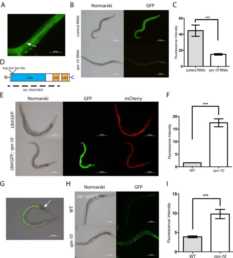
compensate for the absence of RPN-10. To examine the tissue distribution of RPN-10, we con-structed transgenic animals that express an RPN-10::GFP fusion protein from a fosmid clone that has been modified by recombineering to fuse GFP to the C-terminus of RPN-10 [33]. Since the fosmid contains the genomic coding sequence and native promoter, as well as any possible splice variants, the fusion protein is likely both expressed in the proper developmental stages and tissues and trafficked to the correct subcellular locations. The RPN-10::GFP fusion protein was expressed in multiple tissues with the strongest expression seen in the pharynx, intestine, hypodermis, and spermatheca (Fig 1A and 1B). We also observed expression at lower levels in a broader expression pattern, including the excretory cell, body wall muscle, vulva, and somatic gonad, suggesting that the RPN-10 protein might be ubiquitously expressed (S1 Fig). The expression in the spermatheca is consistent with the prior defects noted in sperm development in therpn-10mutant [27]. Additionally, the RPN-10::GFP fusion protein is visi-ble in both the nucleus and cytoplasm (Fig 1AandS1 Fig), suggesting that the proteasome in C.elegansmay play roles in both the cytoplasm and the nucleus as has been noted in other sys-tems [34]. We confirmed that the GFP signal is produced by an RPN-10 fusion protein because treatment of the transgenic worms withrpn-10RNAi reduces the expression of GFP in all tis-sues except for the pharynx (Fig 1B and 1C). The persistent expression of GFP in the pharynx could reflect greater stability of the RPN-10 protein in this tissue compared to others.
Proteasome dysfunction occurs in the
rpn-10
mutant
Despite the efficient knockdown of RPN-10 expression produced by therpn-10RNAi, we failed to see any gross developmental phenotypes in the treated animals (Fig 1B). Similarly, we noted few developmental phenotypes for therpn-10(ok1865)mutant other than the previously described decline in fertility and a mild increase in time needed to reach adulthood [27]. The rpn-10(ok1865)mutation is an 1166 base pair deletion that removes most of the 5’UTR, the entire first, second, and third exons, and a small portion of the fourth exon of therpn-10gene. This deletion entirely removes the coding sequence for von Willebrand factor type A domain (VWA) and the highly conserved DNSE (Asp-Asn-Ser-Glu) sequence, which together are criti-cal for interactions with other proteasome subunits, and the first of two ubiquitin-interacting motifs (UID) that mediate interactions with ubiquitinated target proteins (Fig 1D). Hence the mutation is expected to be a null or strong loss-of-function [35].
subunit genespas-6,pbs-6, orpbs-7starting from egg-hatching result in a robust increase in UbV::GFP expression during larval development compared to therpn-10mutant which peaks later. Further the worms treated withpas-6,pbs-6, orpbs-7RNAi either arrest later in develop-ment or show harmful effects in the adult animal that are absent in therpn-10mutant [26]. The later accumulation of UbV::GFP and lack of the severe detrimental phenotypes suggests that therpn-10mutant produces a graded reduction in proteasome function compared to the inhibition of the 20S catalytic subunit.
Despite the broad expression pattern of the RPN-10 protein, we observed the accumulation of the UbV::GFP fusion protein selectively in the intestine (Fig 1G). Moreover, within the intes-tine we observed regional differences with the group of three cells at the proximal end of the intestine showing little accumulation compared to other intestinal cells, despite the expression of mCherry from thesur-5p::mCherrytransgene at this site (Fig 1E and 1G). The reasons account-ing for the tissue and cell-specific accumulation of UbV::GFP are not clear, but this could reflect either greater demands for proteasome activity or lower proteasome expression in these areas.
To determine if UPS dysfunction could be occurring in tissues besides the intestine, despite the lack of visible UbV::GFP fusion protein accumulation, we examined the expression of the aip-1p::GFPreporter gene. Theaip-1gene encodes an inducible subunit of the proteasome which is selectively activated in the setting of UPS dysfunction produced by a variety of causes [24–26,37]. We found that theaip-1p::GFPreporter was activated in a larger range of tissues in therpn-10(ok1865)mutant, including expression in the pharynx, hypodermis, excretory cell, body wall muscle, intestine, and somatic gonad, which suggests that the limited accumulation of the UbV::GFP fusion protein underestimates the degree of UPS dysfunction (Fig 1H and 1I
andS4 Fig).
Loss of
rpn-10
enhances resistance to oxidative stress, thermal stress,
and mis-folded proteins
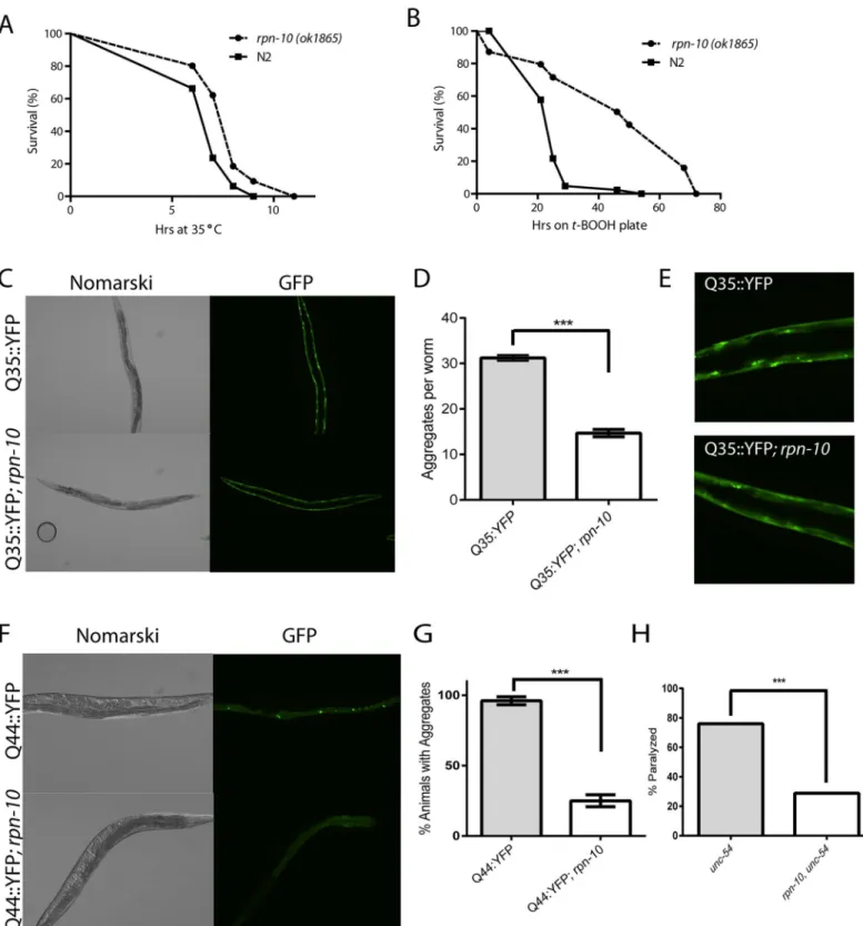
Prior work has demonstrated a role for the UPS system in the response to proteostasis threats including oxidative stress and the expression of mis-folded proteins [38–46]. Given the UPS disruption observed in therpn-10(ok1865)mutant, we tested the ability of this mutant to with-stand thermal stress, oxidative stress, and the expression of an aggregation-prone polygluta-mine fusion protein. We found that therpn-10mutant showed enhanced survival during a 35°C heat-shock compared to the N2 wild-type control (Fig 2A). This finding is consistent with the increased survival of yeast treated with proteasome inhibitors to survive a subsequent heat shock due to the enhanced expression of heat shock factor proteins [47]. To determine if therpn-10mutant showed a differential resistance to thermal stress compared to oxidative stress, we treated the wild-type animals and therpn-10mutant withtert-butyl hydroperoxide (tBHP). While we expected the mutant to be hypersensitive to tBHP, we instead found that the rpn-10mutant animals showed a marked increase in survival when exposed to this source of oxidative stress (Fig 2B). Together these findings suggest that therpn-10mutant is better able to resist acute threats to proteostasis compared to a wild-type animal.
line indicates the coding sequence regions deleted in therpn-10(ok1865)allele, and this line extends beyond the N-terminus of the RPN-10 protein to highlight the extension of the deletion into the 5’UTR of therpn-10gene. (E) Therpn-10(ok1865)mutation disrupts UPS function as shown by the selective accumulation of a UbV::GFP fusion protein in the intestine of the mutant but not wild-type animals. In contrast, therpn-10mutation has no effect on the expression of mCherry driven by the same promoter from a separate transgene in the animals. (F) Quantification of GFP expression from digital images captured as in Panel E.***represents p<0.001 byt-test. (G) The accumulated UbV::GFP fusion protein is localized in the intestine except for the three proximal cells (arrow). (H) In addition to accumulating the UbV::GFP fusion protein, therpn-10mutant induces the expression of theaip-1p::GFPreporter gene in multiple tissues along with the intestine. (I) Quantification of GFP expression from digital images captured as in Panel H.***represents p<0.001 by
t-test.
To investigate whether the resistance extended to long-term proteostasis threats, we used a transgene to express a Q35::YFP fusion protein in the body wall muscles. Previous work has shown this fusion protein to undergo age-dependent aggregation, which can be modified by changes in proteostasis activity in the cell [41]. Particularly, the inhibition of several protea-some subunits via RNAi is known to increase the aggregation of this protein [41]. In contrast to the effects of acute reductions in proteasome activity, we found that the Q35::YFP fusion protein formed fewer aggregates in therpn-10mutant compared to wild-type animals (Fig 2C and 2DandS5 Fig). Furthermore, the aggregates observed tended to be smaller in therpn-10 mutant when observed with fluorescent microscopy (Fig 2E). Since the intestine showed greater evidence of UPS dysfunction than the muscle, we then explored the effects of the rpn-10mutant on the aggregation of a Q44::YFP fusion protein that is expressed in the intestine with a transgene [48]. We found that therpn-10mutation served to protect animals from developing polyglutamine aggregates even in the intestine (Fig 2F and 2GandS6 Fig). These last two phenotypes are in contrast to the effects of proteasome inhibition via RNAi, which could reflect either a difference in the degree of UPS disruption in therpn-10(ok1865)mutant, the activation of one or more compensatory pathways, or perhaps a novel role forrpn-10 out-side of the proteasome.
While the reduced aggregation of these polyglutamine-repeat proteins suggests an improve-ment in proteostasis in therpn-10mutant, a limitation of this experimental approach is that both of the reporters express a non-native protein in the worms. To examine whether the improvement in proteostasis extended to native proteins, we examined the function of the metastable UNC-54 protein that is expressed by theunc-54(e1157)mutant [49]. This protein functions normally at the permissive temperature of 16°C whereas at the non-permissive tem-perature of 25°C, the protein becomes non-functional, presumably due to protein misfolding, and results in the animals becoming paralyzed. We found that therpn-10mutation also pre-vented the loss of UNC-54 activity when larval animals are acutely shifted to the non-permis-sive temperature (Fig 2H), perhaps by promoting the stability or folding of the UNC-54 protein. Together these data suggest that therpn-10mutation enhances cellular proteostasis in multiple tissues of the worms.
Loss of
rpn-10
primes both oxidative and heat-shock responses in
C
.
elegans
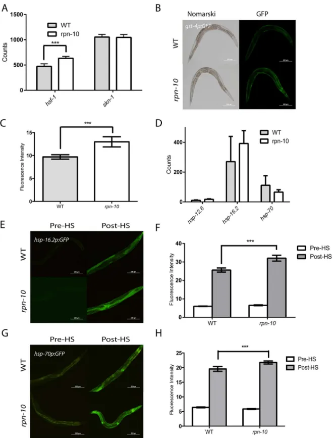
To explore why therpn-10(ok1865)mutant shows increased resistance to oxidative and ther-mal stress, we examined the expression of thehsf-1andskn-1transcription factors that control responses to these stresses via the use of Nanostring. We found that the expression ofskn-1 was unchanged whereas the expression ofhsf-1is increased in therpn-10mutant (Fig 3A) [50,
51]. To examine whether the activity of either transcription factor is changed in therpn-10 mutant animals, we tested effects of the mutation on GFP reporters which are known to be trig-gered by each of the stressors. Thegst-4gene encodes a member of the glutathione-S-transfer-ase family and was identified as being differentially expressed in worms following exposure to oxidative stress [52]. A GFP reporter controlled by thegst-4promoter has similarly been shown to respond to oxidative stress [53]. We found that this reporter is induced in therpn-10 mutant even in the absence of oxidative stress, which could suggest that oxidative stress response pathways are activated even in unstressed animals (Fig 3B and 3C). Since we found (permissive temperature) to 25°C (restrictive temperature) by therpn-10mutation (H) (n = 317 forunc-54(e1157)and n = 284 forrpn-10,unc-54, p<0.0001
by Fisher’s exact test).
hsf-1to be up-regulated in therpn-10mutant animals, we measured the expression of several heat-shock protein genes controlled byhsf-1through the use of Nanostring, and we found a trend towards an increase in the expression of these genes in therpn-10mutant, but the differ-ences failed to reach statistical significance (Fig 3D). We also examined the heat-shock response using GFP reporters for thehsp-16.2andhsp-70genes, which respectively encode an
α-crystalline and the inducible isoform of HSP-70 [54,55]. We found little difference in the expression of eitherhsp-16.2(Fig 3E and 3F) orhsp-70(Fig 3G and 3H) in therpn-10(ok1865) mutant animals compared to control animals. However, we did see increased expression of both reporters in therpn-10mutant compared to wild-type animals during the recovery from a one hour heat shock (Fig 3E, 3F, 3G and 3H), which is consistent with therpn-10mutant serv-ing to prime the heat-shock response. The cause of this primserv-ing is unclear but might reflect the increased expression ofhsf-1or perhaps the delayed clearance of unfolded proteins.
The
rpn-10
mutant shows increased longevity
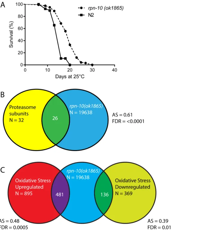
The enhanced proteostasis, oxidative stress responses, and heat shock responses exhibited by therpn-10mutant suggested that these animals could also exhibit an increase in lifespan. How-ever, RNAi studies have demonstrated that the inhibition of most proteasome subunits has a clear detrimental effect on the adult lifespan ofC.elegans[16]. We initially examined the life-span of therpn-10mutant at 20°C but saw only modest effects, so we then repeated the studies at 25°C based on the observation that the over-expression of therpn-6.1subunit only shows a beneficial effect on lifespan at 25°C [56]. At 25°C we observed a consistent increase in the life-span of therpn-10mutant compared to wild-type animals (Fig 4AandS1 Table) with up to almost a 30% increase in mean lifespan observed. This finding suggests that while reductions in proteasome activity typically have an adverse effect on aging, the net effect of the changes in proteasome activity and the subsequent adaptive responses produced by therpn-10mutation can slow aging and enhance longevity [16,18,57].
RNA-seq analysis demonstrates that proteasome subunits and oxidative
stress response genes are over-expressed in
rpn-10
mutants
To identify additional genes controlled by changes in proteasome function, we extracted RNA from three independent samples ofrpn-10(ok1865)mutants and wild-type animals and used the RNA to identify differentially expressed genes through whole transcriptome sequencing (RNA-seq). From these studies, we identified 19638 distinct RNA transcripts, with 111 genes being dif-ferentially up-regulated in therpn-10mutant and 60 genes down-regulated in the mutant (S2 Table). Using the DAVID program to identify themes within the up-regulated and down-regu-lated genes, we found that proteasome subunits were over-represented among the up-regudown-regu-lated genes (50–70 fold enrichment, p<0.001;S3 Table). This finding was not unexpected as a
con-served“bounce-back”response seeks to restore proteasome function in worms or vertebrate cells through the production of additional proteasome complexes [21,22,58]. Analysis of the down-regulated genes with DAVID did not identify any over-represented gene classes.
To complement the analysis using the DAVID program, we also tested if specific gene sets showed differential expression in therpn-10mutant through the use of gene set association anal-ysis (GSAA) [59]. With GSAA we found that 26/32 proteasome subunits showed differential mutant and wild-type animals, therpn-10mutants do not show up-regulation of the heat shock response in the absence of external stressors (p>0.2 for all
three genes). In contrast, therpn-10mutant animals show increased expression of thehsp-16.2p::GFP(E and F) and of thehsp-70p::GFP(G and H) reporters during the recovery from a one hour heat shock. In panel F, n = 10 for all genotypes and conditions and p<0.01 for WT andrpn-10post-heat shock. In panel H, n = 10 for all genotypes and conditions and p<0.05 for WT andrpn-10post-heat shock.
Fig 4. Therpn-10mutant shows increased longevity and increased expression of proteasome and oxidative stress response genes compared to wild-type animals.(A) Therpn-10mutant has an extended lifespan compared to wild-type animal in lifespan studies conducted at 25°C (mean lifespan 15.0 days for WT (n = 121) and 19.4 days forrpn-10(n = 120), p<0.0001 by log-rank test). (B) Shot-gun whole transcriptome sequencing (RNA-seq) was used to
characterize and measure the transcriptome of N2 andrpn-10(ok1865)mutants. From these experiments, a total of 19,638 mRNA and other RNA transcripts were detected. To test for evidence of a proteasome subunit gene expression signature in therpn-10mutant, Gene Set Association Analysis (GSAA) was used. GSAA calculates a differential expression score for each gene in the entire 19,638 gene RNA-seq dataset, and then uses a running weighted Kolmogorov-Smirnov test to examine association of an entire gene set with each phenotypic class. The strength of the association is measured by the association score (AS) where positive scores indicate association of the gene set with the phenotype, and statistical significance is measured by a false discovery rate (FDR) that is adjusted for multiple testing. From 32 proteasome subunit genes, 26 showed association with therpn-10profile. AS represents the association score with positive values indicating association, and FDR represents the false discovery rate for the association. (C) GSAA provides evidence of an oxidative stress response gene signature in therpn-10mutant. From the 895 genes up-regulated in worms exposed to oxidative stress [60], 481 genes show association with therpn-10profile, and among the 369 down-regulated genes, 136 show an association.
expression in therpn-10mutant, and these increases were readily apparent when the expression of individual genes in the RNA-seq data set where examined (Fig 4BandS4 Table). Additionally, our experiments identified enrichment, in therpn-10mutant, of genes previously found to be differentially expressed in worms exposed to oxidative stress produced by hyperbaric oxygen treatment (Fig 4C) [60]. This finding is consistent with the elevated expression of thegst-4::GFP reporter we observed and suggests that the activation of oxidative stress responses extends to a larger number of genes (Fig 3A). To better visualize the effects of therpn-10mutation on the oxi-dative stress and heat-shock responses, we examined the expression changes for individual genes involved in each response within the RNA-seq results, and we found increased expression of multiple gene classes within each group (S4 Table). For example within the heat-shock proteins, we again saw the increased expression ofhsf-1and multiplehsp-16proteins whilehsp-1and mosthsp-12genes showed little change in expression in therpn-10mutant (S4 Table).
Manual inspection of the differentially expressed genes also revealed possible insights into the response of the mutant worms to the reduction in proteasome activity. The up-regulation of proteasome subunits was clearly present withpas-3,pas-6,pas-7,pbs-3,pbs-6,rpt-4,rpn-8, rpn-9, anddss-1, which is theC.elegansortholog of the yeast proteasome regulatory cap pro-tein SEM1, all being overexpressed [61]. We also found that the worm ortholog of NEDD8, ned-8, was up-regulated in therpn-10mutant. NEDD8 is a small protein with a highly similar amino acid sequence to ubiquitin (~60% sequence identity) [62,63]. However, NEDD8 differs functionally from ubiquitin in that NEDD8 is covalently attached selectively to cullin proteins where it promotes the formation of the E2–E3 ligase complex, which then catalyzes the ubiqui-tination of proteins [64,65]. However, under conditions of proteasome dysfunction when free ubiquitin levels are low due to the accumulation of ubiquitinated proteins, NEDD8 can also be activated by the same pathways that act to attach ubiquitin to target proteins [66,67]. The role of NEDD8 in this setting is unclear as this could simply reflect NEDD8 being aberrantly uti-lized by the ubiquitin activating enzymes due to the low levels of ubiquitin. Alternatively, the use of NEDD8 may constitute part of a response pathway to the reduced cellular proteasome activity [67]. Another notable gene found to be up-regulated in therpn-10mutant was atg-16.2, which is one of two worm orthologs of ATG16L1, and participates in autophagy via the recruitment of an ATG5-ATG12 complex to the nascent autophagosome [68,69]. Autophagy and the UPS are known to represent parallel pathways by which protein degradation can occur in the cell, so the responsiveness ofatg-16.2to changes in proteasome function could represent a point of cross-talk between the pathways. Also, among the up-regulated genes is prmt-1/epg-11which encodes an arginine methyltransferase that acts to methylate specific cargo receptor proteins and is essential for their clearance of protein aggregates by autophagy during develop-ment [70]. We also saw further evidence of an increase in the expression of autophagy genes when the expression of individual genes was queried using the RNA-seq data with 14 out of 21 genes selected showing an increase (S4 Table). Among the down-regulated genes, we identified cpi-1, which encodes one of the worm cystatin genes [71]. An important role for cystatins is the inhibition of cathepsins, which are lysosomal proteases that degrade proteins brought to the lysosome via endocytic or autophagic transport pathways [71,72]. Hence, the down-regulation ofcpi-1may suggest changes in lysosomal activity that might facilitate the ultimate degradation of proteins that are engulfed by macro or selective-autophagy.
The
skn-1
transcription factor is required for
rpn-10
mutant survival
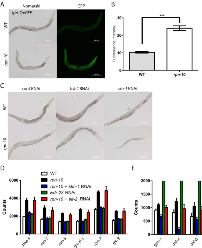
To examine the regulation of proteasome subunit expression, we used anrpn-7p::GFPreporter that expresses GFP under the control of therpn-7promoter. Therpn-7gene encodes the worm ortholog of the human PSMD6 protein, which is a subunit of the 19S regulatory cap [28]. In wild-type worms, therpn-7p::GFPreporter was expressed in multiple tissues including the intestine, pharynx, and hypodermis (Fig 5A), and the level of GFP expression in these tissues was globally increased in therpn-10mutant (Fig 5A and 5B). Hence despite the focal accumu-lation of UbV::GFP in therpn-10mutant, multiple tissues in the animal appear to sense changes in proteasome activity and up-regulate the expression of other subunits.
InC.elegans, the control of oxidative stress responses and the up-regulation of proteasome expression following acute reductions in proteasome activity are both coordinated by the cap’n’collar transcription factorskn-1, which is the ortholog of the Nrf1 and Nrf2 transcription factors from vertebrates [22,50,73]. We have previously shown that an additional aspect of the response to proteasome dysfunction is the induction of theaip-1/AIRAP gene, which encodes an inducible proteasome subunit that enhances proteasome activity and relaxes substrate speci-ficity when bound to the 19S cap, and that this induction requires bothskn-1andhsf-1[24–
26]. Hence we sought to determine ifskn-1and/orhsf-1is required for the activation of protea-some subunit expression in therpn-10mutant. However, when we treated therpn-10mutant withhsf-1andskn-1RNAi, we unexpectedly found that inhibition ofskn-1resulted in animals that were small, sickly, and developmentally arrested prior to adulthood (Fig 5C). The effect of theskn-1RNAi was particularly acute for L3 and L4 larval stage animals becauserpn-10 mutant animals treated withskn-1RNAi from egg hatching appeared similar to the control RNAi treated animals on the second day of treatment but then exhibited the detrimental effects on the third day of treatment (S7 Fig). This could reflect the time needed for full effect of the skn-1RNAi or the presence of a critical developmental period whenskn-1activity is essential. In contrast, treatment withhsf-1RNAi had no effect on worm development (Fig 5C). This finding demonstrates that whileskn-1is usually not essential for larval development,skn-1is essential for development in therpn-10mutant.
To determine if the essential role played byskn-1could be mediated via the control of protea-some subunit expression, we treated wild-type N2 andrpn-10mutant animals with control and skn-1RNAi, and then measured the expression of several proteasome subunit genes using Nanostring analysis [74]. For these studies, we prepared RNA from the RNAi treated animals at 48 hours after synchronization, which is a timepoint prior to the appearance of any visual phe-notypes due toskn-1RNAi (S7 Fig). We found that all of the proteasome subunits, for which we probed, were up-regulated in therpn-10mutant compared to N2, and that treatment withskn-1 RNAi largely prevented this up-regulation (Fig 5DandS5 Table). To determine if SKN-1 activa-tion was sufficient for the increased expression of proteasome subunits, we also treated N2 with wdr-23RNAi. Thewdr-23gene encodes a WD40 repeat protein that binds toskn-1and inhibits its transcriptional activity [75]. We found thatwdr-23RNAi potently increased the expression of oxidative stress response genes (Fig 5EandS5 Table), but had little effect on the expression of proteasome subunits. Conversely, therpn-10mutant showed a greater increase in the expression of proteasome subunits, with a more moderate effect on the activation of oxidative stress response genes (Fig 5D and 5E). Together, these findings suggest thatskn-1is necessary but not sufficient for the activation of proteasome subunit expression, and thatskn-1is capable of mounting distinct responses to oxidative stress and proteasome dysfunction.
elt-2
is required to survive chronic proteasome dysfunction
and contribute to this specificity. To identify such transcription factors, we screened two sepa-rate RNAi libraries consisting of subsets of transcription factors drawn from the Ahringer and Vidal RNAi libraries for clones that produced developmental phenotypes in therpn-10mutant that are similar to those produced byskn-1RNAi (S6 Table). From the two independent screens, we identifiedelt-2, which is a GATA transcription factor and is essential for the expres-sion of most genes expressed in the intestine [76–78]. In addition to its developmental role, elt-2is required for innate immunity, contributes to the response to heavy metal exposure, and contributes to the beneficial effects of changes indaf-2/IGFR signaling on worm lifespan [79–
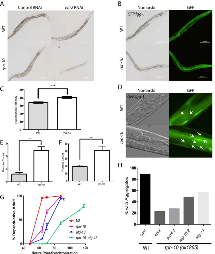
82]. In therpn-10mutant, we found that inhibitingelt-2with RNAi mirrored the effects of skn-1knock-down and produced small, sickly, animals that often arrested during larval develop-ment (Fig 6A).
To determine ifelt-2cooperateswith skn-1to control the expression of either oxidative stress response genes or proteasome subunit genes in therpn-10mutant, we used Nanostring analysis to measure the expression of these genes inrpn-10mutants treated with control or elt-2RNAi. In contrast toskn-1RNAi, we found thatelt-2RNAi did not block the activation of either group of genes (Fig 5D and 5E, andS5 Table). Hence, whileelt-2is similarly required for the viability of therpn-10mutant,elt-2likely acts via a distinct mechanism thanskn-1, and is discussed further below.
Autophagy is activated in the
rpn-10
mutant
As discussed previously, our RNAi-seq studies identified the autophagy genesatg-16.2and prmt-1/epg-11as being up-regulated in therpn-10mutant (S2andS4Tables). This observation is consistent with the activation of autophagy observed when proteasome function is potently reduced via the expression of a dominant-negative proteasome subunit or is chronically reduced in cultured neural cells by long-term exposure to proteasome inhibitors [83,84]. To determine if autophagy is activated in therpn-10mutant, we examined the expression and sub-cellular localization of the LGG-1 protein in wild-type andrpn-10mutant animals via the use of a transgene expressing a GFP::LGG-1 fusion protein [85]. The LGG-1 protein is the worm ortholog of LC3 and has been shown to play an analogous role in the formation of autophago-somes via integration into the autophagosome membrane [85]. We initially found that the expression of the GFP::LGG-1 fusion protein is increased in therpn-10mutant compared to wild-type animals (Fig 6B and 6C), and this increase inlgg-1expression occurs in part at the transcriptional level and in conjunction with the transcriptional up-regulation of the worm beclin orthologbec-1(S8 Fig) [85]. The up-regulation of LGG-1 expression has also been seen in worms with activated autophagy resulting from either removal of the germline via aglp-1 mutation or the inhibition oflet-363/TOR withlet-363RNAi treatment [86]. Based on this and other work, the increased expression of LGG-1 has been therefore suggested to indicate the activation of autophagy [87]. To seek additional evidence of enhanced autophagy in therpn-10 mutant, we looked for the presence of GFP::LGG-1 puncta, which are produced by the integra-tion of LGG-1 into the membrane of developing autophagosomes [85]. We introduced a GFP:: LGG-1 reporter into therpn-10mutant and observed the effects in the intestine and seam cells of the wild-type andrpn-10mutant transgenic animals [86]. We saw an increase in GFP-posi-tive puncta in both the seam cells and intestine of therpn-10mutant animals compared to the subunit expression. (E) In parallel Nanostring studies, therpn-10mutant also shows a smallskn-1dependent, but notelt-2dependent, increase in the expression of the oxidative stress response genes,gcs-1,gst-4, andgst-5. In contrast,wdr-23RNAi produces a marked increase in the expression of these genes (mean expression level ofgcs-1–4,326,gst-4–24,714, andgst-5–4,982). For details of statistical testing for Panel D and Panel E seeS5 Table.
wild-type controls, which suggests an increase of the activity of the autophagy-lysosome path-way in these animals (Fig 6D, 6E and 6F).
The activity of the autophagy pathway is essential for the development
and enhanced proteostasis of the
rpn-10
mutant animals
The increase in autophagy could be involved in the adaptation to the changes in UPS activity in therpn-10mutant and could also contribute to some aspect of the beneficial effects of this mutation on proteostasis. To examine these possibilities we tested the effects of autophagy inhi-bition on both the development and improved proteostasis of therpn-10mutant. For these studies we focused on theepg-1/atg-13,prmt-1/epg-11, andatg-16.2genes based upon either their identification in our RNA-seq studies (S2 Table) or identified role in the clearance of pro-tein aggregates via autophagy [70,88]. During development, we found that loss of theatg-13 gene greatly impaired the development of therpn-10mutant as exhibited by the significantly delayed development of anrpn-10; atg-13mutant compared to either mutant alone (Fig 6G). Notably, almost 20% of therpn-10; atg-13mutants appeared to be permanently arrested during development and failed to reach adulthood even after five days (Fig 6G).
We also tested the role of autophagy in the enhanced resistance to the accumulation of poly-glutamine-repeat protein aggregates in the intestine of animals expressing a Q44::YFP trans-gene by inhibiting theprmt-1/epg-11,atg-16.2, andatg-13genes via the use of RNAi starting on day 1 of adulthood. We began RNAi treatment at this time point to prevent any adverse effects of RNAi treatment from occurring during development. We found that the knock-down of eitheratg-13oratg-16.2produced an increase in the percentage of animals with aggregates compared to the control RNAi treatedrpn-10mutant animals (Fig 6H). In contrast, these RNAi treatments only modestly increased the percentage of wild-type animals with aggregates (S9 Fig). To further explore the effects of these RNAi treatments, we also counted the number of aggregates in each worm in a separate trial. We again found thatatg-13andatg-16.2RNAi treatment had a greater effect on therpn-10mutants compared to the wild-type animals, and additionally nowprmt-1RNAi produced a selective increase in polyglutamine aggregation in therpn-10mutant (S9 Fig). Together these findings show a vital role for autophagy in both promoting the normal development of therpn-10mutant animals, and contributing to the effects of therpn-10mutation on proteostasis.
Lysosome function is essential for the development of the
rpn-10
mutant
The increased autophagic activity seen in therpn-10mutant could act to shuttle proteins to the lysosome for degradation via lysosomal proteases. To examine the role of the lysosomes in the rpn-10mutant, we visualized the intestinal lysosomes through staining with both the Lyso-tracker fluorescent dye, which concentrates in lysosomes due to their low pH, and the use of the Magic Red cathepsin B and cathepsin L substrates [89]. The cathepsin B and L substrates are cell-permeable cresyl violet-conjugated peptides containing either the Arg-Arg or Phe-Arg sequence cleaved by the respective cathepsin inside of the lysosome. These cleavage events relieve the intramolecular quenching of the cresyl violet fluorophore and produces red Measurement of puncta number in the intestine also demonstrates a significant increase in puncta number in therpn-10mutant (n = 14 for WT and = 13 for
rpn-10,***represents p = 0.0024 byt-test). (G) The inhibition of autophagy withan atg-13mutation delays the developmental time of anrpn-10; atg-13
mutant compared to therpn-10oratg-13mutants and only 80% of worms reach adulthood (n = 283 for WT, = 298 forrpn-10, = 282 foratg-13, and = 300 for
rpn-10; atg-13, p<0.0001 for differences betweenatg-13; rpn-10andrpn-10at the 88 hour time point by Fisher’s exact test) (H) The inhibition of autophagy in
rpn-10mutant worms expressing a Q44::YFP transgene in the intestine through treatment withatg-13,atg-16.2, orprmt-1RNAi treatment, starting on day 1 of adulthood, reduces the protective effect of therpn-10mutation. Shown are the percentage of animals with aggregates when scored after 72 hours of RNAi treatment (foratg-13RNAi, n = 50, p = 0.0002 by Fisher’s exact test; foratg-16.2RNAi, n = 50, p = 0.003; andprmt-1RNAi, n = 50, NS).
fluorescence. Our work represents the first application of the Magic Red substrates inC.elegans research as a novel approach to identify lysosomes and quantify their activity. The use of the cathepsin substrates was particularly attractive because the location and degree of fluorescence are directly related to the activity of the cathepsin enzymes [89]. With the Lysotracker dye, we saw staining of intestinal lysosomes, and a similar pattern was observed when the animals were stained with either the cathepsin B or cathepsin L substrates (Fig 7A). Consistent with both Lysotracker and the Magic Red substrates acting to label lysosomes, we observed a high-degree of co-localization when wild-type animals were stained with both Lysosensor Green and the cathepsin B substrate, as evidenced by Pearson’s correlation score or Mander’s overlap score which averaged greater than 0.9 (Pearson average 0.92, n = 15 and Mander average 0.94, n = 15)(S10 Fig) [90].
When the staining of the wild-type andrpn-10mutant animals were compared, we observed an overall decline in both lysosome volume and in the activity of each of the cathepsins as evi-denced by reduced fluorescence in the mutant (Fig 7A and 7B). The decline in overall fluores-cence was likely due to a reduction in lysosome number and volume in therpn-10mutant (Fig 7C, 7D, 7E and 7F). Together our observations could suggest that the cellular lysosome pool is being consumed by an increase in autophagy via the fusion of the lysosomes with the enlarged pool of autophagosomes.
To test the importance of lysosome function in therpn-10mutant, we treated worms with RNAi to inhibitvha-15, which is a part of the vacuolar proton-translocating ATPase and acts to promote the acidification of the lysosomes [91]. If therpn-10mutant relied upon the autop-hagosome-lysosome pathway to compensate for the declines in proteasome activity, we expected these mutants to show enhanced sensitivity to lysosome inhibition withvha-15RNAi compared to wild-type animals. Consistently, we found that therpn-10mutant shows a decrease in body size and developmental rate compared to N2 animals treated in parallel with vha-15RNAi (Fig 7G and 7H). To determine if these effects could result from changes in lyso-some pH, we treated worms with NH4Cl which accumulates in lysolyso-somes and neutralizes the normally acidic pH of the organelle, producing a decrease in proteolytic activity [92]. Consis-tent with the effects ofvha-15RNAi, we observed therpn-10mutant animals to develop slowly following treatment with increasing concentrations of ammonium chloride, while N2 worms treated in parallel showed a lesser effect (Fig 7I and 7J). These data suggest that therpn-10 mutant both exhibits an increase in autophagy and has become dependent on the activity of the autophagy-lysosome pathway for normal development as evidenced by the selective vulner-ability of the mutant to lysosome inhibitors.
elt-2
controls lysosome formation in the gut
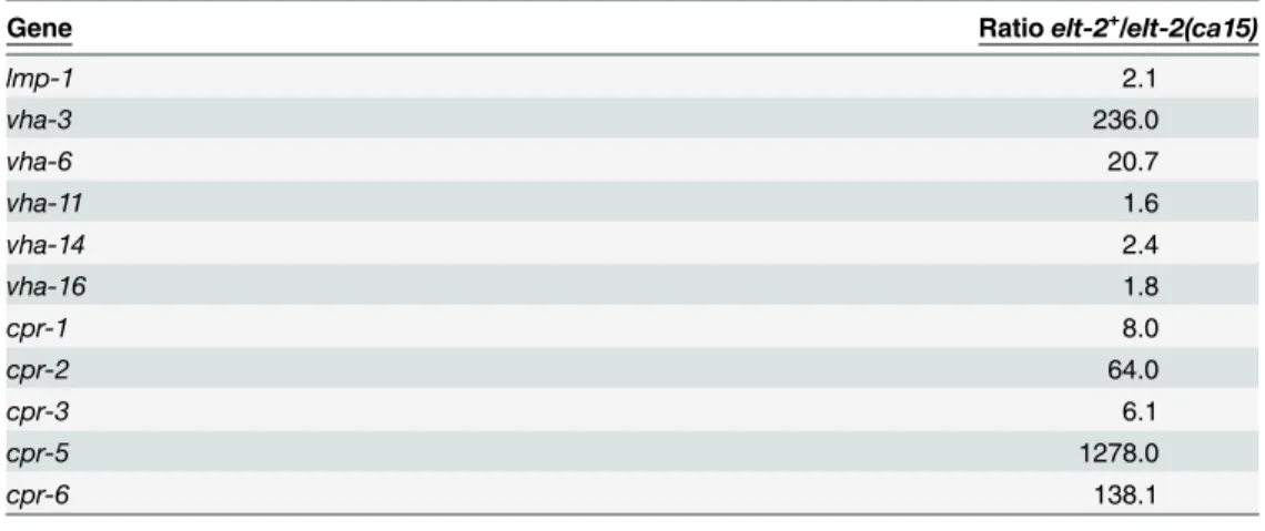
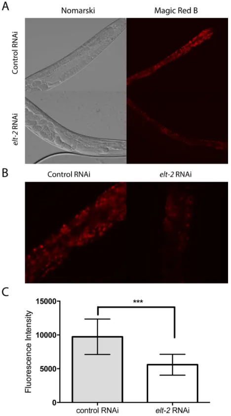
To identify the pathway that could be controlled byelt-2and contribute to the survival of the rpn-10mutant, we explored whetherelt-2might be involved in the generation of lysosomes in the intestine. Initially, we utilized an existing gene expression dataset which was generated using Serial Analysis of Gene Expression, SAGE, to compare gene expression differences in RNA prepared from L1 larvae that either lackedelt-2due to theelt-2(ca15)mutation or con-tained the mutation as well as a rescuing transgene expressingelt-2[77]. In this dataset, we noted that the expression of multiple genes associated with lysosomes, including the lysosome membrane proteinlmp-1, vacuolar proton-translocating ATPase subunits, and cathepsins, all showed decreased expression in theelt-2(ca15)mutants compared to theelt-2+larvae
number of lysosomes present in the RNAi treated worms (Fig 8A, 8B and 8C). The results are consistent with either a reduction in lysosome production or a reduction in the proteolytic activity of the lysosome followingelt-2RNAi treatment.
elt-2
and
skn-1
are limiting in the
rpn-10
mutant
While the inhibition of eitherelt-2orskn-1in therpn-10mutant is harmful, it was less clear to what extent either transcription factor normally acts to promote the growth and survival of the rpn-10mutant. To determine if enhancing the function of either gene is beneficial, we used transgenes to over-express eitherelt-2orskn-1in therpn-10mutant. During the construction of these strains, we observed that the transgenic animals appeared to develop and reproduce faster than the non-transgenic worms. To directly test whether the development of therpn-10 mutant was at least somewhat normalized by the over-expression of either gene, we performed development assays on synchronized animals using successfully reaching reproductive adult-hood as the outcome. We found that the over-expression of either transcription factor led to more rapid development withelt-2over-expression perhaps having a somewhat stronger effect thanskn-1(Fig 9A). This finding suggests that the activities ofelt-2andskn-1are limiting in therpn-10mutant and likely contribute to the slight developmental delay exhibited by these animals. Hence, enhancing the activity of either transcription factor leads to the increased activity of downstream targets and enhances the development of therpn-10mutant. intestine. GFP fluorescence indicates the intestinal nuclei as marked by anelt-2p::GFPreporter gene. (E and F) Images of animals stained with the Magic Red dye specific for cathepsin L, which are cropped similarly to panels C and D; GFP fluorescence indicates the intestinal nuclei. (G) Images showing the effects of treating wild-type andrpn-10mutant animals withvha-15RNAi. (H) Graph quantifying the effects ofvha-15RNAi treatment on wild-type andrpn-10
mutant animals (***represents p = 0.0062 byt-test). (I) Images showing the developmental delay produced by treating therpn-10mutant with 100 mM NH4Cl to neutralize lysosomal pH. (J) Graph showing the developmental effects of increasing NH4Cl doses on wild-type andrpn-10mutant animals.
doi:10.1371/journal.pgen.1005823.g007
Table 1. Control of lysosome gene expression byelt-2.
Gene Ratioelt-2+/elt-2(ca15)
lmp-1 2.1
vha-3 236.0
vha-6 20.7
vha-11 1.6
vha-14 2.4
vha-16 1.8
cpr-1 8.0
cpr-2 64.0
cpr-3 6.1
cpr-5 1278.0
cpr-6 138.1
Ratio of gene sequence counts detected in L1 larval animals either expressing ELT-2 from a transgene (elt-2+) or lackingelt-2due to a genetic mutation (elt-2null). For genes showing a zero count in theelt-2null
condition, a value of 0.1 was used to avoid an error. Data was obtained from the supplemental materials of McGhee, et. al. [77].
Fig 8.elt-2controls lysosome formation in the intestine.(A) Representative images of day 1 adult animals treated with control orelt-2RNAi and stained with the Magic Red dye specific for cathepsin B. (B) Images of animals captured as in panel A, but cropped instead of reduced in scale, to demonstrate details of the intestine. (C) Graph quantifying the effects of control andelt-2RNAi on Magic Red fluorescence (n = 15 for each RNAi treatment, p<0.001 byt-test).
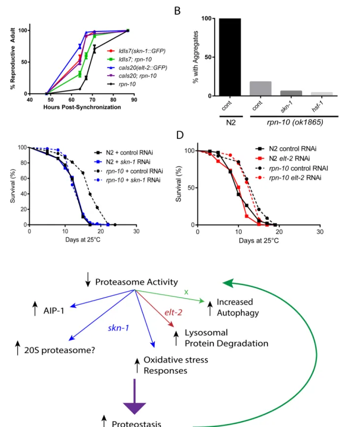
Fig 9. Roles ofskn-1andelt-2in the development, lifespan, and enhanced proteostasis of therpn-10mutant.(A) TheldIs7andcaIs20transgenes express eitherskn-1orelt-2as a GFP fusion protein under the control of the native promoter. Each transgene was crossed into therpn-10mutant, which also has the endogenousskn-1orelt-2genes intact. Development of synchronizedrpn-10and transgenic worms was measured and plotted as shown. (B)rpn-10
Complex roles of
elt-2
and
skn-1
in the beneficial effects of the
rpn-10
mutation
While bothelt-2andskn-1both contribute to the development of therpn-10mutant, it is unclear if either contribute to the improved proteostasis and increased longevity exhibited by therpn-10mutant. We first tested whether the inhibition ofskn-1,elt-2, orhsf-1affected the reduction in Q44::YFP aggregates seen in therpn-10mutant animals by using RNAi to knock-down each gene only in adult animals. We took this approach due to the adverse effects that bothelt-2andskn-1had during development. We found that neitherskn-1norhsf-1was required for the reduction in protein aggregates (Fig 9B). The Q44::YFP transgene was con-trolled by thevha-6promoter, and this promoter appears to be regulated byelt-2because we observed a dramatic decline in YFP fluorescence in either wild-type orrpn-10mutant trans-genic animals treated withelt-2RNAi. We then examined the role ofskn-1andelt-2in the increased longevity exhibited by therpn-10mutant by treating adult wild-type orrpn-10 mutant worms with control,skn-1, orelt-2RNAi and then measuring the effects on lifespan. We found that the increased lifespan of therpn-10mutant requiredskn-1but notelt-2(Fig 9C and 9D). Lastly we examined the role ofskn-1in the enhanced oxidative stress resistance exhib-ited by therpn-10mutant. We were unable to obtain adultrpn-10mutant animals following treatment withskn-1RNAi, so we shifted to using larval animals that had been treated with control orskn-1RNAi for 48 hours. Treatment of these animals with 100μM juglone revealed
that theskn-1RNAi treatment markedly reduced the oxidative stress resistance of therpn-10 mutant (S11 Fig). Together these data suggest thatskn-1is essential for development, oxidative stress resistance, and longevity but not improved proteostasis of therpn-10mutant, whileelt-2 is only essential for development. The role ofelt-2in proteostasis is difficult to judge since many promoters active in the intestine areelt-2target genes [78].
Discussion
Loss of
rpn-10
produces proteasome dysfunction
The inhibition of the majority of the proteasome subunits inC.elegansis clearly detrimental, resulting in phenotypes such as developmental arrest or a marked reduction in body size [26]. Similarly, essentially all of the proteasome subunits with deletion alleles are homozygous lethal and need to be maintained as a balanced heterozygote (see Wormbase for details). Therefore, it was somewhat surprising that two independent viable deletion alleles forrpn-10have been iso-lated. This could have been due to the expression ofrpn-10in a limited pattern in the animal, or the presence of a redundant subunit. Instead, we find thatrpn-10is broadly expressed and produces aspects of proteasome dysfunction in multiple tissues when removed. Despite these findings, the viability of thisrpn-10deletion mutant could imply that the worm proteasome is similar to those of yeast, which are also less dependent on the analogous Mcb1/RPN10 subunit, or that sufficient compensatory pathways can be activated in the wormrpn-10mutant to cope with the reductions in proteasome activity [29]. Our work suggests that the activation of com-pensatory mechanisms, such as the up-regulation of other proteasome subunits via the actions adulthood. Mean lifespan for N2 on control RNAi is 13.0 days (n = 102) and 13.3 days onskn-1RNAi (n = 119). Mean lifespan forrpn-10on control RNAi is 16.7 days (n = 116) and is 13.8 days onskn-1RNAi (n = 126). Comparison ofrpn-10mutant treated with control vsskn-1RNAi, p<0.0001 by log-rank test;
comparison of N2 andrpn-10on control RNAi, p<0.0001 by log-rank test. (D) Treatment withelt-2RNAi has no effect on the extended lifespan of therpn-10
mutant at 25°C. Mean lifespan for N2 on control RNAi is 11.1 (n = 120) and is 10.4 days onelt-2RNAi (n = 113). Mean lifespan forrpn-10on control RNAi is 13.7 days (n = 113) and is 13.3 days onelt-2RNAi (n = 119). Comparison ofrpn-10mutant treated with control vselt-2RNAi, p<0.03 by log-rank test;
comparison of N2 andrpn-10on control RNAi, p<0.0001 by log-rank test. (E) Model summarizing the effects ofskn-1andelt-2in therpn-10mutant and possibly other situations where proteasome function is reduced.
ofskn-1and the increased use of autophagy and lysosomes possibly for protein degradation are an important aspect of the response to the chronic reduction in proteasome activity seen in the rpn-10mutant (Fig 9E). Within this responseelt-2may play a supportive role by maintaining an adequate pool of intestinal lysosomes (Fig 9E). The pathway leading to changes in the expression of autophagy genes and autophagic activity are currently unclear but recent work has shown that the RPN-10 protein serves as an adapter to facilitate the clearance of the protea-some via autophagy inArabidopsis[93]. Perhaps the loss of RPN-10 could stimulate autopha-gic activity via the loss of some form of negative feedback from the proteasome. Alternately, the 19S cap proteasome subunits have been shown to dissociate from the 20S subunit in the set-ting of proteasome dysfunction and then bind to protein aggregates. When localized to the aggregates, the deubiquitinase activity of RPN-11/Poh1 releases ubiquitin chains from the aggregated protein, and these ubiquitin chains then serve to activate autophagy via the HDAC6 protein [94,95]. It could be possible that the loss of RPN-10 destabilizes the 26S proteasome or via other mechanisms promotes the association of RPN-11 and other subunits with cellular protein aggregates to then promote their removal via autophagy. Finally, proteomic studies comparing wild-type and long-liveddaf-2mutants inC.eleganshave made the unexpected finding that thedaf-2mutants have increased amounts of insoluble protein during aging com-pared to wild-type animals [96]. Further, the insoluble fraction includes increased amounts of small heat-shock proteins, which could suggest that the controlled aggregation of proteins is an important mechanism for enhancing proteostasis. Perhaps, therpn-10mutant might act in a similar manner, especially in light of the enhanced heat-shock response observed, with the shunting of UPS substrates into a protected insoluble protein compartment in the cell. Future work could test this possibility via the analysis of amounts and types of insoluble proteins pres-ent in therpn-10mutant.
However, it is also possible that the spectrum of UPS substrates that fail to be degraded in therpn-10mutant differs in an important way from those that accumulate when other subunits are removed via RNAi or genetic mutations. In the Mcb1/RPN10 yeast mutant, UPS substrates that are degraded via the N-end rule pathway are degraded to a similar extent as seen in wild-type yeast, whereas the substrates degraded via the ubiquitin fusion pathway are no longer degraded normally [29]. These differential effects on substrate degradation could reflect the presence of multiple ubiquitin binding proteins in the 19S proteasome subunit or the ability of the 20S proteasome to independently degrade some UPS substrates [30,31,97,98]. Future work can explore this question via the use of proteomic approaches, such as ubiquitin-remnant profiling, to determine if differences in substrate degradation are also observed inC.elegans. In addition, these experiments could determine the identities of substrates that might account for the phenotypic differences observed between therpn-10mutant and mutations or RNAi affect-ing other subunits [66,99,100].
Graded proteasome dysfunction can have beneficial effects on
proteostasis and longevity
consistent with prior work showing that either cultured cells or yeast exposed to proteasome inhibitors show elevated expression of heat shock proteins, and increased resistance to heat shock [47,101,102]. We also find that genes involved in the response to oxidative stress are up-regulated in therpn-10mutant so these two compensatory responses may contribute to the ability of therpn-10mutant to better survive the exposure to heat or oxidative stress.
The reasons accounting for the improved ability of therpn-10mutant to prevent the aggre-gation of the unstable polyglutamine repeat proteins are still somewhat unclear. Data from ver-tebrate cells suggest that proteasome inhibition may occur prior to the development of
polyglutamine protein aggregates, so some aspect of the compensation to proteasome dysfunc-tion, as opposed to proteasome activity alone, may play a key role in determining the timing and levels of protein aggregation [103]. In addition to the UPS, autophagy has been identified as an important pathway for the degradation of polyglutamine repeat proteins. Consequently a cellular environment where there is a high level of autophagic activity may result in low levels of protein aggregation regardless of the level of UPS activity [104]. Our data suggest that this could be at least partially true, because we find that the increased flux in the autophagosome-lysosome pathway in therpn-10mutant contributes to at least some of the reduction in aggre-gation by facilitating the removal of the polyglutamine repeat proteins. Alternately, the increased expression of oxidative stress response genes and priming of the heat-shock response which occur in therpn-10mutant could also act to reduce protein mis-folding and
aggregation.
skn-1
and
elt-2
are required to survive proteasome dysfunction
An important response to the reduction in UPS activity in therpn-10mutant is the activation of theskn-1/Nrf2 transcription factor which then promotes the expression of proteasome sub-units, and accessory factors likeaip-1/AIRAP, as part of a“bounce-back”response [22,25,26]. Importantly we find that therpn-10mutant not only uses this response, but also actually requires the continuous activity of this response in order to develop normally and survive long-term in the presence of chronic reductions in proteasome activity. We also identify a novel role for theelt-2GATA transcription factor in promoting the survival of therpn-10 mutant despite the presence of chronic proteasome dysfunction. This effect could occur through a basal role in determining the level and activity of lysosomes in the worm intestine, or elt-2activity could somehow be enhanced in the setting of proteasome dysfunction. Consis-tently,elt-2has been identified as a UPS substrate, and the UPS-mediated degradation of ELT-2 contributes to the killing of worms by the bacterial pathogenBurkholderia pseudomallei [105]. Furthermore, the vertebrate GATA-1 and GATA-2 transcription factors are known UPS substrates [106–108]. Under normal conditions, GATA-2 is usually rapidly degraded by the UPS, and this event determines the ratio of GATA-1 and GATA-2 bound to target gene pro-moters [106–108]. In worms, we failed to observe significant changes in the expression or local-ization of an ELT-2::GFP fusion protein in therpn-10mutant, butelt-2activity could be altered in other ways in response to changes in UPS activity, such as through post-translational modifications or associations with specific binding partners.
Could similar compensatory pathways also be activated when
proteasome subunits are over-expressed?
the increased expression of the endogenous genes encoding other proteasome subunits [98]. Furthermore, the over-expression ofpbs-5leads to the increased expression of thegst-4p::GFP reporter, suggesting an increase inskn-1activity in this mutant. Consistent with a role for skn-1acting downstream of the increase in PBS-5 expression, the increased lifespan observed in these transgenic worms required the activity ofskn-1[98]. One possible model to account for both the requirement forskn-1and the elevated expression of non-transgene encoded protea-some subunits would be for the initial imbalanced expression of PBS-5 to disrupt rather than enhance the assembly of active proteasomes, which could trigger at least some of the compen-satory mechanisms also utilized by therpn-10mutant in an effort to promote the formation of active proteasomes and maintain proteostasis. The authors examined the expression of protea-some subunit genes following the treatment of the transgenic worms withskn-1RNAi and did see small, but non-significant decreases in subunit expression [98]. These findings could sug-gest thatskn-1only plays a minor role in controlling proteasome subunit expression following increased PBS-5 expression, and that the expression of otherskn-1target genes, such as the oxi-dative stress response or other novel pathways, may be more important in an aging context. It will be interesting to explore whetherelt-2, autophagy, or lysosome activity play roles in the effects of this transgene on stress resistance and aging phenotypes. If so, our work using the rpn-10mutant could identify molecular pathways that could be exploited to improve proteos-tasis via either the augmentation or graded inhibition of proteasome activity.
Materials and Methods
Strains
The strains CB1157 (unc-54(e1157)), CL2166 (dvIs19[pAF15 (gst-4::GFP::NLS)]) [53], CL2070 (dvIs70[hsp-16.2::GFP; rol-6 (su1006)]) [54], DA2123 (adIs2122 [lgg-1p::GFP::lgg-1 + rol-6 (su1006)]) [110], HZ1688 (atg-13(bp414)) [88], MAH236 (sqIs13 [lgg-1p::GFP::lgg-1 + odr-1p:: RFP]) [86], SJ4001 (zcIs1[aip-1::GFP]) [37], and VC1369 (rpn-10(ok1865)) were obtained from theCaenorhabditisGenetics Center which is funded by NIH Office of Research Infrastructure Programs (P40 OD010440). TherrIs1[elt-2::lacZ::GFP]transgene which expresses nuclear localized GFP in the intestine has been previously described [111]. Therpn-10(ok1865)mutant was outcrossed against N2 five times, and one resulting outcrossed homozygous line was used for all subsequent crosses and experiments. The presence of therpn-10(ok1865)allele was determined by single-worm PCR using oligonucleotides designed to amplify the wild-type rpn-10allele (F 5’-AAGAGAACAACGCGCATCTT-3’; R 5’-GTGTGCCCCTTTGAGGAGTA-3’) and to detect the deletion present in therpn-10(ok1865)allele (F 5’-CCCATTCCAATT GTTGCTCT-3’; R 5’-TGCACCAACAACTCCACATT-3’). The strain AM140 (rmIs132[unc-54p::Q35::YFP]) was kindly provided by Dr. Richard Morimoto and previously described by our group [26,112]. The strain AM446 (rmIs223[phsp70::gfp]; pRF4[rol-6(su1006)]) was kindly provided by Dr. Richard Morimoto [55]. The strain PP608(hhIs64[unc-119(+); sur-5::
Transgenic animals
Worms expressing a RPN-10::GFP fusion protein were generated via biolistic bombardment [115]. The 6236103120536928 H12 fosmid clone which contains the entirerpn-10coding sequence in fosmid WRM0618DC02 fused to GFP at the C-terminus as well as>10 kilobases
of 5’and 3’flanking sequences was requested from the TransgenOme project, and the presence and location of the GFP insert was confirmed by PCR and sequencing [33]. The fosmid was purified fromE.coliand used to generate transgenic animals via bombardment using the DP38 (unc-119(ed3)) strain as previously described [115,116]. Transgenic animals were identified via rescue of the mobility and body size defects of theunc-119mutant. This resulted in the iso-lation of the ALF85 (bafEx85) transgenic strain, which was outcrossed with N2 and then used for further study.
RNAi treatment
Thehsf-1,pas-6,pbs-6,pbs-7,rpn-12,skn-1, andwdr-23RNAi clones were previously described [26]. Theatg-13,atg-16.2,elt-2,prmt-1, vha-15, andrpn-10RNAi clones were retrieved from the Ahringer RNAi library and confirmed by sequencing [117]. For RNAi treatment, NGA plates containing 50μg/ml carbenicillin and 0.2%β-lactose (in place of IPTG for dsRNA
induc-tion) were spotted with overnight cultures of RNAi bacteria inoculated from discrete individual colonies [118]. Due to the adverse developmental effects of theelt-2andvha-15RNAi, these clones were typically diluted 1:10 with bacteria containing the empty vector control RNAi clone prior to spotting on the plates. Unless otherwise noted, eggs isolated by hypochlorite treatment were then placed on spotted RNAi plates and incubated at 20°C.
Fluorescence microscopy with GFP reporters
Approximately 50–100 eggs isolated via hypochlorite treatment were placed on NGA plates spotted withE.colistrain OP50-1 and grown to adulthood at 20°C. Digital images of day 1 adult worms were either captured with an Olympus BX51 upright microscope and DP70 cam-era as previously described or with a Nikon Eclipse Ti inverted microscope with a 14-bit Cool-SNAP HQ2 (Photometrics) CCD camera and Nikon Elements software [26,119]. Fluorescence intensity of the respective reporters was then quantified using ImageJ and statistical analysis of the resulting image data was completed in Prism6 (GraphPad Software, San Diego, CA) [120].
To perform heat-shock studies with thehsp-16::GFPandhsp-70::GFPreporters, approxi-mately 20 day 1 adult animals drawn from the same synchronized populations used for the ini-tial baseline imaging were transferred to fresh, spotted NGA plates. These worms were
incubated at 35°C for one hour and allowed to recover at 20°C overnight (approximately 14 hours) before capturing the post-heat shock images.
To assess effects of therpn-10mutation on GFP::LGG-1 expression theadIs2122 [lgg-1p:: GFP::lgg-1 + rol-6(su1006)]transgene was outcrossed into N2 andrpn-10mutant animals via standard crosses. Synchronized day 1 adult animals were mounted and GFP fluorescence was measured via the analysis of digital images with ImageJ. To assess changes in autophagic activ-ity, thesqIs13 [lgg-1p::GFP::lgg-1 + odr-1p::RFP]transgene was outcrossed into N2 andrpn-10 mutant animals via standard crosses. Synchronized day 1 adult animals were mounted and the animals were photographed using a GFP filter set. GFP::LGG-1 puncta in individual seam cells were counted and then analyzed for mean and statistical significance using Prism6 (GraphPad Software, San Diego, CA). GFP::LGG-1 puncta in the intestine were counted by using the
Lifespan assays
Lifespan assays were conducted at 25°C using either NGA or RNAi plates containing 50μM
FUDR as previously described [119]. All worms were synchronized by hypochlorite treatment, hatched, and grown to adulthood at 20°C on NGA plates supplemented with streptomycin (0.2 mg/mL) and spotted withE.colistrain OP50-1. They were transferred to plates containing FUDR (and RNAi when appropriate) on the first day of adulthood and placed at 25°C. The worms were transferred to a second FUDR plate on the second day and left at 25°C for the remainder of the assay. Lifespan assays without RNAi treatment were conducted on NGA plates containing streptomycin (0.2 mg/mL) as well as FUDR (50μM) and spotted with
OP50-1. RNAi treatment lifespans were conducted on NGA plates supplemented with FUDR, carbe-nicillin (50 mg/mL), and isopropylβ-d-thiogalactopyranoside (IPTG,1 mM). These plates were spotted with OP50(xu363) bacteria, which is an OP50-derived bacterial strain that can deliver RNAi to worms, that had been transformed with RNAi-expressing plasmids [121]. Prism6 (Graphpad Software) was used to generate graphs and perform log-rank testing for curve com-parisons. STATA 8 was used to create lifetables and calculate mean survival.
Developmental assays
The worms were synchronized by hypochlorite treatment and eggs were plated on NGA plates spotted withE.colistrain OP50-1 and grown to adulthood at 20°C. Starting at 48 hours after synchronization, worms were scored for development to adulthood by microscopy every 8–16 hours until the entire population had reached adulthood. Three plates of approximately 100 worms each were scored for each genotype.
Stress assays
For heat stress assays, N2 and outcrossedrpn-10(ok1865)worms were synchronized via hypo-chlorite treatment and grown to the L4 stage onE.coliOP50-1-spotted NGA plates at 20°C. Forty L4 individuals of each strain were then transferred to fresh plates in duplicate and incu-bated at 35°C. Scoring for survival was performed every hour beginning six hours after initia-tion of heat stress, with dead worms being identified by lack of responsiveness to gentle prodding with a pick.
Oxidative stress assays usingtert-butyl hydroperoxide (tBHP) were performed as described with 40 L4 animals being transferred toE.coliOP50-1-spotted NGA plates containing 7mM tBHP and scored three times per day using the parameters detailed above until all animals died or were censored [122]. A minimum of two trials with comparable results were performed for each assay.
Oxidative stress assays using 5-Hydroxy-1,4-naphthoquinone (juglone) were performed using WT andrpn-10worms which were treated with control andskn-1RNAi from egg hatch-ing for 48 hours. The worms were then washed from plates and suspended in M9 buffer. Juglone was added to a final concentration of 100μM from a 100X stock made fresh in 100%
ethanol, and the worms were then exposed to juglone for one hour with nutation. The worms were then washed twice with S-basal and returned to NGA. They were scored for survival 48 hours later.