Corinne Jola
INSERM-CEA Cognitive Neuroimaging Unit, NeuroSpin Center, F-91191 Gif-sur-Yvette, France, and School of Psychology, University of Glasgow, Glasgow G12 8QB, UK; e-mail: [email protected]
Phil McAleer
Institute of Neuroscience and Psychology, University of Glasgow, Glasgow G12 8QB, UK; e-mail: [email protected]
Marie-Hélène Grosbras
Institute of Neuroscience and Psychology, University of Glasgow, Glasgow G12 8QB, UK; e-mail: [email protected]
Scott A. Love
Department of Psychological and Brain Sciences, Indiana University, Bloomington, Indiana, USA; e-mail: [email protected]
Gordon Morison
Computer, Communication and Interactive Systems, Glasgow Caledonian University, Glasgow G4 0BA, UK; e-mail: [email protected]
Frank E. Pollick
School of Psychology, University of Glasgow, Glasgow G12 8QB, UK; e-mail: [email protected]
Received 23 April 2012, in revised form 20 February 2013; published online 3 June 2013.
Abstract. The superior temporal sulcus (STS) and gyrus (STG) are commonly identiied to be functionally relevant for multisensory integration of audiovisual (AV) stimuli. However, most neuroimaging studies on AV integration used stimuli of short duration in explicit evaluative tasks. Importantly though, many of our AV experiences are of a long duration and ambiguous. It is unclear if the enhanced activity in audio, visual, and AV brain areas would also be synchronised over time across subjects when they are exposed to such multisensory stimuli. We used intersubject correlation to investigate which brain areas are synchronised across novices for uni- and multisensory versions of a 6-min 26-s recording of an unfamiliar, unedited Indian dance recording (Bharatanatyam). In Bharatanatyam, music and dance are choreographed together in a highly intermodal-dependent manner. Activity in the middle and posterior STG was signiicantly correlated between subjects and showed also signiicant enhancement for AV integration when the functional magnetic resonance signals were contrasted against each other using a general linear model conjunction analysis. These results extend previous studies by showing an intermediate step of synchronisation for novices: while there was a consensus across subjects’ brain activity in areas relevant for unisensory processing and AV integration of related audio and visual stimuli, we found no evidence for synchronisation of higher level cognitive processes, suggesting these were idiosyncratic.
Keywords: intersubject correlation, superior temporal gyrus, audiovisual integration, dance, novice spectators, perception.
1 Introduction
1.1 Audiovisual integration
Day to day, we are exposed to a continuous stream of multisensory audio and visual stimulation. For optimal social interaction, our brain integrates these sensory signals from different modalities into a coherent one. For example, others’ movements, gestures, and emotional expressions are combined with auditory signals, such as their spoken words, to create a meaningful perception. A good illustra-tion for such a cross-modal integraillustra-tion of audiovisual signals (AV) is the McGurk effect (McGurk & MacDonald, 1976), where the resulting percept is a novel creation of the visual and auditory
& Brammer, 2000), sometimes extending into the posterior superior temporal gyrus (STG). Further indication of the crucial role STS plays in AV integration comes from magnetoencephalography (Raij, Uutela, & Hari, 2000), PET (Sekiyama, Kanno, Miura, & Sugita, 2003), and ERP (Reale et al., 2007) studies. Other brain areas showing enhanced activity in AV conditions include the medial tempo-ral gyrus (MTG; Kilian-Hütten, Vroomen, & Formisano, 2011; Li et al., 2011), the insula, the intra parietal sulcus (IPS; see Calvert, 2001), and the pre-central cortex (Benoit, Raij, Lin, Jääskeläinen, & Stuflebeam, 2010). Moreover, many authors agree that most sensory brain areas are multimodal (e.g. Klemen & Chambers, 2011), whereby multimodal describes the responsiveness to more than one sensory modality. However, using fMRI, the requirements to evidence AV integration in multimodal brain areas are more speciic (Calvert, 2001) but also disputed (e.g. Love, Pollick, & Latinus, 2011).
In fMRI, brain activity is localised by task-related increase or decrease in blood-oxygen-level dependence (BOLD). Enhanced activity in response to coherent AV stimulation above the sum of the activation by the unisensory A and V stimuli is one of the criteria for AV integration (i.e. super-additivity). As the initial AV integration principles were based on single neuron animal studies, they do not fully apply to fMRI (Klemen & Chambers, 2011). For instance, superadditivity in fMRI can potentially be missed if individual neurons within a voxel are not integrative and it might thus not be an appropriate means of veriication (e.g. Beauchamp, 2005; Love et al., 2011), as speciically shown by Wright, Pelphrey, Allison, McKeown, and McCarthy (2003) for STS. Furthermore, superadditiv-ity can sometimes be found because of reduced activsuperadditiv-ity to unisensory stimuli: Laurienti et al. (2002) showed overadditive responses for AV stimulation that were based on a deactivation of the auditory and visual cortex by cross-modal visual and auditory stimulation. Hence, despite extensive research in AV integration, the functional processes of multisensory brain areas are still debated. Moreover, the diversity of results may not only lay in the different analyses methods (Beauchamp, 2005; Love et al., 2011) but also in the different experimental designs and stimuli used (see Calvert, 2001).
To identify brain areas where AV integration takes place, most designs involved explicit evaluative behavioural tasks. For instance, in Kreifelts et al. (2007), subjects had to classify the emotion of people speaking single words based on visually (facial expression) and/or auditory (affective speech prosody) information. This approach allows direct association of brain activity with behavioural evidence of AV integration, such as higher classiication hit rates or faster reaction times. In everyday life, however, we do not continuously evaluate others’ expressions into emotional categories yet multisensory stimuli are integrated nevertheless, and largely independent of attentional resources (see Kreifelts et al., 2010). Thus, indings from multisensory integration under passive viewing conditions are a better approxima -tion of mechanisms present in real life and therefore fewer assump-tions made when generalising the results.
2001; Zacks, Speer, Swallow, & Maley, 2010), which enable analyses of fMRI data recorded while presenting stimuli that fulil these criteria (Hasson, Nir, Levy, Fuhrmann, & Malach, 2004).
1.2 Intersubject correlation
ISC is a measure of how similar subjects’ brain activity is over time. In their seminal study, Hasson et al. (2004) found activity in well-known visual and auditory cortices as well as in high-order asso-ciation areas (STS, lateral temporal sulcus, retrosplenial and cingulate cortices) to be correlated when subjects watched a 30-min segment of the ilm “The Good, the Bad and the Ugly.” Some of the areas that Hasson et al. (2004) identiied have not previously been associated with sensory processing of external stimuli. Many subsequent ISC studies further supported the inding that the extent of the cor-relation in the identiied brain areas was indeed determined by the external stimulus that the subjects were exposed to. For example, the areas of ISC were found to be more extended for structured, edited feature ilms than for realistic one-shot, unedited recordings of an everyday life scene (Hasson et al., 2008b). In fact, to explore cortical coherence for AV stimuli within and between different groups of audiences and ilm genres, many of the following ISC studies employed edited feature ilms (Furman, Dorfman, Hasson, Davachi, & Dudai, 2007; Hasson, Furman, Clark, Dudai, & Davachi, 2008a; Hasson, Vallines, Heeger, & Rubin, 2008c, 2009a; Hasson et al., 2004, 2008b; Kauppi, Jääskeläinen, Sams, & Tohka, 2010). Based on the differences found in ISC for speciic ilms, it was even suggested that ISC could be a measure for the ilms’ effectiveness to drive collective audience engagement.
As well as accessing the effects that speciic ilms have on spectators, there are several scientiic reasons to invest in novel approaches such as ISC. First, because of the nature of our environment: long complex moving stimuli are closer to everyday multisensory experiences. Analyses based on the general linear model (GLM) typically require short and repeated presentation of the stimuli in order to conform to the model-based approach. GLM also treats activity in areas that are not task related but consistently activated across subjects as error variance and may not identify these as regions of inter-est (Hejnar, Kiehl, & Calhoun, 2007). Model-based approaches are thus problematic for the analysis of brain responses to natural viewing of complex long stimuli with no a priori assumptions (Bartels & Zeki, 2004). Second, because of the nature of the human brain: measuring functional coherence across spectators by means of data-driven approaches like the ISC is independent of the level of brain activity (Hejnar et al., 2007) and does not rely on estimation of haemodynamic response functions (Jääskeläinen et al., 2008). This is relevant since the haemodynamic response function varies between individuals as well as between brain areas (Aguirre, Zarahn, & D’Esposito, 1998; Handwerker, Ollinger, & D’Esposito, 2004). Hence, ISC is a highly reliable, selective and time-locked activity from natural viewing and allows studying cortical activity without prior assumption on their function-ality (Hasson, Malach, & Heeger, 2009b). Therefore, ISC is a particularly effective method to inves-tigate AV processing as it circumvents a number of issues outlined above: task, stimulus duration, and assumptions on criteria of integration.
1.3 Choreographed but unedited dance stimuli
lapping multisensory areas including STS, temporal parietal junction, and IPS in the left hemisphere. Importantly, however, the two sensory stimuli came from different sources and thus give no indication of ISC in AV integration. In another study, the authors measured the ISC of subjects looking at an unedited recording of a public space and found much less activity than for an edited ilm in the primary visual, auditory and lateral occipital areas than for an edited feature ilm (Hasson et al., 2008b). How-ever, the unedited ilm was a recording of an everyday scene that had no crafted dramatic structure. We predicted that the choreographed dance would show ISC in visual and auditory unisensory, multisen-sory, and AV integration areas despite its unedited format. Finally, as we used a dance form that was unfamiliar to the subjects, we did not expect higher order cognitive and/or motor areas to signiicantly correlate. This prediction was based on the principle of equivalent brain functions in a group of people who are exposed to the same stimuli, up to the level at which consensus on the meaning of the stimuli is given. While the movements and music of the Indian dance are available to all subjects on a sensory level, the associated meaning of the combined sensory information varies between novice spectators (Reason & Reynolds, 2010). Hence, a similar processing between subjects can be expected on a sen-sory level, whereas brain activity at higher levels of cognition is anticipated to be idiosyncratic and thus less correlated. Importantly though, sound and musical gestures were found to jointly enhance an audience’s reception of a piece during music performance (Vines, Krumhansl, Wanderley, & Levitin, 2006; Vines, Krumhansl, Wanderley, Dalca, & Levitin, 2011), similar to enhanced performance for AV stimuli. We thus expected that when dance is accompanied by music, the audience is more likely to understand the narrative of a performance. For example, if people understand a speaker better by integrating his or her verbal expressions with his or her gestural actions, they come to more similar conclusions of what the person is saying based on multisensory integration—and the more coherent a performance is perceived to be, the more neuronal activity is expected to correlate between spectators. We thus expected more correlation for AV than for A or V.
2 Materials and methods
2.1 Subjects
Twelve naïve observers (between 18 and 25 years, all but one from the UK, 50% males) pas-sively watched a video of a woman performing a Bharatanatyam solo (classical Indian dance). All subjects were right handed, had normal or corrected-to-normal vision, and received payment for their participation. The subjects had no hearing problems, no musical training, and were not familiar with Indian dance. The study was approved by the Ethics Committee of the Faculty of Information and Mathematical Sciences, University of Glasgow. All subjects gave their written informed consent prior to inclusion in the study.
2.2 Stimuli
The stimulus material was derived from a standard deinition (720 576 pixels, 25 fps) recording of a solo Bharatanatyam dance performed in appropriate costumes by a semi-professional Indian dancer of 6 min and 26 s (see AV example in Movie 1 or at http://pacoweb.psy.gla.ac.uk/watchingdance, and Figure 1 for an illustration of all three conditions).
Movie 1. AV version.
Bharatanatyam has its roots in south India, in the state of Tamil Nadu. A traditional Bharatanatyam performance lasts about two hours and consists of seven or more sections. We used a “padam” section which is widely regarded as the most lyrical, involving aspects of love such as the love of a mother for a child. In our example, the story was of a woman’s struggle with an especially naughty boy. It is told by use of hand gestures, connotative facial expressions, and spatial shifts in the vertical axis and differ-ent directions of body movemdiffer-ent in space accompanied by typical music that also involves singing in Tamil (e.g. Pillai, 2002). None of our participants understood the language. The padam is also known to be primarily a musical composition, where the dancer represents the music by synchronising the gestures to the melody. The dancer mimes the lyrics using the eyebrows, eyelids, nose, lips, chin and coordinates the movements of the head, chest, waist, hips, and feet with the musical notes in a highly complex manner (Vatsyayan, 1963).
2.3 Conditions
Subjects were presented the complete duration of the dance three times in randomised order: once with both the soundtrack and visual dance, once with just the visual dance, and once with just the soundtrack. During the soundtrack-only condition, a static image that matched the luminance and spatial frequency content of a typical video frame was displayed on screen (Figure 1). Subjects were simply asked to enjoy the performance in all three conditions while their eye movements were moni-tored by the experimenter to make sure they watched the display throughout the experiment. The video was exported to a 540 432 pixels video that covered a ield of view of 20° 17° of the fMRI compatible NNL (Nordic NeuroLab) goggles. The audio was presented via NNL headphones with an average of 75 dB.
2.4 Scanning procedure
Each subject had two scanning sessions of approximately 1 hour separated by a short break. Session 1 was dedicated to another free-viewing experiment and included the acquisition of a high-resolution T1-weighted anatomical scan using a 3D magnetisation prepared rapid acquisition gradient recalled echo (MP-RAGE) T1-weighted sequence (192 slices; 1-mm cube isovoxel; Sagittal Slice; TR 5 1900 ms; TE 5 2.52; 256 256 image resolution). Session 2 consisted of the functional run where T2*-weighted MRIs were acquired continuously (EPI, TR 5 2000 ms; TE 5 30 ms; 32 slices; 3-mm cube voxel; FOV 5 210 mm, 70 70 image resolution) using a 3T Siemens Magnetom TIM Trio scanner with acoustic noise reduced by up to 20 dB compared with other systems and a 12-channel Siemens head coil. Subjects were wearing NNL headphones (www.nordicneurolab.com), which have a further 30 dB passive noise reduction. The functional run was in total 600 volumes and included the three dance videos (193 volumes per presentation) with a 10-s period of black screen with a central white ixation cross between conditions and 10 s at the beginning of the run and 12 s at the end. A short anatomical scan was also included in this session.
2.5 Analysis
A standard pipeline of pre-processing of the functional data was performed for each subject (Goe-bel, Esposito, & Formisano, 2006). For both pre-processing and analysis, we used BrainVoyager QX (Version 2.1, Brain Innovation B.V., Maastricht, the Netherlands). Slice scan time correction was performed using sinc interpolation. In addition, 3D motion correction was performed to detect and correct for small head movements by spatially aligning all the volumes of a subject to the irst vol-ume using rigid-body transformations. Estimated translation and rotation parameters never exceeded 3 mm, or 3 degrees, except for one female subject who was excluded from the data analysis due to excessive head motion during scanning (4 mm). Finally, the functional MR images were high-pass temporal iltered with a cut-off of four cycles (i.e. 0.0033 Hz) and smoothed spatially using a Gauss-ian ilter (FWHM 5 6 mm). The irst ive volumes of the functional scans were excluded to
nate any potential effects of iltering artefacts. The data were then aligned with the AC–PC (anterior commissure–posterior commissure plane) and transformed into Talairach standard space (Talairach & Tournoux, 1988). To transform the functional data into Talairach space, the functional time-series data of each subject were irst co-registered with the subject’s 3D anatomical data of the same run and then co-registered with the anatomical from the irst run, which had been transformed into Talairach space. This step results in normalised 4D volume time-course (VTC) data. Normalisation was performed combining a functional–anatomical afine transformation matrix, a rigid-body AC–PC transforma -tion matrix, and an afine Talairach grid scaling step. The func-tional data were analysed using the “model-free” approach for ISC within and across conditions, and with the GLM RFX approach using conjunction analysis.
For the ISC, we computed a voxel-by-voxel correlation map for each subject in all three condi-tions separately. For this, we chose a more conservative approach than the pairwise correlation (see Kauppi et al., 2010) by modelling the time course of each voxel of a subject with the average time course of the homologue voxel of the remaining subjects in a linear regression analysis for the entire duration of the dance, i.e. sensory condition of 193 volumes. In other words, the ISC used the activ-ity of other subjects’ time courses as the regressors and no other factors were tested, as would be in a conventional GLM with a contrast design (see Friston, 2005). The 11 resulting maps were averaged across subjects using a random-effect analysis. We repeated this analysis across conditions for the cross-modal correlations: AV to V (the time course of one subject in condition AV modelled by the average time course from condition V); AV to A (the time course of one subject in condition AV mod-elled by the average time course from condition A); and A to V.
For the random-effects GLM analysis on the whole run of 600 volumes, each condition was mod-elled as a boxcar function of 386 s, convolved with a haemodynamic response function. We used three contrast models. Parameter estimates of the AV condition were separately contrasted with A (AV A) and V (AV V) to assess regions preferentially responsive to visual and audio processing in a multisensory condition. To identify multisensory regions, we computed the conjunction between the two contrasts (AV V) ∩ (AV A). The conjunction analysis used here tests for the logical AND (conjunction null hypothesis; see Ethofer, Pourtois, & Wildgruber, 2006; Friston, Penny, & Glaser, 2005). AV stimuli are processed by means of their unisensory audio and visual components as well as by multisensory integration. The contrast AV V removes the activity related to unisensory visual processing in AV and reveals audio and multisensory activity. The contrast AV A eliminates the activity related to the unisensory audio processing in AV and reveals visual and multisensory activ-ity. The conjunction analysis of (AV V) ∩ (AV A) thus subtracts unisensory processes, exposing multimodal activity; in particular if the activity is in an area not present in the unisensory conditions (see also Szameitat, Schubert, & Müller, 2011). Each signiicance map (at least p 0.001) was cor-rected for multiple comparisons using a cluster-size threshold at a 0.05 (Forman et al., 1995; Goebel et al., 2006).
3 Results
3.1 ISC within conditions
Table 1. Clusters of signiicant ISC in uni- and multisensory conditions.
BA Area H. Coordinates No. voxels T
ISC within condition A
22 Superior temporal gyrus R 56 211 6 875 4.40
L 261 214 6 1,747 5.29
ISC within condition V
18 Lingual gyrus R 8 271 0 374 3.97
L 213 280 23 270 4.09
37 Middle occipital gyrus R 44 271 3 8,850 6.43
19 Fusiform gyrus R 23 253 26 343 4.15
18 Cuneus L 225 295 1 3,969 5.45
ISC within condition AV
22 Superior temporal gyrus R 59 217 9 5,133 6.36
L 258 214 6 5,864 6.32
18 Lingual gyrus L 213 280 23 6,027 5.16
37 Middle occipital gyrus R 44 268 3 2,156 5.47
L 246 271 3 787 4.06
18 Cuneus R 26 292 0 1,457 4.57
Note. Coordinates 5 peak coordinates; R 5 right; L 5 left; BA 5 Broadmann area; H. = hemisphere; No. voxels 5 cluster size in number of voxels for all p 0.001 (α uncorrected), with a cluster extent of 50 voxels. Signiicance levels are given in T-values (T), all p 0.01, cluster-threshold corrected at α 0.05. BA and area labelling was based on the automated Talairach Daemon system (Lancaster et al., 2000).
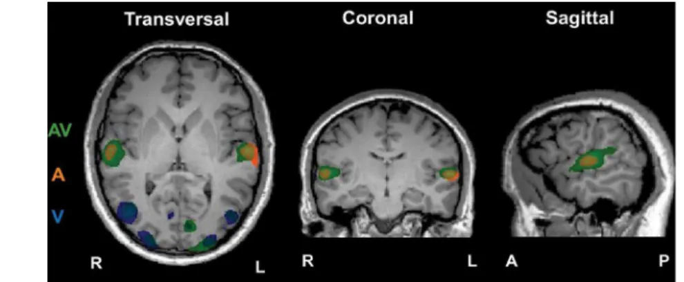
Figure 2. Areas of signiicant correlation between subjects within conditions A (in orange), V (in blue), and
AV (in green). p 0.0001 for all clusters, cluster-level threshold corrected at a 0.05. Slices taken at Talairach
coordinates X5 59, Y5215, and Z5 4 for the sagittal, coronal, and transversal views, respectively.
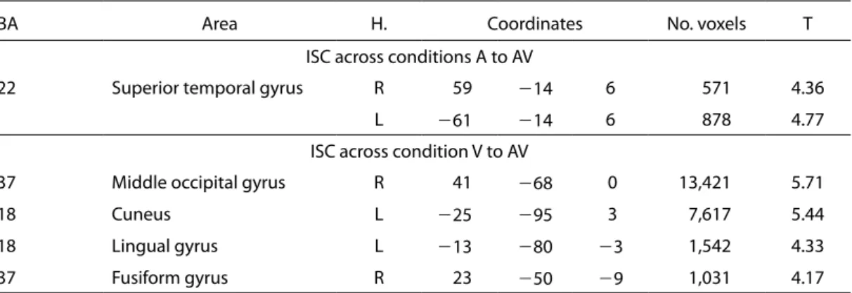
Figure 3. Areas of signiicant correlation between subjects across conditions A to AV (in orange) and V to AV (in
3.2 ISC across conditions
The BOLD time courses across the sensory conditions A and AV showed signiicant correlation in bilateral primary auditory areas while across V and AV showed signiicant correlation in secondary visual areas (see Figure 3 and Table 2). Compared with ISC within conditions (Section 3.1), the extent of the signiicant correlation was reduced in STG, but enhanced in MOG and extrastriate cortex. Nota -bly, V to AV showed correlation only in the left lingual gyrus, consistent with the left lateralisation for this region in ISC AV. Hence, activity in primary and secondary sensory areas in response to a unisen-sory stimulus was partly synchronised with the activity in the same areas produced in response to the multisensory stimulus. This indicates that the processing of the multisensory stimuli at least partly conserved the separate processing characteristics of the unisensory stimuli. No areas were signiicantly synchronised across the two unisensory conditions (A to V).
3.3 GLM-based contrast and conjunction analyses
The GLM-based analyses revealed a pattern of results that was consistent with original expectations and that was supported by the results given in the ISC analysis (see Table 3 and Figure 4). The con-trast AV V represents the involvement of the auditory aspects in AV processing and showed sig-niicant enhanced bilateral activity in the STG, including the primary auditory cortex. The contrast AV A represents the connection of the visual mode within the multisensory condition and showed signiicantly enhanced activity in the right (superior temporal and MOG) and left (MTG, IOG, and cuneus) hemispheres. It is important to note that these contrasts are not revealing brain areas sensitive Table 2. Clusters of signiicant ISC across uni- and multisensory conditions. See the note to Table 1 for columns’ information.
BA Area H. Coordinates No. voxels T
ISC across conditions A to AV
22 Superior temporal gyrus R 59 214 6 571 4.36
L 261 214 6 878 4.77
ISC across condition V to AV
37 Middle occipital gyrus R 41 268 0 13,421 5.71
18 Cuneus L 225 295 3 7,617 5.44
18 Lingual gyrus L 213 280 23 1,542 4.33
37 Fusiform gyrus R 23 250 29 1,031 4.17
Table 3. Signiicant brain activation in GLM analysis for uni- and multisensory contrasts. See the note to Table 1 for columns’ information.
BA Area H. Coordinates No. voxels T
GLM contrast AV V
22 Superior temporal gyrus R 50 28 0 2,783 7.87
L 258 235 12 1,507 7.50
Contrast GLM AV A
22 Superior temporal gyrus R 56 238 12 193 5.79
37 Middle temporal gyrus L 246 262 9 764 8.76
37 Middle occipital gyrus R 44 271 3 1,419 6.60
19 Inferior occipital gyrus L 240 274 23 325 5.31
17 Cuneus L 210 295 3 322 26.20
Conjunction GLM Analysis (AV V) ∩ (AV A)
22 Superior temporal gyrus R 56 232 15 356 4.82
to the unisensory conditions alone; hence, both contrasts manifest aspects of AV: the contrast AV A also showed signiicant activity in areas outside the primary visual cortex (e.g. STG) and the contrast AV V showed enhanced activity over a wider area than was correlated for A. To ind regions associ-ated with multisensory processing, we computed a conjunction analysis (AV V) ∩ (AV A). This analysis revealed bilateral activity in the posterior STS (pSTS) with the signiicant area in the LH being further posterior than the RH.
3.4 Sensory control analysis
To verify that the areas of signiicant ISC were due to sensory processing rather than randomly cor-related activity (e.g. resting state), we conducted an additional control analysis. For this, for each individual subject, the different sensory conditions were randomly assigned to three control groups (R1, R2, and R3). We then calculated the number of voxels synchronised for each control group. The percentage of synchronised voxels of the whole brain was then compared with the percentage of synchronised areas in AV. As visible in Figure 5, the groups containing random assignment of the sensory conditions do not lead to more than 2% of synchronisation across the whole brain.
Figure 4. Signiicant clusters for ISC AV (green) and multisensory conjunction analysis (orange), p 0.001, a
level uncorrected. Slices taken at Talairach coordinates X5 59, Y5234, and Z5 14 for the sagittal, coronal,
and transversal views, respectively.
Figure 5. Percentage of total brain surface signiicantly correlated at p 0.001, a level uncorrected for AV and
three different random assignments of each functional scan to three groups (R1, R2, and R3).
4 Discussion
dance since the narrative and compositional elements are completely unfamiliar and unknown to nov-ices (Reason & Reynolds, 2010; Vatsyayan, 1963) even though the emotional expressions have been found to be universally understood (Hejmadi et al., 2000). A lack of familiarity does not, however, necessarily imply a complete lack of processing the composition. For example, non-signers show competence in understanding the structure of a narrative given by sign language without grasping the meaning (Fenlon, Denmark, Campbell, & Woll, 2007). This is relevant as we studied the level to which sensory processing is correlated between subjects’ brain activity when recognisable gestures are embedded in a novel but structured context that is presented in an unedited format. Unlike an edited feature ilm, we did not expect higher order areas to be correlated; and unlike a recording of a real-life situation (Hasson et al., 2008b), dance is choreographed for movement, sound, and importantly, its combination, and we thus expected enhanced correlation in AV processing across spectators even when presented with an unedited recording.
4.1 Synchronisation and multisensory processing
Brain activity was signiicantly correlated across subjects in several functionally relevant regions for auditory (e.g. Heschl’s gyrus), visual (e.g. lingual gyrus, MOG, cuneus), and multisensory process-ing (e.g. pSTG). Importantly, subjects’ correlation expanded into STG—the area that is frequently reported for processing of AV conditions (Calvert, 2001; Ethofer et al., 2006). Furthermore, the mul-tisensory area pSTS showed enhanced activity as revealed by a GLM conjunction analysis and was partly overlapping with the area that was signiicantly correlated between subjects in the AV condition. Conjunction analysis as a tool to study sensory processing in the human brain has been discussed widely (e.g. Ethofer et al., 2006; Szameitat et al., 2011). It is nevertheless exceptional that the GLM showed similar results to the ISC because we applied GLM in an unconventional block design: a single block for each condition lasting 6 min and 26 s. The GLM does not capture haemodynamic adaptation processes (see Ou, Raij, Lin, Golland, & Hämäläinen, 2009) and thus, normally, repeated presentations of stimuli with short durations are used in order to maximise its power. In light of both ISC and GLM conjunction analyses, our results show that multisensory processing may not only be identiied by enhanced activity but extended synchronisation.
Notably, we did not ind signiicant correlation beyond primary and secondary sensory areas. Our study differs from previous work using ISC on two particular aspects that may explain variation in the results. First, we used a non-edited video of a choreographed dance and, second, we used a more conservative ISC analysis by measuring subject-to-average correlations between several participants. In regard to the former, it is relevant to note that most studies using ISC (e.g. Bartels & Zeki, 2004; Hasson et al., 2004) investigated subjects’ brain response to watching narrative movie sequences that were edited in a particular way, promoting a narrative that maximises the attention of the observer. For instance, Hasson et al. (2004) presented 30 min of the movie “The Good, the Bad, and the Ugly” and reported correlations between subjects over large regions of the brain including higher order areas. Importantly, this ilm is popular and well liked, using highly stylised editing with numerous scene changes and close-ups that draw the spectator into the storyline. Our study explored subjects’ brain responses to long sequences of unedited stimuli which we expected to differ from results obtained from viewing edited ilms.
4.2 The role of STG in sensory processing
parts of STG did not show signiicant correlation between subjects in the audio-only condition. Impor -tantly, the control analyses supported that correlation in pSTS is based on AV correlation, which is unlikely to be observed on the basis of unrelated audio and/or visual stimulation. First, a negligible amount of 2% across the entire brain was correlated randomly. Second, A and V stimulation was present in all conditions (e.g. scanner noise in V, visual control in A), but we found clear differences between correlation for these unisensory conditions and the AV correlation. It is thus very unlikely that the AV correlation in pSTS can be evoked by random, unrelated auditory or visual streams. Finally, many previous publications on AV integration on language and body action processing found that the pSTS activity was enhanced (e.g. Meyer et al., 2011), also when using edited movies (e.g. Wolf et al., 2010). According to Hasson et al. (2008c), who found that the STS was indicative of a coherent pro-gressive narrative by contrasting forward to backward played ilms, we propose that the correlation in STS found here was modulated by the dance narrative, despite its novelty.
While signiicant correlation between subjects in low-level auditory and visual areas has been shown before (e.g. Hasson et al., 2004, 2008c; Lerner, Honey, Silbert, & Hasson, 2011), our study is novel by using a more systematic approach presenting A, V, and AV of the very same dance per-formance, showing evidence for a signiicant correlation of the activity in a multisensory integration area (pSTS) across spectators who watched an unedited recording. This has not been reported before. Hence, ISC seems to be functionally sensitive and has the potential to tackle a number of issues present in AV research when unedited but choreographed complex stimuli are used in their original form. To further investigate why the non-overlapping parts of pSTS showed enhanced activity in a task-related manner (GLM) but were not signiicantly correlated (ISC), additional studies are needed that better fulil the criteria of GLM analysis.
The correlation between subjects in pSTS is indeed interesting considering both continuous audio and visual streams were unfamiliar. Although dance and music in Bharatanatyam are interwoven in such a way that the two arts become one coherent whole (Vatsyayan, 1963), this is not suficient for the novice spectator to either fully comprehend the narrative in such a manner that they would correlate in higher order cognitive areas (see Section 4.4) or to perceive a common cross-modal structure (see Section 4.3). Nevertheless, subjects’ multisensory integration was coherent. In other words, subjects can have a common level of AV integration that leads to idiosyncratic cognitive interpretations. Future studies testing ISC for different levels of disruptions would shed further light on the functional role of STS in the perception of dance structure.
4.3 Other synchronised areas
that is known to be involved in the perception of human form (Downing & Peelen, 2011). In the cur-rent study using movements accompanied by music, this area was bilaterally enhanced (GLM AV A) as well as correlated (ISC AV).
Other laterality differences between correlations for AV and for V were found in the cuneus (right for AV, left for V). Though meta-analyses of previously published work on biological motion percep-tion did reveal lateralisapercep-tion for the RH (Grosbras et al., 2011), it is relevant to note that labelling cortical structures based on group data is not unproblematic, especially for more extensive activations. We used Tailarach Daemon, an automated coordinate-based system (Lancaster et al., 2000) to label the location of the peak activation. However, the cuneus is part of the medial/inferior occipital gyrus as is the lingual gyrus, which is ventral to the cuneus on the lower bank of the calcarine sulcus. One could thus argue that within the scope of a more extended peak activity, that for both, AV and V, the middle occipital and/or the lingual gyri are bilaterally correlated in AV and V. The laterality may thus be an artefact of locating the activity rather than a representation of the actual processes. Hence, the ISC of AV stimuli showed clearly areas that are involved in visual sensory processing but integrating sound with vision led to small but notable changes in the activity across subjects of these visual areas.
Notably, though, the BOLD activity in response to our stimuli did not correlate from sound to vision. Such cross-modal sensory processing has been reported previously but in particular for visual and auditory stimuli that were associated in a more straightforward manner. For instance, visual obser-vation of a light lash evoked an internally generated rhythm (Grahn, Henry, & McAuley, 2011). Fur-thermore, Bidet-Caulet, Voisin, Bertrand, and Fonlupt (2005) found activity in the temporal biological motion area when subjects were listening to a walking human suggesting hierarchical components in processing multisensory stimuli from V5 to posterior STG/STS (see also Beauchamp et al., 2004; Wright et al., 2003). However, our stimuli were much more complex. Since Krumhansl and Schenck (1997) found that music and dance share some common structural patterns, we suggest that our sub-jects did not link these visual and auditory properties due to the novelty and complexity of the stimuli. It is possible that mentally generated images to music as well as mentally evoked music to visual stimulations were present but uncorrelated (i.e. idiosyncratic) across spectators, suggesting that cross-modal synchronisation may only be present for stimuli with low-level correspondences and not for highly complex stimulus material.
Nevertheless, despite the fact that all of our spectators were unfamiliar with the narrative of the Indian dance, a number of unisensory areas and areas of AV integration were correlated. Interest-ingly though, on the one end, subjects’ BOLD response in the lowest level of processing, the primary visual areas, was enhanced (AV A) but sparsely correlated. The lack of extensive correlation in V1 could be due to the free-viewing situation where the focal point can be individual for each subject at each moment in time. Furthermore, on the other end, the activity in higher order areas was neither signiicantly enhanced nor correlated. We argue that this is due to a lack of shared expertise. Though in the case of music, Maess, Koelsch, Gunter, and Frederici (2001) found enhanced cortical activity in higher order areas also for novices. Notably, the authors used classical chords, which are familiar to Westerners.
Hasson and Malach (2006) suggested that ISC allows disentangling the cortex into two systems: areas where subjects process stereotypical responses to the external world and areas that may be linked to individual variation. Similarly, we propose that signs of processing and indices of understanding need to be distinguished. For instance, it is likely that music and/or action are at least partly processed in BA44, but in idiosyncratic ways. In order to link the enhanced cortical activity to shared understand-ing (as in mirror-neuron theories), one would also expect signiicant correlation. For instance, Lerner et al. (2011) found signiicant ISC in the primary auditory cortex on the level of words, whereas the correlation in higher auditory processing areas was sensitive to the length of the intact structure of spoken text. Thus, the more that could be understood (narrative) the higher auditory cortices were cor-related between subjects. Hence, up to a certain level, novices process the stimuli in a similar manner; but irrespective of coherent multisensory integration processes, subsequent higher level processes can be idiosyncratic.
4.4 No correlation in the action observation network
Hamilton, & Grafton, 2006; Orgs, Dombrowski, Heil, & Jansen-Osmann, 2008; Pilgramm et al., 2010). In an earlier transcranial magnetic stimulation study we showed that Bharatanatyam spectators require at least visual experience to enhance muscle-speciic sensorimotor excitement (Jola, Abedian-Amiri, Kuppuswamy, Pollick, & Grosbras, 2012). As stressed earlier, the emotional expressions have been found to be of a universal nature, but the dance and music in which the expressions are embed-ded are highly complex and unfamiliar. It is thus less surprising that the activity in areas associated with motor simulation and emotion recognition was uncorrelated and may be explained by a lack of shared motor or visual expertise between our novices.
It is possible that for synchronisation in the fronto-parietal network, expertise is required. How-ever, Petrini et al. (2011) found reduced activity in areas of AV integration and action–sound represen -tation in expert drummers when compared with novices, including fronto-temporal-parietal regions. Interestingly, while Cross and colleagues (Cross, Hamilton, Kraemer, Kelley, & Grafton, 2009a; Cross, Kraemer, Hamilton, Kelley, & Grafton, 2009b) used music to accompany the movements that dancers learned, none of the studies on dance observation investigated the effect music has on the perception of movement. This is surprising, knowing that the mirror-neuron network is multimodal (e.g. Gazzola, Aziz-Zadeh, & Keysers, 2006; Kohler et al., 2002; Lahav, Saltzman, & Schlaug, 2007) and dance is a complex, multidimensional stimulus consisting of a luid mixture of body movement and sound. As the responses to naturally co-varying sound and actions have been found to be modiied by expertise, future work is required comparing responses of novices and dance experts.
4.5 Merits of ISC
ISC indicates voxels that show signiicant correlation between subjects independent of the level of BOLD activity. ISC is therefore a potential complementary method along with other audio, visual, and AV integration designs (e.g. Beauchamp, 2005; Goebel & van Atteveldt, 2009; Kreifelts et al., 2010; Love et al., 2011). Some known issues from conventional methods however remain while oth-ers are resolved. For instance, current scannoth-ers do not allow capturing individual neuronal activity within a voxel. Thus, neither ISC nor GLM conjunction analysis can distinguish between voxels where unisensory visual and auditory processes coexist and those where AV integration processes take place (Calvert and Thesen, 2004; Szameitat et al., 2011). High-resolution scanning would allow identifying correlated activity of a smaller number of neurons across subjects, but it may reduce ISC as it also increases the effects of anatomical variability. It is thus important to investigate in designs that have greater statistical power but which are still applicable to exploratory approaches such as wavelet cor-relation (Lessa et al., 2011).
Furthermore, the loud scanner noise presents a potential confound of conventional fMRI studies, but it is unlikely that it has signiicantly modiied our results. First and foremost, the scanner noise was most notable in the visual only condition, where we found no auditory areas to be correlated between subjects. Thus, the scanner noise alone was not suficient to drive ISC activation. Furthermore, we found no signiicant correlation in the insula, where activity is reportedly related to scanner noise (Schmidt et al., 2008). Moreover, MacSweeney et al. (2000) showed evidence that the STG activity is independent of scanner noise. In addition, effects of scanner noise on BOLD responses were found to be variable and thus uncorrelated between subjects (Ulmer et al., 1988).
Finally, highly controlled parameterised stimuli may have better allowed contrasting basic physi-cal stimulus properties than the ISC with ecologiphysi-cal valid stimuli. Ecologiphysi-cally valid stimuli indeed consist of a complex mixture of basic feature properties, but are however less arbitrary and closer to real life (less inference steps to be made), less dependent on pre-assumptions (e.g. such as on Haemodynamic Response Function), have fewer additional stimuli effects (e.g. created by artiicial confounds), and prevent acquiring task strategies. We thus argue that natural viewing of complex stimuli of long duration as used here is well suited to studying human perception; that they provide an essential complement to artiicial stimuli and laboratory tasks, and that our indings are a more genu -ine relection of the implicit uni- and multisensory processes in the context of real life. A number of other studies on social interaction (e.g. Risko, Laidlaw, Freeth, Foulsham, & Kingstone, 2012) and AV integration (e.g. de Gelder & Bertelson, 2003; Kreifelts et al., 2010) have recently acknowledged the importance of ecological validity and the potential modiication highly controlled but artiicial stimuli can have on perceptual and cognitive processes. The future challenge is to build a model based on the combination of the two seemingly opposing approaches—the bottom-up approach where models of AV integration are built on results from artiicial stimuli with simple cue combinations and the more top-down approach where AV integration is explored based on naturalistic stimuli.
5 Summary
ISC allows the exploration of sensory areas involved in natural viewing of long stimulus segments, i.e. 6 min. We found a correlation between subjects’ voxel-based time course in previously reported uni- and multisensory areas (occipito-temporal) in early and late stages of sensory processing but we found that subjects’ brain responses were not synchronised in higher order areas relevant for cogni-tion, accogni-tion, and/or emotion. Thus, this study highlights that by presenting a dance form unfamiliar to subjects, correspondences between subjects can be constraint onto the level of sensory AV processing. We also did not ind cross-modal synchronisation (A to V), despite our stimuli showing a narrative of culturally speciic choreographed movements to music. We thus situate our unfamiliar unedited but choreographed dance stimuli between edited feature ilms and random recordings of everyday scenes: we found less correlated areas synchronised than studies that used classically edited movies but more than for non-edited unchoreographed recordings of everyday situations. Our data support the idea that spectators’ visual and auditory processes can be directed to some extent by choreographed movement and music without changes in the visual scene. Furthermore, ISC can show additional indings to conventional GLM analyses and should thus be considered a complementary tool to standard contrast analysis when exploring multisensory integration processes.
Acknowledgments. This study was supported by the Arts and Humanities Research Council (AHRC). Phil McAleer was supported by a grant from the Ministry of Defence. The authors thank Prof. Uri Hasson, Prof. Rainer Goebel, and Jukka-Pekka Kauppi for their helpful analysis suggestions. Furthermore, the authors thank Frances Crabbe for support with the scanning, Jen Todman for the icon picture, and the dance performer Dr Anna Kuppuswamy.
References
Aguirre, G. K., Zarahn, E., & D’Esposito, M. (1998). The variability of human, BOLD hemodynamic responses.
NeuroImage, 8(4), 360–369. doi:10.1006/nimg.1998.0367
Arrighi, R., Marini, F., & Burr, D. (2009). Meaningful auditory information enhances perception of visual biological motion. Journal of Vision, 9(4), 25 (1–7). doi:10.1167/9.4.25
Bläsing, B., Calvo-Merino, B., Cross, E. S., Jola, C., Honisch, J., & Stevens, C. J. (2012). Neurocognitive control in dance perception and performance. Acta Psychologica, 139(2), 300–308. doi.10.1016/j.actpsy
Calvert, G. A. (2001). Crossmodal processing in the human brain: insights from functional neuroimaging studies. Cerebral Cortex, 11(12), 1110–1123. doi:10.1093/cercor/11.12.1110
Calvert, G. A., Bullmore, E. T., Brammer, M. J., Campbell, R., Williams, S. C. R., McGuire, P. K., David, A. S. (1997). Activation of auditory cortex during silent lipreading. Science, 276(5312), 593–596. doi:10.1126/ science.276.5312.593
Calvert, G. A., Campbell, R., & Brammer, M. J. (2000). Evidence from functional magnetic resonance imaging of crossmodal binding in the human heteromodal cortex. Current Biology, 10(11), 649–657. doi:10.1016/ S0960-9822(00)00513-3
Calvert, G. A., & Thesen, T. (2004). Multisensory integration: Methodological approaches and emerging principles in the human brain. Journal of Physiology Paris, 98(1–3), 191–205. doi:10.1027/1618-3169.55.2.121
Calvo-Merino, B., Glaser, D. E., Grèzes, J., Passingham, R. E., & Haggard, P. (2005). Action observation and acquired motor skills: An fMRI study with expert dancers. Cerebral Cortex, 15(8), 1243–1249. doi:10.1093/cercor/ bhi007
Calvo-Merino, B., Grèzes, J., Glaser, D. E., Passingham, R. E., & Haggard, P. (2006). Seeing or doing? Inluence of visual and motor familiarity in action observation. Current Biology, 16(19), 1905–1910. doi:10.1007/s00426-010-0280-9
Calvo-Merino, B., Jola, C., Glaser, D. E., & Haggard, P. (2008). Towards a sensorimotor aesthetics of performing art. Consciousness and Cognition, 17(3), 911–922. doi:10.3389/fnhum.2011.00102
Cross, E. S., Hamilton, A. F., & Grafton, S. T. (2006). Building a motor simulation de novo: Observation of dance by dancers. NeuroImage, 31(3), 1257–1267. doi:10.1016/j.neuroimage.2006.01.033
Cross, E. S., Hamilton, A. F., Kraemer, D. J., Kelley, W. M., & Grafton, S. T. (2009a). Dissociable substrates for body motion and physical experience in the human action observation network. European Journal of Neuroscience, 30(7), 1383–1392. doi:10.1111/j.1460-9568.2009.06941.x
Cross, E. S., Kraemer, D. J., Hamilton, A. F., Kelley, W. M., & Grafton, S. T. (2009b). Sensitivity of the action observation network to physical and observational learning. Cerebral Cortex, 19(2), 315–326. doi:10.1093/cercor/bhn083
de Gelder, B., & Bertelson, P. (2003). Multisensory integration, perception and ecological validity. TRENDS in Cognitive Sciences, 7(10), 460–467. doi:10.1016/j.tics. 2003.08.014
de Gelder, B., Snyder, J., Greve, D., Gerard, G., & Hadjikhani, N. (2004). Fear fosters light: A mechanism for fear contagion when perceiving emotion expressed by a whole body. PNAS, 101(47), 16701–16706. doi:10.1073/pnas.0407042101
Downing, P. E., & Peelen, M. V. (2011). The role of occipitotemporal body-selective regions in person perception. Cognitive Neuroscience, 2(3–4), 186–203. doi:10.1002/mds.23736
Ethofer, T., Pourtois, G., & Wildgruber, D. (2006). Investigating audiovisual integration of emotional signals in the human brain. Progress in Brain Research, 156, 345–361. doi:10.1162/jocn.2009.21099
Fenlon, J., Denmark, T., Campbell, R., & Woll, B. (2007). Seeing sentence boundaries. Sign Language and Linguistics, 10(2), 177–200. doi:10.1075/sll.10.2.06fen
Forman, S. D., Cohen, J. D., Fitzgerald, M., Eddy, W. F., Mintun, M. A., & Noll, D. C. (1995). Improved assessment of signiicant activation in functional magnetic resonance imaging (fMRI): Use of a cluster-size threshold. Magnetic Resonance in Medicine, 33(5), 636–647. doi:10.1002/mrm.1910330508
Friston, K. J. (2005). Models of brain function in neuroimaging. Annual Review of Psychology, 56, 57–87. doi:10.1146/annurev.psych.56.091103.070311
Friston, K. J., Penny, W., & Glaser, D. E. (2005). Conjunction revisited. NeuroImage, 25(3), 661–667. doi:10.1002/hbm.20242
Gauthier, I., Tarr, M. J., Moylan, J., Skudlarski, P., Gore, J. C., & Anderson, A. W. (2000). The fusiform “face area” is part of a network that processes faces at the individual level. Journal of Cognitive Neuroscience,
12(3), 495–504. doi:10.1037/0096-1523.28.2.431
Gazzola, V., Aziz-Zadeh, L., & Keysers, Chr. (2006). Empathy and the somatotopic auditory mirror system in humans. Current Biology, 16(18), 1824–1829. doi:10.1016/j.cub.2006.07.072
Goebel, R., Esposito, F., & Formisano, E. (2006). Analysis of functional image analysis contest (FIAC) data with brainvoyager QX: From single-subject to cortically aligned group general linear model analysis and self-organizing group independent component analysis. Human Brain Mapping, 27(5), 392–401. doi:10.1002/hbm.20249
Goebel, R., & van Atteveldt, N. (2009). Multisensory functional magnetic resonance imaging: A future perspective. Experimental Brain Research, 198(2–3), 153–164. doi:10.1186/1471-2202-11-11
Grahn, J. A., Henry, M. J., & McAuley, J. D. (2011). FMRI investigation of cross-modal interactions in rhythm perception: Audition primes vision, but not vice versa. NeuroImage, 54(2), 1231–1243.doi:10.1016/j. neuroimage.2010.09.033
Grafton, S. T. (2009). Embodied cognition and the simulation of action to understand others. Annals of the New York Academy of Sciences, 1156, 97–117. doi:10.1111/j.1749-6632.2009.04425.x
Grill-Spector, K., & Malach, R. (2004). The human visual cortex. Annual Review of Neuroscience. 27, 649–677. doi:10.1146/annurev.neuro.27.070203.144220.
Grosbras, M. H., Beaton, S., & Eickhoff, S. B. (2011). Brain regions involved in human movement perception: A quantitative voxel-based meta-analysis. Human Brain Mapping, 33(2), 431–454. doi:10.1002/ hbm.21222
Handwerker, D. A., Ollinger, J. M., & D’Esposito, M. (2004). Variation of BOLD hemodynamic responses across subjects and brain regions and their effects on statistical analyses. NeuroImage, 21(4), 1639–1651. doi:10.1016/j.neuroimage.2009.11.014
Hasson, U., Avidan, G., Gelbard, H., Vallines, I., Harel, M. Minshew, N., & Behrmann, M. (2009a). Shared and idiosyncratic cortical activation patterns in autism revealed under continuous real-life viewing conditions. Autism Research, 2(4), 220–231. doi:10.1016/j.visres.2012.11.002
Hasson, U., Furman, O., Clark, D., Dudai, Y., & Davachi, L. (2008a). Enhanced intersubject correlations during movie viewing correlate with successful episodic encoding. Neuron, 57(3), 452–462. doi:10.3389/ fnhum.2012.00248
Hasson, U., Landesman, O., Knappmeyer, B., Vallines, I., Rubin, N., & Heeger, D. (2008b). Neurocinematics: The neuroscience of ilms. Projections: The Journal for Movies and Mind, 2(1), 1–26. doi:10.3167/ proj.2008.020102
Hasson, U., & Malach, R. (2006). Human brain activation during viewing of dynamic natural scenes. Novartis Foundation Symposium, 270, 203–212. doi:10.1155/2012/375148
Hasson, U., Malach, R., & Heeger, D. J. (2009b). Reliability of cortical activity during natural stimulation.
Trends in Cognitive Sciences, 14(1), 40–48. doi:10.1371/journal.pbio.1001462
Hasson, U., Nir, Y., Levy, I., Fuhrmann, G., & Malach, R. (2004). Intersubject synchronization of cortical activity during natural vision. Science, 303(5664), 1634–1640. doi:10.1126/science.1089506
Hasson, U., Yang, E., Vallines, I., Heeger, D. J., & Rubin, N. (2008c). A hierarchy of temporal receptive windows in human cortex. Journal of Neuroscience, 28(10), 2539–2550. doi:10.1523/ JNEUROSCI.3684-10
Hein, G., & Knight, R. T. (2008). Superior temporal sulcus—It’s my area: Or is it? Cognitive Neuroscience,
20(12), 2125–2136. doi:10.3410/f.13254956.14607054
Hejnar, M. P., Kiehl, K. A., & Calhoun, V. D. (2007). Interparticipant correlations: A model free FMRI analysis technique. Human Brain Mapping, 28(9), 860–867. doi:10.1002/hbm.20321
Hejmadi, A., Davidson, R. J., & Paul, R. (2000). Exploring Hindu Indian emotion expressions evidence for accurate recognition by Americans and Indians. Psychological Science, 11(4), 183–187. doi:10.1093/ acprof:oso/9780195373585.003.0029
Jääskeläinen, I. P., Koskentalo, K., Balk, M. H., Autti, T., Kauramäki, J., Pomren, C., & Sams, M. (2008). Inter-subject synchronization of prefrontal cortex hemodynamic activity during natural viewing. Open Neuroimage Journal, 2, 14–19. doi:10.3389/fnhum.2012.00298
Jola, C., Abedian-Amiri, A., Kuppuswamy, A., Pollick, F. E., & Grosbras, M. H. (2012). Motor simulation without motor expertise: Enhanced corticospinal excitability in visually experienced dance spectators.
PLoS One, 7(3), e33343. doi:10.1371/journal.pone.0033343
Jola, C., Davis, A., & Haggard, P. (2011). Proprioceptive integration and body representation: Insights into dancers’ expertise. Experimental Brain Research, 213(2–3), 257–265. doi:10.1016/j.actpsy.2011.12.005.
Kohler, E., Keysers, C., Umiltà, M. A., Fogassi, L., Gallese, V., & Rizzolatti, G. (2002). Hearing sounds, understanding actions: Action representation in mirror neurons. Science, 297(5582), 846–848. doi:10.1073/pnas.1205553109
Kreifelts, B., Ethofer, T., Grodd, W., Erb, M., & Wildgruber, D. (2007). Audiovisual integration of emotional signals in voice and face: An event-related fMRI study. NeuroImage, 37(4), 1445–1456. doi:10.1016/j. neuroimage.2007.06.020
Kreifelts, B., Ethofer, T., Huberle, E., Grodd, W., & Wildgruber, D. (2010). Association of trait emotional intelligence and individual fMRI-activation patterns during the perception of social signals from voice and face. Human Brain Mapping, 31(7), 979–991. doi:10.1002/hbm.20913
Krumhansl, C. L., & Schenck, D. L. (1997). Can dance relect the structural and expressive qualities of music? A perceptual experiment on Balanchine’s choreography of Mozart’s Divertimento no. 15. Musicae Scientiae, 1(1), 63–85. doi:10.1111/j.1469-8986.1969
Laurienti, P. J., Burdette, J. H., Wallace, M. T., Yen, Y., Field, A. S., & Stein, B. E. (2002). Deactivation of sensory-speciic cortex by polymodal stimuli. Journal of Cognitive Neuroscience, 14(3), 420–429. doi:10.1523/JNEUROSCI.0910-09.2009
Lahav, A., Saltzman, E, & Schlaug, G. (2007). Action representation of sound: Audiomotor recognition network while listening to newly acquired actions. Journal of Neuroscience, 27(2), 308–314. doi:10.1523/ JNEUROSCI.4822-06.2007
Lancaster, J. L., Woldorff, M. G., Parsons, L. M., Liotti, M., Freitas, C. S., Rainey, L., Fox, P. T. (2000). Automated Talairach atlas labels for functional brain mapping. Human Brain Mapping, 10(3), 120–131.
Le, T. H., Pardo, J. V., & Hu, X. (1998). 4 T-fMRI study of nonspatial shifting of selective attention: Cerebellar and parietal contributions. Journal of Neurophysiology, 79(3), 1535–1548. doi:10.1016/j. neuroimage.2005.10.032
Lerner, Y., Honey, C. J., Silbert, L. J., & Hasson, U. (2011). Topographic mapping of a hierarchy of temporal receptive windows using a narrated story. Journal of Neuroscience, 31(8), 2906–2915. doi:10.1523/ JNEUROSCI.3684
Lessa, P. S., Sato, J. R., Cardoso, E. F., Neto, C. G., Valadares, A. P., & Amaro, E. (2011). Wavelet correlation between subjects: A time-scale data driven analysis for brain mapping using fMRI. Journal of Neuroscience Methods, 194(2), 350–357. doi:10.1016/j.jneumeth.2010.09.005
Li, Y., Wang, G., Long, J., Yu, Z., Lian, G., Li, Z., & Sun, P. (2011). Reproducibility and discriminability of brain patterns of semantic categories enhanced by congruent audiovisual stimuli. PLoS One, 6(6), e20801. doi:10.1371/journal.pone.0020708
Love, S. A., Pollick, F. E., & Latinus, M. (2011). Cerebral correlates and statistical criteria of cross-modal face and voice integration. Seeing and Perceiving, 24(4), 351–367. doi:10.1371/journal.pone.0019165
Love, S. A., Pollick, F. E., & Petrini, K. (2012). Effects of experience, training and expertise on multisensory perception: Investigating the link between brain and behaviour. Lecture Notes in Computer Science: Proceedings on Cognitive Behavioural Systems, 7403, 304–320. doi:10.1007/978-3-642-34584-5_27
MacSweeney, M., Amaro, E., Calvert, G. A., Campbell, R., David, A. S., McGuire, P., Brammer, M. J. (2000). Silent speechreading in the absence of scanner noise: An event-related fMRI study. Neuroreport, 11(8), 1729–1733.
MacSweeney, M., Campbell, R., Woll, B., Giampietro, V., David, A. S., McGuire, P. K., Brammer, M. J. (2004). Dissociating linguistic and nonlinguistic gestural communication in the brain. NeuroImage, 22(4), 1605–1618. doi:10.1016/j.neuroimage.2004.03.015
McGurk, H., & MacDonald, J. (1976). Hearing lips and seeing voices. Nature, 264(5588), 746–748. doi:10.1073/pnas.0804275105
McNamara, A., Buccino, G., Menz, M. M., Gläscher, J., Wolbers, T., Baumgärtner, A., & Binkofski, F. (2008). Neural dynamics of learning sound–action associations. PLoS One, 3(12), e3845. doi:10.1016/j. physbeh.2008.06.011
Meyer, G. F., Greenlee, M., & Wuerger, S. (2011). Interactions between auditory and visual semantic stimulus classes: Evidence for common processing networks for speech and body actions. Journal of Cognitive Neuroscience, 23(9), 2291–2308. doi:10.1162/jocn.2010.21575
Möttönen, R., Calvert, G. A., Jääskeläinen, I. P., Matthews, P. M., Thesen, T., Tuomainen, J., & Sams, M. (2006). Perceiving identical sounds as speech or non-speech modulates activity in the left posterior superior temporal sulcus. NeuroImage, 30(2), 563–593. doi:10.1016/j.neuroimage.2005.10.002
Nath, A. R., & Beauchamp, M. S. (2011). Dynamic changes in superior temporal sulcus connectivity during perception of noisy audiovisual speech. Journal of Neuroscience, 31(5), 1704–1714. doi:10.1523/ JNEUROSCI.2605-11.2011
Navarra, J., & Soto-Faraco, S. (2005). Hearing lips in a second language: Visual articulatory information enables the perception of second language sounds. Psychological Research, 71(1), 4–12. doi:10.1016/j. actpsy.2008.08.004
Orgs, G., Dombrowski, J. H., Heil, M., & Jansen-Osmann, P. (2008). Expertise in dance modulates alpha/beta event-related desynchronization during action observation. European Journal of Neuroscience, 27(12), 3380–3384. doi:10.1111/j.1467-7687.2010.00991.x
Ou, W., Raij, T., Lin, F.-H., Golland, P., & Hämäläinen, M. (2009). Modeling adaptation effects in fMRI analysis. Medical Image Computing and Computer-Assisted Intervention, 12(Pt 1), 1009–1017. doi:10.1007/978-3-642-04268-3_124
Petrini, K., Pollick, F. E., Dahl, S., McAleer, P., McKay, L. S., Rocchesso, D., Puce, A. (2011). Action
expertise reduces brain activity for audiovisual matching actions: An fMRI study with expert drummers.
NeuroImage, 56(3), 1480–1492. doi:10.1016/j.neuroimage.2011.03.009
Pilgramm, S., Lorey, B., Stark, R., Munzert, J., Vaitl, D., & Zentgraf, K. (2010). Differential activation of the lateral premotor cortex during action observation. BMC Neuroscience, 11, 89. doi:10.1186/1471-2202-11-89
Pillai, S. (2002). Rethinking global Indian dance through local eyes: The contemporary Bharatanatyam scene in Chennai. Dance Research Journal, 34(2), 14–29. doi:10.1080/14647890903568305
Raij, T., Uutela, K., & Hari, R. (2000). Audiovisual integration of letters in the human brain. Neuron, 28(2), 617–625. doi:10.1016/S0896-6273(00)00138-0
Reale, R. A., Calvert, G. A., Thesen, T., Jenison, R. L., Kawasaki, H., Oya, H., Brugge, J. F. (2007). Auditory– visual processing represented in the human superior temporal gyrus. Neuroscience, 145(1), 162–84. doi:10.1016/j.bandl.2008.10.005
Reason, M., & Reynolds, D. (2010). Kinesthesia, empathy, and related pleasures: An inquiry into audience experiences of watching dance. Dance Research Journal, 42(2), 49–75. doi:10.3366/drs.2011.0019
Risko, E. F., Laidlaw, K., Freeth, M., Foulsham, T., & Kingstone, A. (2012). Social attention with real versus reel stimuli: Toward an empirical approach to concerns about ecological validity. Frontiers in Human Neuroscience, 6: 143. doi:10.3389/fnhum.2012.00143
Rizzolatti, G., & Sinigaglia, C. (2010). The functional role of the parieto-frontal mirror circuit: Interpretations and misinterpretations. Nature Review Neuroscience, 11(4), 264–274. doi:10.1038/nrn2805
Schmidt, C. F., Zaehle, T., Meyer, M., Geiser, E., Boesiger, P., & Jancke, L. (2008). Silent and continuous fMRI scanning differentially modulate activation in an auditory language comprehension task. Human Brain Mapping, 29(1), 46–56. doi:10.1371/journal.pone.0054273
Sekiyama, K., Kanno, I., Miura, S., & Sugita, Y. (2003). Auditory–visual speech perception examined by fMRI and PET. Neuroscience Research, 47(3), 277–287. doi:10.1016/S0168-0102(03)00214-1
Sevdalis, V., & Keller, P. E. (2011). Captured by motion: Dance, action understanding, and social cognition.
Brain and Cognition, 77(2), 231–236. doi:10.3389/fnhum.2011.00102
Szameitat, A. J., Schubert, T., & Müller, H. J. (2011). How to test for dual-task-speciic effects in brain imaging studies—an evaluation of potential analysis methods. NeuroImage, 54(3), 1765–1773. doi:10.1037a0025816
Szycik, G. R., Tausche, P., & Münte, T. F. (2008). A novel approach to study audiovisual integration in speech perception: Localizer fMRI and sparse sampling. Brain Research, 1220, 142–149. doi:10.1016/j. brainres.2007.08.027
Talairach, J., & Tournoux, P. (1988). Co-planar stereotaxic atlas of the human brain: 3-dimensional proportional system—an approach to cerebral imaging. New York: Thieme Medical Publishers.
doi.org/10.1016/S1361-8415(01)00043-3
Ulmer, J. L., Biswal, B. B., Yetkin, F. Z., Mark, L. P., Mathews, V. P., Prost, R. W., Daniels, D. L. (1998). Cortical activation response to acoustic echo planar scanner noise. Journal of Computer Assisted Tomography, 22(1), 111–119.
Vatsyayan, K. (1963). Notes on the relationship of music and dance in India. Ethnomusicology, 7(1), 33–38. doi:10.2307/924145
Copyright 2013 C Jola et al.