Prevalence of brain calcifications
in a Brazilian cohort
A retrospective study in radiology services
Matheus Fernandes de Oliveira1, Edison Barros e Silva2, João Ricardo Mendes de Oliveira2
ABSTRACT. Brain calcifications may be present in a wide range of neuropsychiatric, infectious traumatic, neoplastic and endocrine conditions, associated with motor and cognitive symptoms. They are often considered “physiologic” or pathologic. Objective: To determine the prevalence of brain calcifications in a Brazilian cohort. Methods: 1898 consecutive patients underwent skull CTs, comprising 836 men and 1062 women. Results: Among all 1898 patients, 333 (197 women and 136 men) presented with brain calcifications, representing an overall prevalence of 17.54%. The prevalence in the female group was 18.54% versus 16.26% in the male group. Conclusions: A growth in incidental findings on CT scans are likely as these exams become more widely available. However, a current debate between radiologists and regulators is set to redefine the CT usage, especially in the United States and the United Kingdom, considering new norms for use and radiation dosage per exam.
Key words: brain calcinosis, basal ganglia calcification, neuroimaging, neuroepidemiology.
PREVALÊNCIA DAS CALCIFICAÇÕES CEREBRAIS EM UMA COORTE BRASILEIRA: ESTUDO RETROSPECTIVO EM SERVIÇOS DE RADIOLOGIA
RESUMO. Calcificações cerebrais podem estar presentes em uma grande variedade de condições médicas, incluindo doenças infecciosas, endocrinológicas, traumáticas, neoplásicas, psiquiátricas e neurológicas, associados a diferentes manifestações clínicas. Elas são muitas vezes consideradas “fisiológicas” ou patológicas. Objetivo: Determinar a prevalência de calcificações cerebrais em uma coorte brasileira. Métodos: 1898 pacientes consecutivos realizaram tomografia de crânio simples. 836 eram homens e 1062 eram mulheres. Resultados: Entre todos os 1.898 pacientes, 333 (197 mulheres e 136 homens) apresentaram calcificações cerebrais. Assim, a prevalência total foi de 17,54%. No grupo feminino a prevalência foi de 18,54% e nos homens, 16,26%. Conclusões: Provavelmente os achados incidentais em tomografias computadorizadas (TC) serão crescentes, uma vez que estes exames estão mais disponíveis. No entanto, um debate atual entre radiologistas provavelmente deverá redefinir o uso de TC muito em breve, especialmente nos Estados Unidos e Reino Unido.
Palavras-chave: neuroepidemiologia, calcinose cerebral, neuroimagem.
INTRODUCTION
B
rain calciications may be present in a wide range of neuropsychiatric, infectious and endocrine conditions, associated with motor and cognitive symptoms.1-9.hese BCs are often considered “physio-logic” or pathologic. Intracranial physiological calciications are disconected of a demonstra-ble pathological cause and, especially in older
subjects, are represented mainly by pineal gland, habenula and choroid plexus calcinosis. Other sites such as the vasculature or paren-chyma are more often found in the context of clinical manifestations such as parkinsonism, dementia, psychosis and mood disorders.1-11
Physiological calciications, such as in pi-neal gland, habenula and choroid plexus of lateral ventricles can appear in up to 50-70%
1MD, Neurosurgery Residency Program. Hospital do Servidor Público Estadual de São Paulo, São Paulo SP, Brazil. 2MD, PhD, Neuropsychiatric Department - Federal
University of Pernambuco (UFPE), Recife PE, Brazil.
Matheus Fernandes de Oliveira. Department of Neurosurgery, Hospital do Servidor Público Estadual de São Paulo – Rua Pedro de Toledo, 1800 - 04029-000 São Paulo SP - Brazil. E-mail: [email protected]
of patients.4-9 Basal ganglia calciications (BGC) are also
well described in the context of idiopathic and second-ary causes, having been demonstrated in various studies with a prevalence ranging from 0.3% to 12%.1,11-14
Brain calcinosis is usually found in patients over 30 years old, increasing progressively with age. Given the wider use of neuroimaging techniques, particularly to-mography, BCs are increasingly viewed even in asymp-tomatic patients.1,2,11-17
Reports also depend on the proile of the health fa-cility where data is gathered and analyzed because most can be highly biased towards a given level of severity, gender, age range or medical area of expertise such as Neurology, Psychiatry, Radiology or Geriatrics.
During a one-year period, 4219 consecutive comput-ed tomograms (CT) were reviewcomput-ed for basal ganglia cal-ciications and 14 patients harboring calcal-ciications were identiied. Calciications on CT scans were bilateral in 12 cases and unilateral in 2 cases. he globus pallidus was the site of calciication in 13 of the 14 patients. Bilateral dentate nucleus calciication was seen in one patient.1,18
Ostling et al. (2003) studied the cross-sectional re-lationship between psychotic symptoms and BGC in a population sample of non-demented subjects, all of whom were 85 years old: 86 mentally healthy, 11 psy-chotic, 21 with mood disorders, and 20 subjects with anxiety disorders. Basal ganglia calciications on CT were observed in 19% of the mentally healthy and 64% of the non-demented individuals with hallucinations or delusions. he authors concluded that basal ganglia calciication is strongly associated with psychotic symp-toms in old age.18
Eskandary et al. (2005) found 3 cases of abnormal calciication, in the pineal region, basal ganglia, and temporal horn area, respectively, amongst 3000 CTs of head trauma patients with a mean age of 32±17.76 years attended at an emergency facility.19
Radaideh et al. (2012) studied a total of 1040 CT scans, observing an overall prevalence of basal ganglia calciications of 1.25%. he prevalence increased with age; being 0.6% in younger age group vs. 2.4% in sub-jects older than 60 years. Elevated parathyroid hormone was found in 6 patients; of whom only one proved to have low vitamin D3 level.20
Calciications in pediatric radiology became a much more common inding after the pandemic of children infected with HIV, most of whom acquired the condition from their infected mother.1
Few studies have speciically addressed the preva-lence of other sites of brain calciications and tend to associate their indings with speciic symptoms. he
ob-jective of the present study was to determine the preva-lence of brain calciications in a Brazilian cohort.
METHODS
A total of 1898 consecutive patients submitted to Skull computerized tomography (CT) had their CTs evaluated in order to determine the prevalence of intracranial cal-ciications. hese images were consecutively performed at two diferent medical institutions (Hospital das Clíni-cas da Universidade Federal de Pernambuco e Instituto de Medicina Integral de Pernambuco) from April 2006 to April 2007. A total of 332 exams were collected at the Clinical Hospital of the Federal University of Pernambu-co (HC-UFPE) and 1566 at the Instituto de Medicina In-tegral de Pernambuco (IMIP). his project was approved by the Research and Ethics Committee of the Federal University of Pernambuco.
All exams were evaluated by a single radiologist in order to determine the presence of pathological brain calciications and their anatomical site (vascular and parenchymal). CTs were performed with a standardized protocol, consisting of slices of 3 mm in posterior fossa and 10 mm in the supratentorial space.
When evaluating age distribution, the WHO (World Health Organization) classiication was employed, which considers the pediatric group as all patients un-der 20 years old; adults from 20 to 59 years old and el-derly over 60 years old.
Statistics. he numerical data were expressed as mean ± standard deviation. he categorical data were expressed as percentages. Student’s t-test was used for unpaired groups. he signiicance level was established as p<0.05.
RESULTS
Overall, 1898 consecutive patients had their skull CTs evaluated. A total of 332 exams were conducted at the Hospital das Clínicas da Universidade Federal de Per-nambuco (HC-UFPE) and 1566 at Instituto de Medici-na Integral de PerMedici-nambuco (IMIP) involving 836 men and 1062 women. he mean age in the IMIP group was 34.25±24.82 years whereas the mean age in the HC-UF-PE group was 47±23.27 years. here was a statistically signiicant diference between age proile in the two in-stitutions (p<0.05).
Table 1. Characteristics of brain calcifications.
Site Patients Prevalence Mean age (years) Age extremes Gender predominance*
Vascular Carotid 244 12.85% 66 1-102 F>M
Vertebral 35 1.84% 68 1-93 F>M
Basilar 17 0.89% 69 63-93 M>F
Parenchymal Basal Ganglia 46 2.42% 50 1-88 F>M
Parietal 26 1.36% 36 1-84 F>M
Occipital 10 0.52% 36 7-79 F>M
Cerebral Falx 10 0.52% 54 30-51 F>M
Frontal 8 0.42% 38 14-68 F=M
Cerebellar 7 0.36% 32 3-72 F>M
Temporal 7 0.36% 41 11-68 F>M
Brainstem 2 0.01% 45 40-50 F=M
Cerebellar falx 2 0.01% 26 3-50 F=M
*Without statistical significance (p<0.05).
100
90
80
70
60
50
40
30
20
10
0
< 5 5-10 11-20 21-30 31-40 41-50 51-60 61-70 71-80 81-90 > 90
Age (years) 11 11
6 10 9 30
51
87 85
31
2
Number of patients with brain calcifications according to age
Figure 1. Number of patients with BC according to age group.
Figure 2. Prevalence of brain calcinosis across different age groups.
70
60
50
40
30
20
10
0
5,16
Prevalence of brain calcifications (%)
5,39 2,71 5,46 4,28
12,29
22,76 41,82
62,5 65,95
40
< 5 5-10 11-20 21-30 31-40 41-50 51-60 61-70 71-80 81-90 > 90
Among all 1898 patients, 333 (197 women and 136 men) presented with brain calciications correspond-ing to an overall prevalence of 17.54%. he prevalence in the female group was 18.54% whereas among males the prevalence was 16.26%. he absolute number of pa-tients with BC, and prevalence of BC according to age, are shown in Figures 1 and 2.
Among intracranial vascular calciications, the carot-id artery was the main site afected with a prevalence of 12.85%, followed by the vertebral artery at 1.84% and the basilar artery at 0.89%. here was no statistically signiicant diference between males and females.
Among parenchymal calciications, the basal ganglia was the main site afected with a prevalence of 2.42%, followed by parietal at 1.36%, occipital at 0.52%, ce-rebral falx at 0.52%, frontal at 0.42%, cerebellum at 0.36%, and temporal at 0.26% of patients. Brainstem and cerebellar falx each accounted for 2 patients. hala-mus and sella represented 1 patient each (Table 1).
DISCUSSION
Due to growing use of neuroimaging techniques, par-ticularly CTs, brain calciications are detected more of-ten.1,14 Indeed, even the standardized protocol applied
in most reference centers as well as in this study may underestimate the prevalence of calciications. Multiple detector scanners are able to perform 0.5 mm thickness slices and thus calciications smaller than this size may go undetected.
An intriguing consequence is the incidental inding of BC during the investigation of milder symptoms, some being transient, in asymptomatic subjects with massive calciications and positive family history, in emergency rooms or even randomly during the inves-tigation of other pathologies afecting the patient´s face.1,14
For example, a pediatric case in which a 12-year-old girl presented transient symptoms who recovered completely after symptomatic treatment. A CT screen-ing showed substantial calcinosis on her brain and also found in her other asymptomatic siblings.21 Other
au-thors unexpectedly found three generations of asymp-tomatic carriers of brain calcinosis after investigating the kindred of a psychotic patient.22
Our analysis detected a prevalence of brain calciica-tions of 17.54% among 1898 CTs collected at two medi-cal facilities, from subjects between 3 months of age and 103 years old. hese two facilities had diferent proiles with one biased towards a younger population of chil-dren and the other focused on older adults and seniors. None of these facilities worked as emergency rooms
and both focused equally on inpatient and outpatient caregiving. Intracranial vascular calciications were the main indings, followed by basal ganglia calciications. Although a female predominance was observed in most calciication types, statistical analysis failed to reveal a signiicant diference (p>0.05).
Interestingly, the hospital oriented for the adult group had higher rates of calciication. A signiicantly higher presence of basal ganglia calciication was de-tected in subjects over 60 years old, suggesting a prob-able link between calcinosis and the processes of aging and neuronal death. he prevalence of brain calcinosis across the diferent age groups is given in Figure 2 which also shows a progressive increase across diferent age groups when pooling together all sites of calciication detected, not only in basal ganglia but also including the cerebellum, white matter, pineal and vascular deposits.
Curiously, a dip in prevalence was observed after 90 years old. We hypothesized that there is a selection efect induced by calciication in the older old, with a smaller number of carriers after this cut-of, character-ized by elder subjects with less prior calcinosis better brain health and consequently less calciication.
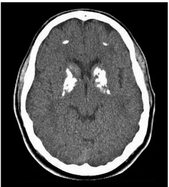
We also identiied unusual images from subjects with massive brain, calcinosis, yet only mild symptoms, suggesting a high level of resilience against brain calci-nosis (Figure 3).
Intracranial vascular calciications are usually asso-ciated with higher cardiovascular and ischemic cerebro-vascular disease risk and may even represent a sign in the context of the atherosclerosis process.23-25 In
chil-dren, vascular calciications are also associated with obesity and cardiovascular diseases while chronic renal failure must also be excluded, since parathormone dis-turbances lead to ectopic calciications.26,27,28
Parenchymal calciications, on the other hand, rep-resent a challenge, given they are found in a myriad of situations. Moreover, other sites of brain calciications have also become the focus of recent attention, no longer merely as an incidental inding but as a predic-tive marker of prognosis in other neurodegenerapredic-tive conditions. Using computed tomography, Mahlberg et al. (2008) found that the degree of pineal calciication in patients with AD was signiicantly higher than in patients with other types of dementia, depression or among controls.29 Another interesting fact is the already
known association of pineal calciications and psychotic syndromes, such as schizophrenia.30 hus,
mineraliza-tion remains a challenge in modern neuroscience, test-ing cerebral resilience and promottest-ing the discussion over diferences between physiological and pathological conditions.
In the reported sample, very few calciications were depicted in the pineal gland, choroid plexus and ha-benula and this underreporting reveals the potential bias concerning conditions considered physiological or pathological. Although pathological calciications are duly highlighted in radiological reports, physiological
calciications are sometimes not even included, based on the common sense premise that such indings do not represent harmful events and thus do not need further characterization. Considering the studied population and current scientiic data, these calciications would have involved up to 70% of our patients, well above the 0.02% encountered.
Undoubtedly, this unexpected inding may guide fu-ture discussions towards standardization of radiological protocols since a growth of incidental indings on CT scans are likely, as they become more widely available. Following the growing suspicion that some of the pre-viously reported physiological calciications may indeed have been pathological, many important observations and diagnosis are not being adequately conducted. Ad-ditionally, a current debate between radiologists and regulators is set to redeine CT usage, especially in the United States and the United Kingdom, considering new norms for use and radiation dosage per exam.31
In conclusion, brain calciications are progressively more diagnosed and cited in the literature and new as-pects about their clinical signiicance and implications are becoming clearer. here is an established association with advancing age and some of the previously consid-ered physiological calciications may indeed represent as-pects of diferent nosologic conditions, thus stimulating discussion and standardization of radiological protocols.
While a thorough comprehension of patient features remains paramount, further basic science and clinical reports are expected to elucidate many unanswered questions.
REFERENCES
1. Manyam BV. What is and what is not ‘Fahr’s disease’. Parkinsonism Relat Disord 2005;11:73-80.
2. Oliveira MF, Oliveira JRM. A Comorbid Case of Familial Idiopathic Basal Ganglia Calcification (“Fahr´s Disease”) Associated with Post-Polio Syn-drome. J Neuropsychiatry Clin Neurosci 2012;24:E14-5.
3. Oliveira MF, Steinberg SS, Oliveira JRM. The challenging interpretation of genetic and neuroimaging features in basal ganglia calcification. Gen Hosp Psychiatry 2013;35:210-211.
4. Daghighi MH, Rezaei V, Zarrintan S, et al. Intracranial physiological cal-cifications in adults on computed tomography in Tabriz, Iran. Folia Mor-phol (Warsz) 2007;66:115-119.
5. Kwak R, Takeuchi F, Ito S, Kadoya S. Intracranial physiological calcifica-tion on computed tomography (Part 1): Calcificacalcifica-tion of the pineal region. No To Shinkei 1988;40:569-574.
6. Kwak R, Takeuchi F, Yamamoto N, Nakamura T, Kadoya S. Intracra-nial physiological calcification on computed tomography (Part 2): Cal-cification in the choroid plexus of the lateral ventricles. No To Shinkei 1988;40:707-711.
7. Uduma FU, Pius F, Mathieu M. Computed tomographic pattern of phys-iological intracranial calcifications in a city in central Africa. Glob J Health Sci 2011;4:184-191.
8. Chew AP, Gupta G, Alatakis S, Schneider-Kolsky M, Stuckey SL. Hip-pocampal calcification prevalence at CT: a retrospective review. Radiol-ogy 2012;265:504-510.
9. Doyle AJ, Anderson GD. Physiologic calcification of the pineal gland in children on computed tomography: prevalence, observer reliability and association with choroid plexus calcification. Acad Radiol 2006;13: 822-826.
10. Turgut AT, Karakas HM, Ozsunar Y, et al. Age-related changes in the in-cidence of pineal gland calcification in Turkey: A prospective multicenter CT study. Pathophysiology 2008;15:41-48.
11. Gomille T, Meyer RA, Falkai P, Gaebel W, Königshausen T, Christ F. Prevalence and clinical significance of computerized tomography verified idiopathic calcinosis of the basal ganglia. Radiologe 2001;41: 205-210.
12. Geschwind DH, Loginov M, Stern JM. Identification of a locus on chro-mosome 14q for idiopathic basal ganglia calcification (Fahr disease). Am J Hum Genet 1999;65:764-72.
13. Oliveira JRM, Sobrido MJ, Spiteri E, et al. Analysis of Candidate Genes at the IBGC1 Locus Associated with Idiopathic Basal Ganglia Calcifica-tion (“Fahr’s Disease”). J Mol Neurosci 2007;33:151-154.
14. Oliveira JR, Steinberg SS. Far from rare: revisiting the relevance of idio-pathic basal ganglia calcifications. Neurol Sci 2010;31:679.
15. Lemos RR, Oliveira MF, Oliveira JRM. Reporting a new mutation at the SLC20A2 gene in familial idiopatic basal ganglia calcification. Eur J Neurol 2013;20:e43-e44.
17. Oliveira JRM. Managing Idiopathic Basal Ganglia Calcification (“Fahr’s Disease”). Nova Publishing, New York, 2012.
18. Ostling S, Andreasson LA, Skoog I. Basal ganglia calcification and psychotic symptoms in the very old. Int J Geriatr Psychiatry 2003;18: 983-987.
19. Eskandary H, Sabba M, Khajehpour F, Eskandari M. Incidental findings in brain computed tomography scans of 3000 head trauma patients. Surg Neurol 2005;63:550-553; discussion 553.
20. Radaideh AM, Jaradat DM, Haddad FH. Prevalence of incidental basal ganglia calcification on routine brain computed tomography. RMJ 2012; 37:1-9.
21. Negrón D, Colón-Castillo L, Morales-Melecio I, Correa-Rivas M. Asso-ciation of extensive brain calcifications, myelofibrosis, and retinopathy in a 12-year-old child. Pediatr Dev Pathol 2008;11:148-151.
22. Shirahama M, Akiyoshi J, Ishitobi Y, et al. A young woman with visual hallucinations, delusions of persecution and a history of performing ar-son with possible three-generation Fahr disease. Acta Psychiatr Scand 2010;121:75-77.
23. Bugnicourt JM, Leclercq C, Chillon JM, et al. Presence of intracranial artery calcification is associated with mortality and vascular events in patients with ischemic stroke after hospital discharge: a cohort study. Stroke 2011;42:3447-3453.
24. Koton S, Tashlykov V, Schwammenthal Y, et al. Cerebral artery
calcifica-tion in patients with acute cerebrovascular diseases: determinants and long-term clinical outcome. Eur J Neurol 2012;19:739-745.
25. Chung PW, Park KY, Moon HS, et al. Intracranial internal carotid artery calcification: a representative for cerebral artery calcification and as-sociation with white matter hyperintensities. Cerebrovasc Dis 2010;30: 65-71.
26. Gilardini L, Pasqualinotto L, Di Matteo S, et al. Factors associated with early atherosclerosis and arterial calcifications in young subjects with a benign phenotype of obesity. Obesity (Silver Spring) 2011;19: 1684-1689.
27. Koch B, Blackham A, Jones B. Incidental internal carotid artery calcifica-tions on temporal bone CT in children. Pediatr Radiol 2007;37:141-144. 28. Oh J, Wunsch R, Turzer M, et al. Advanced coronary and carotid ar-teriopathy in young adults with childhood-onset chronic renal failure. Circulation 2002;106:100-105.
29. Mahlberg R, Walther S, Kalus P, et al. Pineal calcification in Alzheimer’s disease: an in vivo study using computed tomography. Neurobiol Aging 2008;29:203-209.
30. Bersani G, Garavini A, Taddei I, Tanfani G, Nordio M, Pancheri P. Com-puted tomography study of pineal calcification in schizophrenia. Eur Psychiatry 1999;14:163-166.