Increased oxidative stress markers may be a
promising indicator of risk for primary ovarian
insufficiency: a cross
‐
sectional case control study
Marcadores de estresse oxidativo aumentados podem ser um
indicador de risco promissor para insuficiência ovariana
primária: um estudo transversal caso-controle
Gülçin yildirim esmA sArikAyA1 mehmet çinAr1 nihAl BoğdAycioğlu2 FAtmA meriç yilmAz2 nAFiye yilmAz1
Abstract
PURPOSE: The aim of this study was to evaluate serum levels of inducible nitric oxide synthase (INOS), myeloperoxidase (MPO), total antioxidant status (TAS), and total oxidative status (TOS) in women with primary ovarian insuficiency (POI) and to compare them with healthy fertile women. We also examined the possible risk factors associated with POI.
METHODS: This cross-sectional case control study was conducted in Zekai Tahir Burak Women’s Health Education and Research Hospital. The study population consisted of 44 women with POI (study group) and 36 healthy fertile women (control group). In all patients, serum levels of INOS, MPO, TAS, and TOS were determined. INOS and MPO levels were measured by enzyme-linked immunosorbent assay whereas colorimetric method was used for evaluating TAS and TOS levels. Age, body mass index (BMI), obstetric history, smoking status, family history, comorbidities, sonographic indings, complete blood count values, C-reactive protein and baseline hormone levels were also analyzed. Student’s t-test or Mann-Whitney U test was used to compare continuous variables between the groups; categorical data were evaluated by using Pearson χ2 or Fisher exact test, when appropriate. Binary logistic regression method was used to
identify risk factors for POI. RESULTS: We found signiicantly elevated levels of INOS (234.1±749.5 versus 133.8±143.0; p=0.005), MPO (3,438.7±1,228.6 versus 2,481.9±1,230.1; p=0.001), and TOS (4.3±1.4 versus 3.6±1.4; p=0.02) in the sera of the study group when compared to the BMI-age matched control group. However, difference in serum levels of TAS were not signiicant between the 2 groups (1.7±0.2 versus 1.6±0.2; p=0.15). Logistic regression method demonstrated that BMI <25 kg/m2, nulliparity, family history of POI, smoking, and elevated serum levels of
INOS, MPO, and TOS were independent risk factors for POI. CONCLUSION: We found an increase in INOS, MPO, and TOS in women with POI. These serum markers may be promising in early diagnosis of POI. Further large-scale studies are required to determine whether oxidative stress markers have a role in diagnosing POI.
Resumo
OBJETIVO: Avaliar os níveis séricos da sintetase nítrica induzível (INOS), da mieloperoxidase (MPO), do estado antioxidante total (EAT) e do estado oxidante total (EOT) em mulheres portadoras de insuiciência ovariana primária (IOP) e compará-las às mulheres férteis. Também examinamos os possíveis fatores de risco associados à IOP. MÉTODOS:
Trata-se de estudo transversal caso-controle desenvolvido no Zekai Tahir Burak Women’s Health Education and Research Hospital. A população de estudo abrangeu 44 mulheres portadoras de IOP (grupo de estudo) e 36 mulheres férteis hígidas (grupo controle). Em todas as pacientes, foram determinados os níveis séricos de INOS, MPO, EAT e EOT. Os níveis de INOS e MPO foram determinados com o uso do teste ELISA e os níveis de EAT e EOT foram determinados mediante método colorimétrico. Analisou-se também a idade, o índice de massa corporal (IMC), os antecedentes obstétricos, tabagismo, histórico familiar, comorbidades, achados sonográicos, valores completos do hemograma, proteína C-reativa e níveis hormonais basais. O teste t de Student ou o teste U de Mann-Whitney foi utilizado para comparar as variáveis contínuas entre os grupos; os dados categóricos foram avaliados pelo teste do χ2 de Pearson
ou o teste exato de Fisher, conforme o caso. O método de regressão logística binária foi utilizado para identiicar os fatores de risco para IOP. RESULTADOS: Encontramos níveis signiicativamente elevados de INOS (234,1±749,5 versus
Zekai Tahir Burak Women’s Health Education and Research Hospital – Ankara, Turkey.
1Department of Obstetrics and Gynecology, Zekai Tahir Burak Women’s Health Education and Research Hospital – Ankara, Turkey. 2Department of Biochemistery, Ankara Numune Training and Research Hospital – Ankara, Turkey.
Conlict of interests: none.
Keywords
Oxidative stress Primary ovarian insuficiency Risk factors
Palavras-chave
Estresse oxidativo Insuiciência ovariana primária Fatores de risco
Correspondence
Aytekin Tokmak Zekai Tahir Burak Women’s Health Education and Research Hospital Talatpaşa Bulvarı, Hamamönü, Altındağ 06230 Ankara, Turkey
Received
05/19/2015
Accepted with modiications
06/25/2015
Introduction
Premature ovarian failure is deined as the cessation of the ovarian function under the age of 40 years and is characterized by the development of hypergonadotrophic hypogonadism. A follicle stimulating hormone (FSH) level greater than 40 mIU/mL is indicative of ovarian failure. Recently, it has been suggested that the term premature ovarian failure should be replaced by the term primary ovarian insuficiency (POI) since its course can be long, variable and reversible1.The incidence of spontaneous POI was found to be 1/100 under the age of 40 years2. Although the exact etiology is unknown in 90% of the cases with POI, genetic factors play a key role, with family history reported in 10–15% of the cases3. Women with POI are confronted with a variety of health problems such as cardiovascular dis-eases which were caused by decreased estrogen during their lifetime4. One of the most important problems is infertility in the majority of these women. Although different assisted reproductive methods were deined for the treatment, oocyte donation still remains the most successful method5.
Oxidative stress had been proposed to be a determi-nant for apoptosis in a reproductive cell system6. Increased reactive oxygen species (ROS) levels inhibit follicle growth in antral follicles and antioxidants such as N-acetyl sistein restores ROS levels and protect ovaries from damaging mediated by free oxygen radicals7. The production of ROS, although important for physiologic processes, may also induce pathologic conditions. The cyclical production of ROS may result in cumulative overlong periods and contributes to increased risk of ovarian diseases such as primary ovarian insuficiency8.
In this study, we aimed to evaluate serum levels of some oxidative stress markers including inducible nitric oxide synthase (INOS), myeloperoxidase (MPO), total anti-oxidant status (TAS), and total oxidative status (TOS) in women with POI and to compare them with healthy fertile women. We also examined the possible risk factors associated with POI.
Methods
This study was designed as a cross-sectional case control and performed between October 2013 and March
2014 in Zekai Tahir Burak Women’s Health Education and Research Hospital, Ankara, which is a tertiary refer-ral hospital in the middle region of Turkey. The study group was comprised of 44 women who were referred to the infertility clinic and diagnosed with POI. The con-trol group included 36 healthy fertile women who were referred to the family planning clinic for contraception. The institutional review board approved the study, and the universal principles of the Helsinki Declaration were applied. All women in the study gave written Informed Consent to participate.
The POI group selection criteria were as follows: female aged between 20 and 40 years who had no menses or experienced irregular menses within the last 4 months, FSH levels >40 mIU/mL recorded during at least 2 readings 1 month apart, no history of pelvic surgery, no exposure to chemotherapy or radiotherapy that could lead to ovarian dysfunction or failure, a normal female 46,XX karyotype in women ≤30 years of age, and no estrogen, progestogen, or other steroid hormonal therapy for at least 4 months. The inclusion criteria for the control group were age and body mass index (BMI) matched women with regular menstrual cycles who presented to our hospital requesting contraception on the third day of their menstrual cycle, no use of hormonal contraceptive methods, having had at least two children and with no history of infertility. The patients with acute and chronic infections, rheumatic and other inlammatory diseases, malignancies, metabolic disorders, and cardiovascular diseases were excluded from the study. We also excluded women who reported use of gonadotropin-releasing hormone agonists/antagonists, anti-androgens, oral anti-diabetics, and multivitamin containing pills.
Gynecological and general histories were obtained, and demographic and obstetrical characteristics were recorded for each woman. The main parameters recorded were: age, BMI, gravidity, parity, smoking status, fam-ily history of POI, comorbidities, sonographic indings, complete blood count values, baseline hormone levels, and serum levels of some oxidative stress markers includ-ing INOS, MPO, TAS, and TOS. A complete physical and pelvic examination was performed in all women. Uterine and ovarian structures were evaluated using transvaginal ultrasound (ALOKA Prosound 4 Ultrasound device with a 5–6.5-MHz endovaginal probe). Blood samples were taken after an overnight fasting from
133,8±143,0; p=0,005), MPO (3.438,7±1.228,6 versus 2.481,9±1.230,1; p=0,001) e EOT (4,3±1,4 versus 3,6±1,4; p=0,02) nos soros do grupo estudo em relação ao grupo controle pareado por IMC e idade. Entretanto, as diferenças entre os níveis séricos de EAT nos dois grupos não foram signiicantes (1,7±0,2 versus 1,6±0,2; p=0,15). O método de regressão logística demonstrou que IMC <25 kg/m2, nuliparidade, histórico familiar de
the antecubital vein of each participant. Hematologic parameters were immediately investigated using the Coulter LH-780 hematology blood analyzer (Beckman Coulter Inc., Brea, CA). Serum samples were evaluated for some hormonal and blood parameters; then they were separated by centrifugation at 3,000 rpm for 10 minutes. C-reactive protein (CRP) levels were measured with a nephelometric method using an IMAGE 800 analyzer (Beckman Coulter Inc.) and serum baseline hormone levels were measured by electrochemiluminescence immunoassay (Roche Diagnostics GmbHx, Mannheim, Germany) on the same day after blood samples were taken. The rest of the serum samples was illed into polypropylene tubes and immediately frozen in aliquots at -80°C until analysis.
Analysis of oxidative stress markers
Serum MPO levels were analyzed by enzyme linked immunosorbent assays (ELISA) method using MPO ELISA kit (Insight Genomics Co., VA, USA) — measurement range: 312 to 20,000 pg/mL). Serum INOS levels were also analyzed by ELISA method using INOS/NOS2 ELISA kit (Hangzhou Eastbiopharm Co., Ltd., Zhejiang, PRC) — measurement range: 0.2 to 60 ng/mL). Colorimetric method was used for determining serum TAS and TOS levels. TAS measurements were performed by application of TAS assay kit (Rel Assay Analysis Diagnostics Co., Gaziantep, TR) on Beckman Coulter AU680 auto-analyzer (measurement range: 1.2 to 1.5 mmol/L). TOS measure-ments were also performed by application of TOS assay kit (Rel Assay Analysis Diagnostics Co., Gaziantep, TR) on Beckman Coulter AU680 auto-analyzer (measurement range: 4 to 6 µmol/L).
Statistical analysis
Statistical Package for the Social Sciences version 21 (SPSS Inc., Chicago, IL, USA) and Medcalc 9 (Acacialaan 22, B-8400 Ostend, Belgium) software were used for sta-tistical analysis. Compliance with the normal distribution of data was analyzed considering the Kolmogorov-Smirnov test and the Shapiro-Wilk test. Parametric methods were used to analyze the variables with normal distribution whereas in the analysis of non-normally distributed variables non-parametric methods were used. In order to compare the two independent groups, independent-samples of Student’s t-test and Mann-Whitney U (exact) test were used. Differences between categorical data were evaluated using Pearson χ2 and Fisher exact test. Receiver operating characteristic (ROC) analysis of the area under the curve (AUC) was used to determine the discriminative values of INOS, MPO, and TOS. Binary logistic regres-sion method was used to identify risk factors for POI. Mean±standard deviation and median±interquartile range
for quantitative data, as well as numbers and percentages for qualitative data were computed. Data were examined in the 95% conidence level, and statistical signiicance was set at p<0.05.
Results
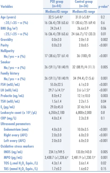
A total of 80 women (44 POI cases and 36 BMI-age matched healthy controls) were included in the study. There was a signiicant difference between the groups in terms of obstetric characteristics. Women with POI had signiicantly lower values for gravidity and parity. Nulliparity was also more common among the POI group (p<0.001). Sonographic indings including endometrial thickness and antral follicle count (AFC) were signiicantly lower in the POI group than in the control group (p=0.001). No signiicant difference in hematologic parameters was found between the groups. There was also no signiicant difference in mean serum CRP levels between the study group and the control group. We found signiicantly elevated levels of INOS (234.1±749.5 versus 133.8±143.0; p=0.005), MPO (3,438.7±1,228.6 versus 2,481.9±1,230.1; p=0.001), and TOS (4.3±1.4 versus 3.6±1.4; p=0.02) in the sera of the study group compared to the control group. But serum levels of TAS were statistically insigniicant between the 2 groups (1.7±0.2 versus 1.6±0.2; p=0.15). The compari-son of demographics, clinical and laboratory features of the groups are presented in Table 1. The smoking habit was more common among the patients in the study group (40.9 versus 11.1%, p=0.005). A history of POI in the mother or sister was found in 18 (40.9%) cases in the POI group and 2 (5.6%) cases in the control group (p=0.001). The only comorbidity detected in the his-tory was thyroid disorder (3 versus 2), but none of these patients was receiving any medical treatment. The most frequent causes of previous surgery were cesarean, ap-pendectomy and hernia repair, respectively. There were no signiicant differences between the groups in terms of history of surgery and comorbidity.
ROC analyses demonstrated that increased INOS, MPO, and TOS were the statistically signiicant discrimi-native factors for POI cases. According to the highest Youden’s index, cut-off values were found to be 178 at 65.9% sensitivity and 69.4% speciicity for INOS. For MPO, the cut-off value was calculated as 2,768.2 at 77.3% sensitivity and 66.7% speciicity. The cut-off value for TOS was determined to be 3.6 with 55.6% sensitivity and 77.3% speciicity. The AUCs and their standard errors were 0.680±0.059 (p=0.002), 0.708±0.057 (p<0.001), and 0.643±0.063 (p=0.022), respectively.
POI, smoking, and higher serum levels of INOS, MPO, and TOS than the calculated threshold values were found to be independent risk factors for POI (Table 2).
Discussion
The exact pathophysiologic mechanism underlying POI still remains unclear. It is an indisputable fact that further investigations are required for early detection and treatment of POI. In this study, we aimed to evaluate serum levels of some oxidative stress markers including INOS, MPO, TAS, and TOS in women with POI and to compare them with healthy fertile women. The main inding of this study is that serum levels of INOS, MPO, and TOS, but not TAS, are signiicant discriminative markers for POI. We also found that a BMI less than 25, nulliparity, fam-ily history of POI, smoking, and increased serum levels of INOS, MPO, and TOS are independent risk factors for POI.
Apoptosis or programmed cell death is an essential phenomenon to human physiology, occurring at all periods of life. The rate of apoptosis should be balanced by the proliferation rate of the cell division. This mechanism has a greater importance for ovarian physiology; from the settlement of primordial cells in the genital cresta up to the cessation of ovarian activity at menopause, ovarian germ cells and follicle somatic cells are eliminated by activation of apoptotic processes9. A cell may initiate intracellular apoptotic signaling in response to a stress. Both intracellular and extracellular factors may cause this stress. Most of the morphological and functional changes seen in apoptotic cells can be accounted for by a family of enzymes which become activated in a reaction cascade10.
Reactive oxygen and nitrogen species are a family of antimicrobial molecules derived from nitric oxide and super-oxide anions generated during normal metabolic reactions11. However, if these molecules and their metabolites required for many systems in the body, particularly the immune sys-tem, are not suficiently neutralized, they may pose a serious threat to cellular viability. Proteins in the body undergo a variety of post-translational modiications. Among these modiications, oxidative modiications are substantially involved in aging and certain diseases12. The transduction pathways that link toxic oxidant accumulation to apoptosis include peroxidative perturbations in membrane phospho-lipid dynamics, cytosolic calcium accretion, microskeletal disruption, DNA (deoxyribonucleic acid) damage, and endonuclease activation. MPO is a biomarker of oxidative stress causing halogenations in activated immune cells such as neutrophils13. INOS is derived from nitric oxide and can be a marker indicating activation of macrophages. Although it was primarily identiied in macrophages, the expression of the enzyme can be virtually stimulated in any cell or tis-sue, provided that the appropriate agents get deployed for inducing its synthesis14.
The function of oxidative stress in pathogenesis of POI has not been investigated extensively. It has been reported that female reproductive system maintains a delicate balance
Table 2. Logistic regression method of risk factors for primary ovarian insuficiency
Group (study) p-value Odds Ratio (95%CI)
TOS (3.6) 0.002 4.6 (1.71–12.35)
INOS (178) 0.002 4.3 (1.7–11.29)
MPO (2,768.2) <0.001 6.8 (2.5–18.27)
BMI (<25) 0.008 3.5 (1.3–8.83)
Parity (no) <0.001 12.7 (3.8–42.33)
Smoker (yes) 0.005 0.1 (0.05–0.60)
Family history (yes) 0.001 11.7 (2.5–55.31)
Logistic regression (Method = Enter).
95%CI: conidence interval of 95%; TOS: total oxidative status; INOS: inducible nitric oxide synthase; MPO: myeloperoxidase; BMI: body mass index.
Table 1. Comparison of demographics, clinical characteristics and laboratory parameters between the groups
Variables
POI group (n=44)
Control group
(n=36) p-value* Median±IQ range Median±IQ range
Age (years) 32.5 (±4.4)# 31.0 (±5.8)# 0.2
≤35/<35 – n (%) 16 (36.4)/28 (63.6) 11 (30.6)/25 (69.4) 0.6
BMI (kg/m2) 24.2±4.1 26.6±5.9 0.06
≤25/<25 – n (%) 16 (36.4)/28 (63.6) 24 (66.7)/12 (33.3) 0.01
Gravidity 0.0±2.0 2.0±1.0 0.002
Parity 0.0±2.0 2.0±0.5 <0.001
Nulliparity
17 (38.6)/27 (61.4) 36 (100)/0 <0.001
No/yes – n (%) Smoker
26 (59.1)/18 (40.9) 32 (88.9)/4 (11.1) 0.005
No/yes – n (%) Family history
26 (59.1)/18 (40.9) 34 (94.4)/2 (5.6) 0.001
No/yes – n (%)
FSH (mIU/mL) 55.0±22.5 6.1±2.0 <0.001
LH (mIU/mL) 29.7 (±14.1)# 3.6 (±1.5)# <0.001
Prolactin (ng/mL) 8.0±4.2 12.1±10.0 0.003
TSH (mIU/mL) 1.5±1.4 2.2±1.5 0.04
E2 (pg/mL) 29.0±45.0 37.4±14.4 0.06
Leukocyte count (x 103/µL) 6,350±2,100 6,800±2,000 0.8
CRP (mg/L) 4.0±2.4 2.3±2.8 0.1
Ultrasound parameters
Endometrium (mm) 4.0±3.0 10.0±3.5 <0.001 Right ovary (AFC) 2.0±3.0 6.0±2.0 <0.001 Left ovary (AFC) 2.0±3.0 6.0±2.0 <0.001 Oxidative stress markers
INOS (ng/mL) 234.1±749.5 133.8±143.0 0.005 MPO (pg/mL) 3,438.7 (±1,228.6)# 2,481.9 (±1,230.1)# 0.001
TOS (μmol H2O2 Equiv./L) 4.3±1.4 3.6±1.4 0.02 TAS (mmol H2O2 Equiv./L) 1.7±0.2 1.6±0.2 0.15
#mean (±standard deviation); *p<0.05 is considered as statistically signiicant.
IQ: interquartile; POI: primary ovarian insuficiency; BMI: body mass index; FSH: follicle stimulating hormone; LH: luteinizing hormone; TSH: thyroid stimulating hormone; E2: estradiol; CRP: C-reactive protein; AFC: antral follicle count; INOS:
of pro- and anti-oxidants to minimize oxidative stress15. In a recent study, it was reported that administration of coen-zyme Q having antioxidant properties in POI patients with high ROS levels improves the embryo quality16.We know that the development of oocyte occurs under the hypoxic condition in the ovarian cortex, however exposure to supra-physiological concentrations of ROS are detrimental to developing oogonia. The mammalian oocyte and embryo are very sensitive to oxidative stress; physiological levels sub-serve several important functions whereas high levels impair oocyte maturation17. In another study, it was suggested that increased production of ROS contributes to oophoritis associated with POI8. Kumar et al.18 found an increase in the number of nucleotide alterations in mitochondrial DNA and supra-physiological ROS levels in POF cases compared to age-matched controls. They concluded that high levels of ROS lead to mitochondrial DNA damage results in mito-chondrial dysfunction and increase apoptosis. This situation could lead to less adenosine triphosphate production due to impaired oxidative phosphorylation and thus to deteriorated oogenesis, low oocyte number and POI.
Increased endogenous hormone levels in obese women may lead to faster follicle consumption due to persistent stimulation of follicular growth. However, previous epidemiological studies on the relationship between increased BMI and natural menopausal age have disclosed no conclusive results19. A recent study reported that low BMI is associated with earlier onset of natural menopause20. However, the same authors could not ind such a risk for POI cases. In the present study, we found no signiicant difference between the two groups in terms of mean BMI. On the other hand, when we clas-siied groups according to their BMI as overweight and normal weight, having a lower BMI, it was found as an independent risk factor for POI.
Although the association between smoking and POI has not been established yet deinitely, studies indicate that smoking is toxic for ovarian germ cells and cigarette smoke contains polycyclic hydrocarbons that exert a negative impact on follicle development. Hayatbakhsh et al.21 reported that smoking during reproductive age was associated with earlier menopause. In addition, smoking was associated with de-creased Antimüllerian hormone (AMH)22, AFC and increased
serum FSH23 levels in fertile women. Consistent with the literature, our study showed that smoking frequency was higher in the POI group than the control group, and that smoking was an independent risk factor for POI.
Several studies have shown evidence of a strong re-lationship between the menopausal age of mothers and their daughters and genetic factors in the determination of menopause. In a case series including 71 idiopathic POI cases, familial transmission was found in 31% of the patients24. A genetic etiology is suggested by the occurrence of families with two or more affected females. Although they assumed the mode of inheritance is autosomal reces-sive in some POI cases, Davis et al.25 have proposed the X chromosome defects as the main reason for POI. The pres-ent study showed that POI history was more common in families of women with POI than the control group, even despite excluding major chromosomal anomalies such as fragile X syndrome. In a previous study evaluating risk factors for POI, it was found that nulliparity is associated with an increased risk for POI26.Similarly, Harlow and Signorello27 showed that multiparous women had a later age at natural menopause in their study. In our study, we also found nulliparity to be an important risk factor for the development of POI.
In conclusion, predicting POI before development or the early detection of POI could be useful for developing appropriate treatment options that would solve infertil-ity problem. To the best of our knowledge, the present study is the irst to suggest that the INOS, MPO, and TOS may be signiicant markers of POI. However, serum levels of TAS were not signiicantly different from healthy controls in women with POI. This study indicates that the balance between prooxidants and antioxidants has shifted in favor of the prooxidants in women with POI. While the etiologic factors of disease are being evaluated, analysis of serum markers of oxidative stress may also be included in the initial work up of a woman with POI. If these cases are diagnosed at an early stage, appropriate treatment, such as antioxidants, which prevents DNA damage and slows down germ cell apoptosis, can be initi-ated. Further studies with more participants are required to determine whether these oxidative stress markers have a role in diagnosing POI.
References
1. Welt CK. Primary ovarian insuficiency: a more accurate term for premature ovarian failure. Clin Endocrinol (Oxf). 2008;68(4):499-509. 2. Tong ZB, Sullivan SD, Lawless LM, Vanderhoof V, Zachman K,
Nelson LM. Five mutations of mitochondrial DNA polymerase-gamma (POLG) are not a prevalent etiology for spontaneous 46,XX primary ovarian insuficiency. Fertil Steril. 2010;94(7):2932-4.
3. van Kasteren YM, Hundscheid RD, Smits AP, Cremers FP, van Zonneveld P, Braat DD. Familial idiopathic premature ovarian failure: an overrated and underestimated genetic disease? Hum Reprod. 1999;14(10):2455-9.
5. Mendoza N, Juliá MD, Galliano D, Coronado P, Díaz B, Fontes J, et al. Spanish consensus on premature menopause. Maturitas. 2015;80(2):220-5.
6. Limoli CL, Hartmann A, Shephard L, Yang CR, Boothman DA, Bartholomew J, et al. Apoptosis, reproductive failure, and oxidative stress in Chinese hamster ovary cells with compromised genomic integrity. Cancer Res. 1998;58(16):3712-8.
7. Wang W, Craig ZR, Basavarajappa MS, Hafner KS, Flaws JA. Mono-(2-ethylhexyl) phthalate induces oxidative stress and inhibits growth of mouse ovarian antral follicles. Biol Reprod. 2012;87(6):152.e1-10.
8. Behrman HR, Kodaman PH, Preston SL, Gao S. Oxidative stress and ovary. J Soc Gynecol Investig. 2001;8(1 Suppl):S40-2. 9. Rolaki A, Drakakis P, Millingos S, Loutradis D, Makrigiannakis
A. Novel trends in follicular development, atresia and corpus luteum regression: a role for apoptosis. Reprod Biomed Online. 2005;11(1):93-103.
10. Forges T, Monnier-Barbarino P, Leheup B, Jouvet P. Pathophysiology of impaired ovarian function in galactosaemia. Hum Reprod Update. 2006;12(5):573-84.
11. Fang FC. Antimicrobial reactive oxygen and nitrogen species: concepts and controversies. Nat Rev Microbiol. 2004;2(10):820-32. 12. Naito Y, Masaichi-Chang-il L, Kato Y, Nagai R, Yonei Y. Oxidative
stress markers. Anti-Aging Med. 2010;7(5):36-44.
13. Vita JA, Brennan ML, Gokce N, Mann SA, Goormastic M, Shishehbor MH, et al. Serum myeloperoxidase levels independently predict endothelial dysfunction in humans. Circulation. 2004;110(9):1134-9. 14. Nagpal L, Panda K. Characterization of calmodulin-free murine
inducible nitric-oxide synthase. PLoS One. 2015;10(3):e0121782. 15. Agarwal A, Aponte-Mellado A, Premkumar BJ, Shaman A, Gupta
S. The effects of oxidative stress on female reproduction: a review. Reprod Biol Endocrinol. 2012;10:49.e1-31.
16. Bentov Y, Esfandiari N, Burstein E, Casper RF. The use of mitochondrial nutrient to improve the outcome of infertility treatment in older patients. Fertil Steril. 2010;93(1):272-5.
17. Harvey AJ, Kind KL, Thompson JG. Redox regulation of early embryo development. Reproduction. 2002;123(4):479-86. 18. Kumar M, Pathak D, Kriplani A, Ammini AC, Talwar P, Dada
R. Nucleotide variations in mitochondrial DNA and supra-physiological ROS levels in cytogenetically normal cases of premature ovarian insuficiency. Arch Gynecol Obstet. 2010;282(6):695-705.
19. Beser E, Aydemir V, Bozkaya H. Body mass index and age at natural menopause. Gynecol Obstet Invest. 1994;37(1):40-2. 20. Yasui T, Hayashi K, Mizunuma H, Kubota T, Aso T, Matsumura
Y, et al. Factors associated with premature ovarian failure, early menopause and earlier onset of menopause in Japanese women. Maturitas. 2012;72(3):249-55.
21. Hayatbakhsh MR, Clavarino A, Williams GM, Sina M, Najman JM. Cigarette smoking and age of menopause: a large prospective study. Maturitas. 2012;72(4):346-52.
22. Fuentes A, Escalona J, Céspedes P, Repetto V, Iñiguez G. [Effects of smoking on plasma antimullerian hormone concentrations among infertile women]. Rev Med Chil. 2013;141(1):23-7. Spanish. 23. Caserta D, Bordi G, Di Segni N, D’Ambrosio A, Mallozzi
M, Moscarini M. The inluence of cigarette smoking on a population of infertile men and women. Arch Gynecol Obstet. 2013;287(4):813-8.
24. Vegetti W, Grazia Tibiletti M, Testa G, de Lauretis Yankowski, Alagna F, Castoldi E, et al. Inheritance in idiopathic premature ovarian failure: analysis of 71 cases. Hum Reprod. 1998;13(7):1796-800. 25. Davis CJ, Davison RM, Payne NN, Rodeck CH, Conway GS.
Female sex preponderance for idiopathic familial premature ovarian failure suggests an X chromosome defect: opinion. Hum Reprod. 2000;15(11):2418-22.
26. Progetto Menopausa Italia Study Group. Premature ovarian failure: frequency and risk factors among women attending a network of menopause clinics in Italy. BJOG. 2003;110(1):59-63. 27. Harlow BL, Signorello LB. Factors associated with early menopause.