in HER2-positive breast carcinomas
Perda da expressão do PTEN e ativação da
AKT em carcinomas mamários HER2-positivos
FiLoMena Marino carvaLho4Abstract
PURPOSE: To examine the expression of AKT and PTEN in a series of HER2-positive primary invasive breast tumors using immunohistochemistry, and to associate these expression proiles with classic pathologic features such as tumor grade, hormone receptor expression, lymphatic vascular invasion, and proliferation. METHODS: A total of 104 HER2-positive breast carcinoma specimens were prepared in tissue microarrays blocks for immunohistochemical detection of PTEN and phosphorylated AKT (pAKT). Original histologic sections were reviewed to assess pathological features, including HER2 status and Ki-67 index values. The associations between categorical and numeric variables were identiied using Pearson’s chi-square test and the Mann-Whitney, respectively. RESULTS: Co-expression of pAKT and PTEN was presented in 59 (56.7%) cases. Reduced levels of PTEN expression were detected in 20 (19.2%) cases, and these 20 tumors had a lower Ki-67 index value. In contrast, tumors positive for pAKT expression [71 (68.3%)] were associated with a higher Ki-67 index value. CONCLUSION: A role for AKT in the proliferation of HER2-positive breast cancers was conirmed. However, immunohistochemical detection of PTEN expression did not correlate with an inhibition of cellular proliferation or control of AKT phosphorylation, suggesting other pathways in these mechanisms of control.
Resumo
OBJETIVOS: Avaliar a expressão imuno-histoquímica de AKT e PTEN em uma série de carcinomas mamários invasivos HER2-positivos, e associar seus padrões de expressão com variáveis anatomopatológicas clássicas, como grau histológico, expressão de receptores hormonais, embolização vascular linfática e atividade proliferativa. MÉTODOS: Um total de 104 amostras de carcinomas mamários invasivos HER2-positivos foram preparadas em blocos de microarranjos de tecido para detecção imuno-histoquímica de PTEN e AKT fosforilada (pAKT). Cortes histológicos originais foram revistos para avaliação das características anatomopatológicas, incluindo o estado do HER2 e a avaliação da expressão de Ki-67. As associações entre as variáveis categóricas e as numéricas foram feitas com o uso dos testes do chi-quadrado de Pearson e Mann-Whitney, respectivamente. RESULTADOS: Co-expressão de pAKT e PTEN foi identiicada em 59 (56,7%) casos. Expressão reduzida de PTEN foi detectada em 20 (19,2%) casos, e esses 20 tumores mostraram menores valores de Ki-67. Por outro lado, tumores positivos para pAKT [71 (68,3%)] apresentaram células positivas para valores mais altos de Ki-67. CONCLUSÕES: O papel de AKT na proliferação de carcinomas mamários HER-2 positiva foi conirmada. Entretanto, a detecção imuno-histoquímica de PTEN não se correlacionou com inibição da proliferação celular ou controle da fosforilação de AKT, sugerindo outras vias nesses mecanismos de controle.
Department of Obstetrics and Gynecology, Faculdade de Medicina, Universidade de São Paulo – USP – São Paulo (SP), Brazil.
1 Department of Obstetrics and Gynecology Postgraduation Program, Faculdade de Medicina, Universidade de São Paulo – USP – São
Paulo (SP), Brazil.
2 Patology Consultancy – Botucatu (SP), Brazil.
3 Department of Obstetrics and Gynecology, Faculdade de Medicina, Universidade de São Paulo – USP – São Paulo (SP), Brazil. 4 Department of Pathology, Faculdade de Medicina, Universidade de São Paulo – USP – São Paulo (SP), Brazil.
Conlict of interests: none.
Keywords Genes, erbB-2 Breast neoplasms Immunohistochemistry PTEN phosphohydrolase Oncogene protein v-akt
Palavras-chave Genes, erbB-2 Neoplasias da mama Imuno-histoquímica PTEN fosfo-hidrolase Proteína oncogênica v-akt
Correspondence
Filomena Marino Carvalho Department of Pathology, Faculdade de Medicina, Universidade de São Paulo Avenida Doutor Arnaldo, 455 – room 1149 Zip code: 01246-903 São Paulo (SP), Brazil
Received
05/26/2014
Accepted with modiications
07/04/2014
DOI: 10.1509/SO100-720320140005034
Introduction
Breast cancer is a heterogeneous disease including distinct entities according to clinical behavior, pathologi-cal features, and molecular subtypes. Prognosis is largely dependent on intrinsic molecular subtype based on gene expression: luminal A, luminal B, HER2 overexpressing,
and basal-like1. Approximately 20–25% of breast cancer
cases involve overexpression of human epidermal growth
factor receptor 2 (HER2)2, a transmembrane receptor
with tyrosine kinase activity that regulates several cel-lular processes such as cell proliferation, differentiation,
adhesion, survival, and migration3. One of the most potent
signaling pathways promoted by HER2 over expression is the phosphatidylinositol 3-kinase (PI3K)/AKT signaling cascade which affects cell cycle progression and can inhibit
apoptosis3. The PI3K enzyme is a heterodimer composed
of a regulatory subunit (p85) and a catalytic subunit (p110). Upon activation by tyrosine kinase receptors, PI3K phosphorylates phosphatidylinositol-4,5-biphosphate (PIP2) to produce phosphatidylinositol-3,4,5-triphosphate
(PIP3)4. PIP3 then recruits the serine/threonine kinase,
AKT, to the plasma membrane. Upon phosphorylation of Ser473 of AKT, several kinases are activated, including the mammalian target of rapamycin (mTOR), an impor-tant molecule that regulates cell growth, p21 and p27, as well as several other molecules that inhibit apoptosis,
such as Bad and caspase proteins5-7. It has been reported
that activated AKT can be an indicator of poor prognosis,
possibly by promoting cell survival8. To regulate AKT
activity, PI3K is opposed by PTEN (phosphatase and ten-sin homolog deleted on chromosome 10), which converts PIP3 back to PIP2, thus preventing phosphorylation and activation of AKT. As a result, cellular proliferation is
inhibited and tumor formation is suppressed9.
PTEN is a tumor-suppressor gene that is located on
chromosome 10q23.310. Mutations in the PTEN gene,
some of which lead to loss of PTEN protein expression, are related to a variety of human cancers, including pros-tatic and endometrial carcinomas. Among breast cancers, lack of PTEN protein expression is mainly attributed to loss of heterozygosity or promoter methylation of the PTEN gene10,11. In addition, activation of the PI3K pathway, as
a result of low levels, or the absence, of PTEN expression, has been associated with resistance to trastuzumab, a recombinant humanized monoclonal antibody that
rec-ognizes the extracellular domain of HER212-14. Therefore,
reduced PTEN expression in breast carcinomas may relect
a more aggressive biologic behavior15. Correspondingly,
several studies have demonstrated that deregulation of the PI3K/AKT/PTEN pathway is associated with poor prognosis, an increased incidence of disease recurrence,
and a shorter disease-free survival period15-18. In this study,
immunoexpression of pAKT and PTEN were assayed in a series of HER2-positive primary invasive breast tumor specimens. These expression proiles were then compared with clinicopathologic features of the tumors, including proliferative activity determined by expression of Ki-67.
Methods
Selection of the samples
This project was approved by the Scientiic Committee of the Department of Obstetrics and Gynecology, and the Department of Pathology, of the Faculdade de Medicina da Universidade de Sao Paulo (Brazil), and also by the Ethical Committee for Research Projects of the Hospital das Clinicas da Faculdade de Medicina da Universidade de Sao Paulo, Brazil (CAPPesq) (protocol 0756/08).
Formalin-ixed, parafin-embedded tissue specimens from 104 consecutive patients with HER2-positive pri-mary breast carcinomas diagnosed in 2007 and 2008 were selected from the iles of the Consultoria em Patologia (Botucatu, São Paulo), a major reference laboratory in Brazil that receives approximately 6,000 breast specimens per year. Inclusion criteria were: the availability of adequate tissue material, lack of any previous treatment, and HER2 positivity according to the guidelines of the American Society of Clinical Oncology (ASCO) and the college of
American Pathologists (CAP)19. The mean age of the
patients included in this study was 54.0±12.4 years old
(range 30–91).
Pathological examination and tissue microarray construction
Two observers (F.M.L.L. and F.M.C) reviewed all slides, and the histologic type was determined based on the World
Health Organization classiication criteria20. Ninety-six
(92.3%) tumors were classiied as ductal, 3 (2.9%) as mucinous, 2 (1.9%) as micropapillary, 2 (1.9%) as signet ring cell, and 1 (0.9%) as pleomorphic invasive lobular carcinoma. Other histologic features evaluated included
grade according to the Nottingham criteria21, nuclear grade,
presence of necrosis (absent or present), and peritumoral vascular invasion. Histologic and nuclear grades 1 and 2 were combined for statistical purposes, since we had only one case histologic and nuclear grade. Immunohistochemical detection of estrogen receptor (ER), progesterone receptor (PR), Ki67, and HER2 were performed using original whole histologic sections. All of the immunohistochemistry assays were performed in the same laboratory, using the same protocol, by the same pathologist.
in tissue sections stained with hematoxylin/eosin, and these sections were marked in the corresponding paraf-in donor blocks. One cylparaf-inder of material (2.0 mm paraf-in diameter) was punched from each case and was mounted into recipient parafin blocks at 2 mm intervals using a precision microarray instrument (Beecher Instruments, Silver Spring, MD). A grid system was established such that each core had an x- and y-coordinate refer-ence for sample identiication. Blocks were sealed at 60°C for 10 minutes. Sections (3 µm) from each TMA block were then prepared using standard techniques
and mounted on Starfrost® slides. The irst
histologi-cal sections cut were stained with hematoxylin-eosin to ensure that the appropriate sections of the tumor had been obtained.
Immunohistochemistry and scoring
The antigens employed were ER (clone SP1, Dako, Carpinteria, USA, dilution 1:1000); PR (clone PgR 636, Dako, Carpinteria, USA, dilution 1:600); HER2 (clone
SP3, Neomarkers, Fremont, USA, dilution 1:300); Ki-67 (clone MIB1, Dako, Carpinteria, USA, dilution 1:4800); pAKT (clone LP18, Novocastra, Richmond, USA, dilution 1:200), and PTEN (clone 28H6, Novocastra, Richmond, USA, dilution 1:800). Pressure cooker was the epitope retrieval method for all markers, except for PTEN, for which microwave oven was used. The bound antibodies were detected by goat anti-rabit or anti-mouse horserad-ish peroxidase-labelled polymerized secondary antibodies (DAKO EnVision TM System). Peroxidase activity was visualized with diamminobenzidine (Dako).
HER2 expression was conirmed on TMA slides and the samples were considered positive if they received a
3+ score according to guidelines from ASCO and CAP19
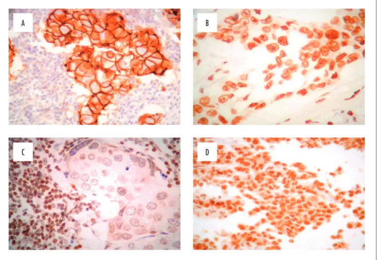
(Figure 1A).
Tumors were considered positive for ER and PR ex-pression if at least 10% of the cells were stained, according
to the criteria of Park et al.22. Ki-67 index values were
expressed as the percentage of positive cells present among at least 500 tumor cells in hot spot areas. The highest
Figure 1. (A) HER2 positive carcinoma with complete and strong membrane expression of the protein in more than 30% of cells; (B) PTEN present characterized by nuclear expression in 100% of neoplastic cells; (C) PTEN reduced characterized by negative reaction contrasting with positive lymphocyte; (D) AKT diffusely positive in the neoplastic cells
A
C
B
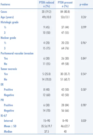
Table 1. Clinicopathological features of the HER2-positive breast carcinomas examined according to PTEN expression
Features PTEN reduced n (%) PTEN present n (%) p-value
Cases 20 (19.2) 84 (80.8)
Age (years) 49±10.0 53±13.1 0.26a
Histologic grade
½ 9 (45) 37 (44) 0.99b
3 10 (50) 47 (55)
Nuclear grade
½ 4 (20) 20 (23) 0.96 b
3 15 (75) 64 (76)
Peritumoral vascular invasion
Yes 6 (30) 26 (30) 0.84 b
No 11 (55) 49 (58)
Tumor necrosis
Yes 5 (25.0) 30 (35.7) 0.54 b
No 14 (70.0) 51 (60.7)
ER
Positive 8 (40) 42 (50) 0.58 b
Negative 12 (60) 42 (50)
PR
Positive 6 (30) 28 (84) 0.98 b
Negative 14 (70) 56 (66)
Ki-67
Interval 15–90 0–90 0.03a
Mean ± SD 35.5±19.7 46±22.7
Median 37.5 40
aMann-Whitney test; bPearson´s chi-square test; ER: estrogen receptor; PR: progesterone receptor.
Table 2. Clinicopathological features of the HER2-positive breast carcinomas examined according to pAKT expression
Features pAKT positive
n (%)
pAKT negative
n (%) p-value
Cases 71 (68.3) 33 (31.7)
Age (years) 53.2±13.2 54.9±11.9 0.62a
Histologic grade
½ 37 (52.8) 9 (27.2) 0.03b
3 33 (47.1) 24 (72.7)
Not assessed 1 0
Nuclear grade
½ 18 (25.7) 6 (18.1) 0.55 b
3 52 (74.2) 27 (81.8)
Not assessed 1 0
Lymphatic vascular invasion
Yes 20 (31.7) 12 (41.3) 0.50 b
No 43 (68.2) 17 (58.6)
Not assessed 8 4
Tumor necrosis
Yes 20 (28.5) 15 (50.0) 0.07 b
No 50 (71.4) 15 (50.0)
Not assessed 1 3
ER
Positive 39 (54.9) 11 (33.3) 0.06 b
Negative 32 (45.0) 22 (66.6)
PR
Positive 27 (38.0) 7 (21.2) 0.14 b
Negative 44 (61.9) 26 (78.7)
Ki-67
Interval 0–95 15–80 0.22a
Mean ± SD 45.6±22.1 40.4±21.5
Median 40 30
aMann-Whitney test; bPearson´s chi-square test; ER: estrogen receptor; PR: progesterone receptor.
value obtained in TMA sections or whole sections was the reported score. Tumors were classiied according to the expression of PTEN. “PTEN present” indicated tumors with diffuse and strong positivity in 100% of the nuclei (Figure 1B), while “PTEN reduced” indicated tumors with less than 100% of nuclear positivity (Figure 1C).
The pAKT expression was assessed based on the percentage of nuclear and/or cytoplasmic positive cells in a sample. Tumors with at least 10% of the stained cells positive for pAKT expression were classiied as positive
according to the criteria of Gori et al.23 (Figure 1D).
Statistical analysis
Associations between categorical variables were identiied using Pearson’s chi-square test, while the Mann-Whitney test was used to compare groups according to numeric variables (e.g., patient age, Ki-67 index values).
A p-value less than 0.05 was considered signiicant. Statistical analysis were performed using MedCalc for Windows (version 11.5.0.0; MedCalc Software, Mariakerke, Belgium).
Results
Associations between PTEN expression and clini-copathologic features of the cohort studied are listed in Table 1. The only statistically signiicant difference between PTEN- reduced and PTEN-present tumors was the proliferative activity detected with Ki-67 staining. Furthermore, samples with diffuse expression of PTEN exhibited higher levels of proliferation.
Expression of pAKT was found to be associated with a low histological grade (p=0.03). In addition, an associa-tion between expression of pAKT, a lower tumor necrosis, and an ER-positive status were observed. However, these associations were not statistically signiicant. None of the other clinicopathological features studied were associated with pAKT expression (Table 2). The Ki-67 index values were higher for tumors expressing pAKT compared with
40.4±21.5), although the difference was not statistically signiicant. Moreover, diffuse PTEN expression was ob-served in 59/71 (83.1%) of tumors positive for pAKT, and in 25/33 (75.7%) of tumors negative for pAKT, although the difference was not signiicant (p=0.06; chi-square=0.4).
Discussion
In this study, expression of PTEN and pAKT was assayed in 104 HER2-positive primary breast tumors. Since these molecules have previously been shown to mediate antagonistic mechanisms, their associations with clinicopathologic features were examined.
Cowden’s disease is a hereditary cancer predisposition syndrome that is associated with an elevated risk of breast and thyroid cancer. Germ line mutations in PTEN have been identiied in patients with Cowden’s disease, and the frequency of sporadic breast cancer with loss of PTEN
expression has been reported to range from 30-40%4,18,24-26.
In the present study, the frequency of breast cancer with loss of PTEN expression was 19.2%. However, loss of PTEN expression was evaluated using immunohistochemistry, and this method may underestimate the proportion of invasive breast tumors that contain an inactivated version of PTEN. For example, in a retrospective study conducted by Bose
et al.18, loss of PTEN expression in a set of 34 sporadic
invasive breast tumors was also evaluated using immuno-histochemistry. The molecular status of these tumors had previously been established, including the PTEN genotype. For 13 cases with loss of heterozygosity (LOH) at the 10q23 locus, only 6 exhibited lower levels of PTEN expression in immunohistochemistry assays. The remaining seven cases exhibited no loss of PTEN expression. These data suggest that immunohistochemistry assays may not have suficient sensitivity to detect the decrease in PTEN levels associated with LOH, and this is consistent with the indings of the present study.
Except for proliferative activity, none of the clini-copathological parameters examined was found to be as-sociated with the PTEN results obtained. While similar
results have been reported in other studies11,16,27, signiicant
associations between certain clinicopathological charac-teristics and PTEN loss have also been reported.
For ex-ample, Lee et al.15 found that reduced PTEN expression
is correlated with lymph node status, tumor grade, and TNM stage. However, there was no signiicant difference between PTEN expression and patient age, tumor size, or
invasion of the lymphovascular space15. Bose et al.18 also
demonstrated that reduced expression of PTEN was associ-ated with a high tumor grade. On the other hand, though,
Pérez-Tenorio et al.4 demonstrated that loss of PTEN was
associated with an ER positive status (p=0.001), small tumor size (p=0.02) and low levels of HER2 expression
(p=0.01), characteristics that have been associated with a less aggressive phenotype and low proliferative activity.
The results of Pérez-Tenorio et al.4 with the proliferative
activity were similar to ours. In addition, it cannot be ruled out that methodological factors and/or the number of cases examined may be responsible for these discrepancies.
Surprisingly, Ki-67 index values were found to be higher for tumors with strong, diffuse expression of PTEN compared to PTEN reduced tumors, or tumors negative for PTEN expression. We hypothesize that high PTEN expression represents a positive feedback signal for prolifera-tive activity, which has previously been described for the endometrium. Immunohistochemistry was used to evaluate the expression of PTEN in samples of normal endometrium obtained during different phases of the menstrual cycle.
In their study, Mutter et al.28 demonstrated that
endome-trial expression of PTEN varies during the proliferative and secretory phases, with levels of PTEN being higher during the irst one. This inding was unexpected at the time, and the explanation proposed was that a functional requirement for PTEN-mediated tumor suppressor activity was speciic for a highly mitotic, estrogenic environment, while progestin-dominated conditions would inhibit cell division and PTEN activity would be reduced. Although breast tissue and endometrium tissue are quite different, it is possible that this mechanism may be common to both. For example, PTEN positivity in breast tumors that exhibit higher proliferative activity may involve a compensatory mechanism that aims to control cell proliferation to main-tain the delicate balance of the intracellular environment.
In the series examined, the proportion of breast cancers positive for pAKT expression (71/104, 68.3%) was similar
to that reported in other studies6,23. The AKT has been
found to mediate protumorigenic effects of hormones and growth factors, and the anchorage-mediated survival of epithelial cells. Accordingly, activation of AKT stimulates glucose metabolism, cell cycle progression, and survival through the phosphorylation of multiple substrates,
in-cluding GSK3, BAX, and mTOR among others29. In the
present study, a signiicant inverse relationship between pAKT positivity and tumor histological grade was observed (p=0.02). Additional inverse relationships between pAKT positivity and lymphatic vascular invasion and tumoral necrosis were also observed, yet these were not statistically signiicant. These indings are consistent with those of other
studies6,27, yet are in contrast with accumulating evidence
that AKT harbors anti-apoptotic properties and promotes
tumor progression and tumor growth29,30. In addition, other
studies have demonstrated a direct relationship between
pAKT and tumor histologic grade30.
1. Perou CM, Sorlie T, Eisen MB, van de Rijn M, Jeffrey SS, Rees CA, et al. Molecular portraits of human breast tumours. Nature. 2000;406(6797):747-52.
2. Ravdin PM, Chamness GC. The c-erbB-2 proto-oncogene as a prognostic and predictive marker in breast cancer: a paradigm for the development of other macromolecular markers--a review. Gene. 1995;159(1):19-27.
3. Barros FF, Powe DG, Ellis IO, Green AR. Understanding the HER family in breast cancer: interaction with ligands, dimerization and treatments. Histopathology. 2010;56(5):560-72.
4. Pérez-Tenorio G, Alkhori L, Olsson B, Waltersson MA, Nordenskjöld B, Rutqvist LE, et al. PIK3CA mutations and PTEN loss correlate with similar prognostic factors and are not mutually exclusive in breast cancer. Clin Cancer Res. 2007;13(12):3577-84. 5. Al-Bazz YO, Underwood JC, Brown BL, Dobson PR. Prognostic
signiicance of Akt, phospho-Akt and BAD expression in primary breast cancer. Eur J Cancer. 2009;45(4):694-704.
6. Aleskandarany MA, Rakha EA, Ahmed MA, Powe DG, Ellis IO, Green AR. Clinicopathologic and molecular signiicance of phospho-Akt expression in early invasive breast cancer. Breast Cancer Res Treat. 2011;127(2):407-16.
7. Bose S, Chandran S, Mirocha JM, Bose N. The Akt pathway in human breast cancer: a tissue-array-based analysis. Mod Pathol. 2006;19(2):238-45.
8. Pérez-Tenorio G, Stål O; Southeast Sweden Breast Cancer Group. Activation of AKT/PKB in breast cancer predicts a worse outcome among endocrine treated patients. Br J Cancer. 2002;86(4):540-5. 9. Wang L, Zhang Q, Zhang J, Sun S, Guo H, Jia Z, et al. PI3K pathway activation results in low eficacy of both trastuzumab and lapatinib. BMC Cancer. 2011;11:248.
10. Hollander MC, Blumenthal GM, Dennis PA. PTEN loss in the continuum of common cancers, rare syndromes and mouse models. Nat Rev Cancer. 2011;11:289-301.
11. Bakarakos P, Theohari I, Nomikos A, Mylona E, Papadimitriou C, Dimopoulos AM, et al. Immunohistochemical study of PTEN and phosphorylated mTOR proteins in familial and sporadic invasive breast carcinomas. Histopathology. 2010;56(7):876-82. 12. Dave B, Migliaccio I, Gutierrez MC, Wu MF, Chamness GC,
Wong H, et al. Loss of phosphatase and tensin homolog or phosphoinositol-3 kinase activation and response to trastuzumab or lapatinib in human epidermal growth factor receptor 2-overexpressing locally advanced breast cancers. J Clin Oncol. 2011;29(2):166-73.
13. Calabrich A, Fernandes GS, Katz A. Trastuzumab: mechanisms of resistance and therapeutic opportunities. Oncology (Willist Park, N.Y.). 2008;22(11):1250-8.
14. Razis E, Bobos M, Kotoula V, Eleftheraki AG, Kalofonos HP, Pavlakis K, et al. Evaluation of the association of PIK3CA mutations and PTEN loss with efficacy of trastuzumab therapy in metastatic breast cancer. Breast Cancer Res Treat. 2011;128(2):447-56.
15. Lee JS, Kim HS, Kim YB, Lee MC, Park CS, Min KW. Reduced PTEN expression is associated with poor outcome and angiogenesis in invasive ductal carcinoma of the breast. Appl Immunohistochem Mol Morphol. 2004;12(3):205-10.
16. Capodanno A, Camerini A, Orlandini C, Baldini E, Resta ML, Bevilacqua G, et al. Dysregulated PI3K/Akt/PTEN pathway is a marker of a short disease-free survival in node-negative breast carcinoma. Hum Pathol. 2009;40(10):1408-17.
References
that a complex network of cross-talk occurs between the
HER2 activation pathway and ER mediated signaling31-33.
For example, in an in vitro model, Stoica et al.34 demonstrated
that estradiol can activate the PI3K/AKT pathway in MCF-7 breast cancer cells, and that this effect was ErbB2 dependent. While these results remain to be conirmed in other breast
cancer cells in vitro and in vivo, they are consistent with the
positive correlation observed between expression of ER and
pAKT in our study, and in other studies6,27.
Regarding PR expression, Tokunaga et al.35
previ-ously showed that LOH at the PTEN locus and HER2 overexpression enhance activation of AKT. Since acti-vated AKT has been shown to repress transcription of PR independent of ER expression, loss of PR expression
can be induced even in ER-positive breast carcinomas35.
Accordingly, in the present study, PR expression was lower in tumors positive for pAKT expression, although this correlation was not signiicant.
An unexpected result was that PTEN expression tended to correlate with pAKT expression. Similarly, in a study
by Bose et al.7 with 145 invasive breast cancers and 140
pure ductal carcinomas in situ, AKT and its downstream
proteins were found to be activated in approximately 30% of the cancers analyzed independent of PTEN loss. This inding suggests that other mechanisms of AKT activation exist in addition to the activation of AKT by growth factor receptors with tyrosine kinase activity.
17. Saal LH, Johansson P, Holm K, Gruvberger-Saal SK, She QB, Maurer M, et al. Poor prognosis in carcinoma is associated with a gene expression signature of aberrant PTEN tumor suppressor pathway activity. Proc Natl Acad Sci U S A. 2007;104(18):7564-9. 18. Bose S, Crane A, Hibshoosh H, Mansukhani M, Sandweis L,
Parsons R. Reduced expression of PTEN correlates with breast cancer progression. Hum Pathol. 2002;33(4):405-9.
19. Wolff AC, Hammond ME, Schwartz JN, Hagerty KL, Allred DC, Cote RJ, et al. American Society of Clinical Oncology/College of American Pathologists guideline recommendations for human epidermal growth factor receptor 2 testing in breast cancer. J Clin Oncol. 2013;31(31):3997-4013. 20. Tavassoli FA, Devilee A. Pathology and genetics: tumours of breast
and female genital organs. Lion: IARC Press; 2003.
21. Elston CW, Ellis IO. Pathological prognostic factors in breast cancer. I. The value of histological grade in breast cancer: experience from a large study with long-term follow-up. Histopathology. 1991;19(5):403-10.
22. Park S, Koo J, Park HS, Kim JH, Choi SY, Lee JH, et al. Expression of androgen receptors in primary breast cancer. Ann Oncol. 2010;21(3):488-92.
23. Gori S, Sidoni A, Colozza M, Ferri I, Mameli MG, Fenocchio D, et al. EGFR, pMAPK, pAkt and PTEN status by immunohistochemistry: correlation with clinical outcome in HER2-positive metastatic breast cancer patients treated with trastuzumab. Ann Oncol. 2009;20(4):648-54.
24. Depowski PL, Rosenthal SI, Ross JS. Loss of expression of the PTEN gene protein product is associated with poor outcome in breast cancer. Mod Pathol. 2001;14(7):672-6.
25. Perren A, Weng LP, Boag AH, Ziebold U, Thakore K, Dahia PL, et al. Immunohistochemical evidence of loss of PTEN expression in primary ductal adenocarcinomas of the breast. Am J Pathol. 1999;155(4):1253-60.
26. Gonzalez-Angulo AM, Ferrer-Lozano J, Stemke-Hale K, Sahin A, Liu S, Barrera JA, et al. PI3K pathway mutations and PTEN levels in primary and metastatic breast cancer. Mol Cancer Ther. 2011;10(6):1093-101.
27. Panigrahi AR, Pinder SE, Chan SY, Paish EC, Robertson JF, Ellis IO. The role of PTEN and its signalling pathways, including AKT, in breast cancer; an assessment of relationships with other prognostic factors and with outcome. J Pathol. 2004;204(1):93-100. 28. Mutter GL, Lin MC, Fitzgerald JT, Kum JB, Eng C. Changes in
endometrial PTEN expression throughout the human menstrual cycle. J Clin Endocrinol Metab. 2000;85(6):2334-8.
29. Riggio M, Polo ML, Blaustein M, Colman-Lerner A, Lüthy I, Lanari C, et al. PI3K/AKT pathway regulates phosphorylation of steroid receptors, hormone independence and tumor differentiation in breast cancer. Carcinogenesis. 2012;33(3):509-18.
30. Gershtein ES, Scherbakov AM, Shatskaya VA, Kushlinsky NE, Krasil’nikov MA. Phosphatidylinositol 3-kinase/AKT signalling pathway components in human breast cancer: clinicopathological correlations. Anticancer Res. 2007;27(4A):1777-82.
31. Kato S, Endoh H, Masuhiro Y, Kitamoto T, Uchiyama S, Sasaki H, et al. Activation of the estrogen receptor through phosphorylation by mitogen-activated protein kinase. Science. 1995;270(5241):1491-4.
32. Miller TW, Hennessy BT, González-Angulo AM, Fox EM, Mills GB, Chen H, et al. Hyperactivation of phosphatidylinositol-3 kinase promotes escape from hormone dependence in estrogen receptor-positive human breast cancer. J Clin Invest. 2010;120(7):2406-13.
33. Miller TW, Balko JM, Arteaga CL. Phosphatidylinositol 3-kinase and antiestrogen resistance in breast cancer. J Clin Oncol. 2011;29(33):4452-61.
34. Stoica GE, Franke TF, Wellstein A, Czubayko F, List HJ, Reiter R, et al. Estradiol rapidly activates Akt via the ErbB2 signaling pathway. Mol Endocrinol. 2003;17(5):818-30.