ISSN 1414-431X
www.bjournal.com.br
www.bjournal.com.br
Volume 45 (12) 1102-1340 December 2012
Braz J Med Biol Res, December 2012, Volume 45(12) 1334-1340
10.1590/S0100-879X2012007500163
doi:
Clinicopathological significance of PTPN12 expression in human
breast cancer
Yuan Xunyi, Yuan Zhentao, Jiang Dandan and Li Funian
Institutional Sponsors
The Brazilian Journal of Medical and Biological Research is partially financed by
Faculdade de Medicina de Ribeirão Preto Campus
Ribeirão Preto
Explore High - Performance MS Orbitrap Technology In Proteomics & Metabolomics
analiticaweb.com.br S C I E N T I F I C
BIOMEDICAL SCIENCES
AND
Clinicopathological significance of PTPN12
expression in human breast cancer
Yuan Xunyi
1*, Yuan Zhentao
2*, Jiang Dandan
1and Li Funian
1*
1Breast Disease Diagnosis and Treatment Centre, Affiliated Hospital of Medical College,
Qingdao University, Qingdao, Shandong Province, China
2Department of Anesthesiology, Shengli Oilfield Central Hospital, Dongying, Shandong Province, China
Abstract
Protein tyrosine phosphatase non-receptor type 12 (PTPN12) is a recently identified tumor suppressor gene (TSG) that is fre
-quently compromised in human triple-negative breast cancer. In the present study, we investigated the expression of PTPN12 protein by patients with breast cancer in a Chinese population and the relationship between PTPN12 expression levels and patient clinicopathological features and prognosis. Additionally, we explored the underlying down-regulation mechanism from the perspective of an epigenetic alteration. We examined PTPN12 mRNA expression in five breast cancer cell lines using semi-quantitative reverse-transcription PCR, and detected PTPN12 protein expression using immunohistochemistry in 150 primary invasive breast cancer cases and paired adjacent non-tumor tissues. Methylation-specific PCR was performed to analyze the promoter CpG island methylation status of PTPN12. PTPN12 was significantly down-regulated in breast cancer cases (48/150) compared to adjacent noncancerous tissues (17/150; P < 0.05). Furthermore, low expression of PTPN12 showed a significant positive correlation with tumor size (P = 0.047), lymph node metastasis (P = 0.001), distant metastasis (P = 0.009), histological grade (P = 0.012), and survival time (P = 0.019). Additionally, promoter CpG island hypermethylation occurs more frequently in breast cancer cases and breast cancer cell lines with low PTPN12 expression. Our findings suggest that PTPN12 is potentially a methylation-silenced TSG for breast cancer that may play an important role in breast carcinogenesis and could potentially
serve as an independent prognostic factor for invasive breast cancer patients.
Key words: Breast cancer; Tumor suppressor gene; PTPN12; Prognosis; Methylation
Introduction
Correspondence: Li Funian, Breast Disease Diagnosis and Treatment Centre, Affiliated Hospital of Medical College, Qingdao University, Qingdao, Shandong Province, 266003, China. E-mail: [email protected]
*These authors contributed equally to this study.
Received July 7, 2012. Accepted August 29, 2012. Available online October 15, 2012. Published December 17, 2012.
Approximately one million new cases of breast cancer
are diagnosed each year worldwide and it is the most
common neoplastic disease in women (1). Triple-negative breast cancer (TNBC), characterized by a lack of ER, PR, and HER2 expression, accounts for approximately 15~20% of the cases of all breast cancer types (2). TNBC
is an aggressive malignancy that exhibits a poor prognosis
and whose treatment is limited and often ineffective. The molecular mechanisms underlying TNBC development
are not clear.
PTPN12, located in 7q11.23, is a member of the protein tyrosine phosphatase (PTP) family. Accumulating evidence indicates that there is a correlation between PTPN12 and
tumor development, including ovarian cancer, colon cancer, and prostate cancer (3-5). Recent reports have
demon-strated that PTPN12 inactivation leads to HER2/EGFR hyperactivity and cellular transformation in HER2-negative
breast cancer and that PTPN12 is frequently compromised in human TNBC, indicating that it is potentially a new tumor suppressor gene (TSG) of TNBC (6).
The mechanisms underlying TSG inactivation primar -ily include two aspects: genetic change and epigenetic
change. Aberrant methylation of promoter CpG islands
is one of the most important epigenetic mechanisms that
causes TSG silencing and leads to transcription repressor
binding, compressed chromatin, and transcription
silenc-ing in the early stages of tumor genesis (7,8). Currently, research concerning the mechanisms underlying PTPN12
inactivation has been limited to genetic changes and, to
our knowledge, there are no reports to date concerning PTPN12 gene promoter methylation. In the present study, we hypothesized that the silencing of PTPN12 could be
caused by methylation.
PTPN12 expression in human breast cancer 1335
potential TSG for breast cancer and a candidate that may serve as a prognostic factor in breast cancer. The pres
-ent study is the first to examine the expression profile of PTPN12 in a comparatively large group of Chinese breast cancer patients with sufficient follow-up data, and the first to analyze the association of PTPN12 expression with clini -copathological and prognostic characteristics. Furthermore,
we investigated whether promoter CpG island methylation is correlated with PTPN12 inactivation mechanisms using methylation-specific PCR (MSP).
Material and Methods
Cell lines and tumor samples
Five human breast cancer cell lines (HCC1937, MDA-MB-231, MCF-7, BT-474, and SK-BR-3) were purchased from the China Center for Type Culture Collection, Chinese Academy of Sciences. HCC1937 and MDA-MB-231 are basal-like subtype, MCF-7 is luminal A subtype, BT-474 is luminal B subtype, and SK-BR-3 is HER2 subtype. The cells were routinely maintained in RPMI or DMEM medium with 10% fetal bovine serum (Hyclone, USA) at 37°C in a humidified atmosphere of 5% CO2. Formalin-fixed,
paraffin-embedded samples of 150 primary invasive breast
cancer tissues and paired adjacent non-tumor tissues were
obtained from the Qingdao University Affiliated Hospital from January to December 1999. Our research was ap
-proved by the Institutional Ethics Committee of Qingdao University Affiliated Hospital and informed written consent
was obtained from all patients.
Cases and clinical data
The median age of the patients was 50.5 years (range: 29-87 years). The duration of follow-up ranged from 2 to 134 months (median: 85.7 months); all patients underwent a follow-up of over 36 months unless they died of any
cause. Cases lost to follow-up were excluded from the
study. Pathological diagnoses were routinely assessed by pathologists in the Pathology Department, Qingdao Univer
-sity Affiliated Hospital. None of the patients had received prior radiotherapy or neo-adjuvant therapy. All patients
received conventional postoperative treatment, depending
on the extent of disease. Patients without axillary lymph
node involvement were treated with surgical treatment alone, whereas patients with axillary lymph node involve-ment received six courses of adjuvant chemotherapy with
a cyclophosphamide/methotrexate/fluorouracil regimen. Patients with positive nodes or tumor size ≥5 cm received postoperative radiation. Patients with ER+/PR+ tumors
were treated for 2-5 years with tamoxifen.
RNA extraction and semi-quantitative reverse-transcription PCR
Total RNA was extracted from each sample using Trizol (Invitrogen, USA) and reverse transcribed into cDNA us
-ing the PrimeScript RT-PCR kit (TaKaRa Bio Inc., Japan) according to manufacturer recommendations. The primers for the human PTPN12 gene were designed with Primer Premier 5.0 (Premier Biosoft, USA) and the sequences were sense: 5’-AAATACTGCAGCCACCGGAAC-3’, antisense: 5’-GCAACACTGGCTTTGGATGG-3’; the amplicon size was 126 bp. GAPDH was used as the internal control with the spe
-cific primers - sense: 5’-TCATGGGTGTGAACCATGAGAA-3’, antisense: 5’-GGCATGGACTGTGGTCATGAG-3’; the amplicon size was 150 bp. The primers cited above were synthesized by Shanghai Sangon Biological Engineering Technology & Services Co. Ltd. (China). The PCR products
were analyzed by 1% agarose gel electrophoresis.
Immunohistochemistry
Expression levels of HER2, ER, PR, and PTPN12
were investigated in 150 primary breast cancer tissue specimens and paired adjacent non-tumor tissues using
immunohistochemistry. Antibodies to ER, PR, and HER2 were purchased from ZSGB-BIO (ZSGB, China). Rabbit
polyclonal antibody to PTPN12 was purchased from Abcam
(Abcam plc, UK). The PTPN12 protein was detected primar
-ily in the cytoplasm. ER and PR were positive primar-ily in the cell nucleus, whereas HER2 was detected in the cell
membrane and cytoplasm.
To determine PTPN12 levels, each sample was assigned
to 5 categories and the proportion of positive-stained cells
was rated as follows: 0 (0-4%), 1 (5-24%), 2 (25-49%), 3 (50-74%), or 4 (75-100%). The intensity of immunostaining was categorized as 0 (no stain), 1+ (weak stain), 2+ (medium stain), and 3+ (strong stain). In addition, an immunoreac -tive score was calculated by multiplying the percentage of positive cells and the stain intensity. In instances of heterogeneous staining intensity within one sample, each component was scored independently and the results were summed. For statistical analysis, the criteria were
com-bined as follows: 0 = negative; 1 to 4 = weakly positive; 5 to 8 = moderately positive, and 9 to 12 = strongly positive (9). In the present study, low expression was defined as negative and weakly positive, and high expression was defined as moderately positive and strongly positive. The Allred scoring system (10) was used for ER and PR stain
-ing interpretation and HER2 was evaluated accord-ing to ASCO guidelines (11).
Methylation-specific PCR
Genomic DNA was extracted from cells and tissues using standard phenol-chloroform extraction. EpiScope Methylated HeLa gDNA (TaKaRa Bio Inc.) was used as a positive control for methylation analysis. Placenta tis
unmethylation primers, which were designed with Methyl
Primer Express v1.0 (ABI, USA). Methylation primer -sense: 5’-TCGTTTGTGAAGAAGGTATTTC-3’, anti-sense: 5’-AAAAAACGAAACGCTTCCTA-3’. Unmethylation primer - sense: 5’-GTTGTTTGTGAAGAAGGTATTTT-3’, antisense: 5’-AAAAAAACAAAACACTTCCTA-3’. The PCR product sizes were both 104 bp. Methylation-specific PCR for the PTPN12 promoter was conducted in a total PCR volume of 20 µL. MSP primers were tested previously to ensure that no unbisulfited DNA was amplified and to confirm that our MSP system is specific. The PCR products were
analyzed by 1% agarose gel electrophoresis.
Statistical analysis
Statistical analyses were performed using the SPSS statistical software (SPSS, Inc., USA). The correlations between PTPN12 expression and clinicopathological vari
-ables were analyzed using Pearson chi-square analysis. The same method was used to test for associations of PTPN12 with ER, PR, and HER2. Kaplan-Meier survival
analysis and univariate analysis of the differences between
the survival curves were tested using the log-rank test. Uni
-variate and multi-variate survival analyses were performed
using Cox proportional hazards regression. A value of P < 0.05 indicated a statistically significant result.
Results
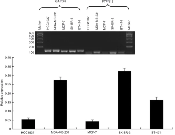
PTPN12 mRNA transcription was down-regulated in one of the triple-negative breast cancer cell lines (HCC1937) as well as in two other types of cell lines (MCF-7, BT-474; Figure 1). PTPN12 protein immunohistochemistry (Figure 2) demonstrated that PTPN12 protein expression was down-regulated (negative and weak expression) in 32.0% of tumor specimens (48/150) and 11.3% of paired adjacent non-tumor tissues (17/150), with a significant difference (P = 0.00009). The PTPN12 protein exhibited low expression in 34.7% of TNBC specimens (26/75) and 29.3% of non-TNBC specimens (22/75) with no significant difference (P = 0.556; Table 1).
As shown in Table 1, significant positive correlations were found between low-level expression of the PTPN12 protein and tumor size (P = 0.047), lymph node metastasis (P = 0.001), histological grade (P = 0.012), and distant
Figure 1. Expression of protein tyrosine phosphatase non-receptor type 12(PTPN12) mRNA in five breast can
-cer cell lines detected by RT-PCR. PTPN12 transcription was down-regulated in HCC1937, MCF-7, and BT-474. GAPDH = glyceraldehyde-3-phosphate dehydrogenase.
HCC1937
Marker MDA-MB-231 MCF-7 SK-BR-3 BT
-474
HCC1937 MDA-MB-231 MCF-7 SK-BR-3 BT Marker
-474
600 500 400 300 200
100
0.40
0.35
0.30
0.25
0.20
0.15
0.05
0 0.10
Relative expression
HCC1937 MDA-MB-231 MCF-7 SK-BR-3 BT-474
PTPN12 expression in human breast cancer 1337
metastasis (P = 0.009). However, there was no significant correlation between PTPN12 expression and age, and no significant correlation between PTPN12 expression and can
-cer recurrence (local/regional) by statistical analysis. There was no statistically significant difference between PTPN12 expression levels and HER2 or EGFR (P > 0.05).
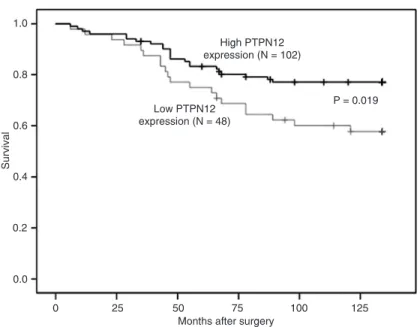
Kaplan-Meier analysis demonstrated significant differ -ences in overall survival between patients with high-level
PTPN12 protein expression and patients with low-level PTPN12 expression. Moreover, low-level PTPN12 expres
-sion was associated with decreased overall survival (P = 0.019; Figure 3). Cox univariate survival analysis demon -strated that tumor size, lymph node status, histological
grade, and PTPN12 expression levels were statistically significant risk factors that affected the outcome of patients
with breast cancer. In multivariate survival analysis, lymph
node status, histological grade, tumor size, and PTPN12 ex
-pression were independent prognostic factors (Table 2). Methylation was observed in two of three PTPN12 down-regulated cell lines (MCF-7 and BT-474; Figure 4). We then examined PTPN12 promoter methylation in 150
formalin-fixed paraffin-embedded primary breast cancer
tumor specimens and their paired adjacent non-tumor
tissues using MSP. A total of 39.6% PTPN12 low protein expression specimens (19/48) exhibited methylation (Figure 4) whereas none exhibited methylation in the PTPN12 high
protein expression specimens and non-tumor specimens
(P < 0.05).
Discussion
PTPN12 is among the PTPs that regulate the equilib -rium of tyrosine phosphorylation and play a prominent role
in tumor suppression. Loss of PTPN12 leads to malignant
transformation of human mammary epithelial cells, and
res-toration of PTPN12 expression in PTPN12-deficient breast
cancer cells inhibits their proliferation, tumorigenicity, and
metastatic potential (6). The present study demonstrates for the first time the relationship between the characteristics of PTPN12 expression and patient prognosis in human
breast cancer and investigated the mechanisms
underly-ing down-regulation of PTPN12 expression through the
Figure 2. Representative immunohistochemistry of protein tyrosine phosphatase non-receptor type 12 (PTPN12) in breast cancer tissues. A, Negative. B, Weakly positive (+). C, High expression of PTPN12 in tumor (++ to +++). D, High
examination of epigenetic alterations.
Sun et al. (6) reported that PTPN12 was undetectable in 37% of invasive breast cancer cases, 9.1% of HER2-amplified tumors, and 60.4% of TNBC cases. These findings indicate that PTPN12 is a new tumor suppressor
gene for TNBC. In the present study, down-regulation of PTPN12 mRNA was detected in three of five cell lines by RT-PCR and not only in TNBC. To further elucidate the correlations of PTPN12 expression levels in breast cancer tissues, we investigated PTPN12 protein expression in
Table 1. Association of protein tyrosine phosphatase non-receptor type 12 (PTPN12) expression with clinicopathological parameters.
Characteristics N PTPN12 P Χ2
Low expression High expression Cases
Tumor tissues 150 48 (32.00) 102 (68.00) 0.000 18.88 Non-tumor tissues 150 17 (11.33) 133 (88.67)
Age (years)
≤50 66 26 (39.39) 40 (60.61) 0.085 2.961 >50 84 22 (39.47) 62 (60.53)
Tumor size
≤2 cm 74 18 (24.32) 56 (75.68) 0.047 3.954
>2 cm 76 30 (39.47) 46 (60.53)
Lymph node metastasis
Positive 57 32 (56.14) 25 (43.86) 0.001 11.84
Negative 93 26 (27.96) 67 (72.04)
Distant metastasis
Present 31 16 (51.60) 15 (48.40) 0.009 6.908
Absent 119 32 (26.90) 87 (73.10) Recurrence (local/regional)
Present 24 7 (29.20) 17 (70.8) 0.745 0.105
Absent 126 41 (32.5) 85 (67.5)
Histological grade
I 16 4 (25.00) 12 (75.00) 0.012 8.884
II 65 12 (33.85) 43 (66.15) III 69 32 (46.38) 37 (53.62) Molecular subtype
Luminal A 36 10 (27.78) 22 (61.12) 0.311 3.575
Luminal B 18 3 (16.67) 15 (83.33)
HER2 20 9 (45.00) 11 (55.00)
Basal (TNBC) 76 26 (34.21) 50 (65.79) Luminal A subtype
Yes 36 10 (27.78) 26 (72.22) 0.814 0.055 No 114 34 (29.82) 80 (70.18)
Luminal B subtype
Yes 18 3 (16.67) 15 (83.33) 0.137 2.210 No 132 45 (34.09) 87 (65.91)
HER2 subtype
Yes 20 9 (45.00) 11 (55.00) 0.276 1.189 No 130 39 (32.23) 81 (67.77)
Basal subtype
Yes 76 26 (34.21) 50 (76.92) 0.556 0.346 No 74 22 (29.73) 52 (70.27)
PTPN12 expression in human breast cancer 1339
150 breast cancer cases. Our findings indicated that PTPN12
protein expression decreased in 32.0% cases of breast cancer
and only in 11.3% paired adjacent non-tumor tissues. Our study confirms that PTPN12 is an important suppressor in breast cancer. Nevertheless, no statistically significant differ
-ences were observed here between TNBC and non-TNBC,
HER2-positive or not. The different conclu -sions drawn from these results may be due to the variety of racial gene structure and to the
experimental methodology. PTPN12 protein expression can be influenced by multiple fac
-tors. The underlying regulatory mechanisms
remain unclear.
It is understood that the loss of cell-cell adhesion increases invasion and metasta-sis, which is an important step in carcinoma
progression. The PTPN12 protein is thought
to act as an important regulator in control-ling cell adhesion, motility, and metastasis by interacting with and inhibiting multiple
oncogenic tyrosine kinases (12). Silencing of PTPN12 has been shown to enhance
migration in ovarian cancer (3) and colon cancer cells (4), and may cause metastasis
in breast cancer (6). Analysis of the associa
-tion of PTPN12 expression with clinicopatho -logical characteristics indicated that low-level
PTPN12 expression was strongly associated
with worse prognosis, including increased tumor size, histological stage progression,
and appearance of lymph node and/or distant metastasis. However, other clinicopathologi -cal characteristics such as age and lo-cal or regional recurrence exhibited no difference
between low- and high-level PTPN12 protein expression. Low PTPN12 expression was significantly associated with
poor survival in patients with breast cancer according to
Kaplan-Meier analysis and was a statistically significant
independent poor prognostic factor by multivariate Cox
survival analysis. Thus, the outcomes after surgery are
Figure 4. Analysis of protein tyrosine phosphatase non-receptor type 12
(PTPN12) methylation by methylation-specific PCR (MSP). A, MSP results of
five breast cancer cell lines. B, MSP results of breast cancer samples (T1-2) and
their matched normal renal tissues (N1-2). LanesM = MSP for methylated pro
-moter; lanesU = MSP for unmethylated promoter.
Figure 3. Correlation between protein tyrosine phosphatase non-receptor type 12(PTPN12) expression and cumulative survival rate in breast cancer patients,
as determined by the Kaplan-Meier method. The survival of patients with a low expression of PTPN12 was worse than that of patients with high PTPN12 ex
-pression (P = 0.019).
0.8 1.0
0.6
0.4
0.2
0.0
Survival
25 0
Months after surgery Low PTPN12
expression (N = 48)
P = 0.019 High PTPN12
expression (N = 102)
50 75 100 125
Table 2. Univariate and multivariate Cox survival analysis.
Variable P
Univariate
Age 0.472
Tumor size 0.017 Lymph node status 0.025
Histological grade 0.001
PTPN12 expression 0.020 Multivariate
Tumor size 0.022 Lymph node status 0.034
Histological grade 0.003
PTPN12 expression 0.022
PTPN12 = protein tyrosine phosphatase
significantly poorer in patients with low PTPN12 expression than in those with high expression, indicating that PTPN12
inactivation may result in aggressive proliferation of tumors
and can be used as a key marker for the assessment of
prognosis in breast cancer.
Interactions of genetic and epigenetic alterations play an
important role in tumor genesis. Promoter methylation that
causes tumor suppressor gene inactivation is an important
epigenetic phenomenon. Aberrant DNA methylation occurs
frequently in human breast cancer, is associated with the loss of expression of numerous tumor-suppressor genes, and is correlated with clinical outcomes (13). We observed
that promoter CpG island hypermethylation occurred much more frequently in cell lines or specimens with low PTPN12
expression, indicating that it is potentially an important
mechanism underlying PTPN12 down-regulation. We hy
-pothesized that demethylation drugs potentially up-regulate
PTPN12 expression, which needs to be rigorously tested
in further experiments.
Our study confirms that PTPN12 is a tumor suppressor gene of breast cancer. PTPN12 is down-regulated in several
breast cancer cell lines and in Chinese primary invasive
breast cancer patients. The evidence does not indicate that PTPN12 is a TSG only for TNBC in Chinese people. The present findings are the first to suggest that methylation of the PTPN12 promoter region potentially provides a suitable biomarker for PTPN12 diagnosis and therapy.
Acknowledgments
Research supported by the Natural Science Foundation
of Shandong China Grant (#Y2007C124).
References
1. Hoque MO, Prencipe M, Poeta ML, Barbano R, Valori VM, Copetti M, et al. Changes in CpG islands promoter methy -lation patterns during ductal breast carcinoma progression. Cancer Epidemiol Biomarkers Prev 2009; 18: 2694-2700.
2. Carey LA, Perou CM, Livasy CA, Dressler LG, Cowan D, Con -way K, et al. Race, breast cancer subtypes, and survival in the Carolina Breast Cancer Study. JAMA 2006; 295: 2492-2502.
3. Villa-Moruzzi E. Tyrosine phosphatases in the
HER2-directed motility of ovarian cancer cells: Involvement of
PTPN12, ERK5 and FAK. Anal Cell Pathol 2011; 34: 101-112.
4. Espejo R, Rengifo-Cam W, Schaller MD, Evers BM, Sastry SK. PTP-PEST controls motility, adherens junction assem
-bly, and Rho GTPase activity in colon cancer cells. Am J Physiol Cell Physiol 2010; 299: C454-C463.
5. Sahu SN, Nunez S, Bai G, Gupta A. Interaction of Pyk2 and PTP-PEST with leupaxin in prostate cancer cells. Am J Physiol Cell Physiol 2007; 292: C2288-C2296.
6. Sun T, Aceto N, Meerbrey KL, Kessler JD, Zhou C, Migliaccio I, et al. Activation of multiple proto-oncogenic tyrosine ki
-nases in breast cancer via loss of the PTPN12 phosphatase.
Cell 2011; 144: 703-718.
7. Jones PA, Baylin SB. The fundamental role of epigenetic
events in cancer. Nat Rev Genet 2002; 3: 415-428.
8. Jones PA, Baylin SB. The epigenomics of cancer. Cell 2007;
128: 683-692.
9. Milde-Langosch K, Bamberger AM, Rieck G, Kelp B, Löning T. Over expression of the p16 cell cycle inhibitor in breast
cancer is associated with a more malignant phenotype. Breast Cancer Res Treat 2001; 67: 61-70.
10. Chae BJ, Bae JS, Yim HW, Lee A, Song BJ, Jeon HM, et al. Measurement of ER and PR status in breast cancer using the QuantiGene2.0 assay. Pathology 2011; 43: 248-253.
11. Wolff AC, Hammond ME, Schwartz JN, Hagerty KL, Allred DC, Cote RJ, et al. American Society of Clinical Oncology/ College of American Pathologists guideline recommenda -tions for human epidermal growth factor receptor 2 testing in breast cancer. J Clin Oncol 2007; 25: 118-145.
12. Streit S, Ruhe JE, Knyazev P, Knyazeva T, Iacobelli S, Peter S, et al. PTP-PEST phosphatase variations in human can -cer. Cancer Genet Cytogenet 2006; 170: 48-53.
13. Feng W, Shen L, Wen S, Rosen DG, Jelinek J, Hu X, et al. Correlation between CpG methylation profiles and hormone
receptor status in breast cancers. Breast Cancer Res 2007;