Computed tomography-guided transthoracic needle biopsy
of pulmonary nodules
*
Biópsia transtorácica de nódulos e massas pulmonares dirigida por tomografia computadorizada
Rubens Chojniak1, Paula Nicole Vieira Pinto2, Chiang Jeng Ting2, Marcela Pecorah Cohen3,
Marcos Duarte Guimarães2, Liao Shin Yu2, Almir Galvão Vieira Bitencourt4
Computed tomography-guided needle biopsy has been widely utilized as an effective and safe diagnostic procedure in many clinical settings. In the lungs, transthoracic needle biopsy has become one of the primary choices to investigate nodules and mass lesions. The procedure versatility allows access to either peripheral or central lesions at almost any site, even in cases of small nodules. In this article, indications, technical aspects of the procedure, expected success and complication rates of computed tomography-guided transthoracic needle biopsy of pulmonary nodules and masses are discussed.
Keywords: Needle biopsy; Pulmonary nodules; Computed tomography.
Biópsia percutânea dirigida por tomografia computadorizada tem sido amplamente utilizada como um procedimento efetivo e seguro para obtenção de diagnóstico histológico em muitas situações clínicas e em diversos órgãos. No pulmão, a biópsia percutânea tornou-se uma das principais escolhas para investigação de nódulos e massas. Sua versatili-dade permite o acesso de lesões nas diversas localizações do pulmão, podendo ser utilizada para lesões periféricas e profundas mesmo de pequenas dimensões. Discutiremos as indicações, os aspectos técnicos do procedimento e os índices esperados de sucesso e complicação das biópsias percutâneas de nódulos e massas pulmonares.
Unitermos: Biópsia por agulha; Nódulo pulmonar; Tomografia computadorizada.
Abstract
Resumo
* Study developed at Hospital A. C. Camargo, São Paulo, SP, Brazil.
1. PhD, MD, Radiologist, Director, Imaging Department of Hospital A. C. Camargo, São Paulo, SP, Brazil.
2. Masters, MDs, Radiologists, Imaging Department of Hos-pital A. C. Camargo, São Paulo, SP, Brazil.
3. PhD, MD, Radiologist, Imaging Department of Hospital A. C. Camargo, São Paulo, SP, Brazil.
4. Fellow PhD degree, MD, Radiologist, Hospital A. C. Ca-margo, São Paulo, SP, Brazil.
Mailing Address: Dr. Rubens Chojniak. Hospital A. C. Camargo. Rua Professor Antônio Prudente, 211, Liberdade. São Paulo, SP, Brazil, 01509-010. E-mail: [email protected]
Received March 28, 2011. Accepted after revision May 24, 2011.
Chojniak R, Pinto PNV, Ting CJ, Cohen MP, Guimarães MD, Yu LS, Bitencourt AGV. Computed tomography-guided transthoracic needle biopsy of pulmonary nodules. Radiol Bras. 2011 Set/Out;44(5):315–320.
In the present article the authors discuss indications, technical aspects of the proce-dure and expected success and complica-being utilized both for peripheral and deep
lesions, even in cases where such lesions are rather small(2) (Figure 1).
INTRODUCTION
Computed tomography-guided trans-thoracic needle biopsy has been widely utilized as an effective and safe diagnostic procedure in many clinical settings and in different organs(1).
In the lungs, percutaneous biopsy by means of fine needle aspiration biopsy (FNAB) or by cutting needle (CNB) has become one of the main choices for the investigation of nodules and masses. The versatility of such a method allows the ac-cess to lesions in different lung locations,
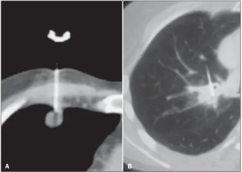
Figure 1. Computed tomography-guided transthoracic needle biopsy of pulmonary nodules. Lesions, needles and their interaction have excellent visualization at computed tomography. A: Peripheral pleural-based nodular lesion measuring 10 mm. B: Central lesion measuring 20 mm.
Chojniak R et al. Transthoracic needle biopsy of pulmonary nodules
tion rates of percutaneous biopsies of lung nodules and masses.
DISCUSSION
1. Indications
Percutaneous biopsies of lung nodules and masses are performed in various clini-cal settings, preferably in cases of periph-eral lesions not easily accessible at bron-choscopy or in cases of central lesions in patients who have previously undergone non diagnostic bronchoscopy.
Pulmonary nodules
Pulmonary nodules may be either inci-dentally identified or identified at routine assessments and be considered suspicious for primary lung cancer based on the pa-tients’ clinical history and radiological findings. In such cases, the biopsy may be useful in the approach to the disease in various different ways. In cases of lesions with low malignancy probability, the docu-mentation of benignity by means of percu-taneous biopsy can spare the patient more invasive procedures, such as thoracoscopy or thoracotomy. In cases of lesions highly suggestive of malignancy, biopsy is most frequently utilized for diagnostic documen-tation in patients who either refuse or do not present conditions to tolerate more in-vasive procedures.
Pulmonary nodules identified in pa-tients with primary malignant neoplasias in other organs may be submitted to percuta-neous biopsy whenever it is necessary to document metastatic disease, immunohis-tochemical study to support the definition of the therapeutic approach definition, or in cases where there is the possibility of the pulmonary nodule being a second primary or secondary neoplasm(3).
Pulmonary masses
In determined clinical contexts, pulmo-nary masses with certain radiological char-acteristics are strongly suggestive of pri-mary lung neoplasia, and, as such lesions are often followed by unequivocal signs of locally advanced or metastatic disease, percutaneous biopsy may be performed to determine the histologic type of the neo-plasm to allow the adoption of the appro-priate non surgical treatment.
Occasionally, percutaneous biopsy is requested in order to define the nature of radiological findings that are suspicious for tumor recurrence, or to establish the mo-lecular profile of a previously diagnosed neoplasm, with the purpose of refining the indication of chemotherapy agents or tar-geted therapies(3).
2. Technical aspects
In most of centers, percutaneous biopsy is performed by radiologists or interven-tional radiologists. The patients referred for the procedure are generally under clinical care by physicians or surgeons who at some point request the percutaneous biopsy.
Preprocedural assessment
Some aspects are important for the as-sessment of the patient candidate for per-cutaneous biopsy as well as of the pulmo-nary lesion to be approached. Ideally, such an assessment should be performed during a preceding consultation. In such an oppor-tunity it is possible to provide the patient with information on the procedure’s inher-ent risks and characteristics.
Clinical issues that should be assessed previously to the procedure include: gen-eral patient status, consciousness level, pulmonary functional reserve, coagulation activity and the patient ability to follow instructions regarding breathing and breath hold(3,4).
Small pulmonary lesions are hardly ac-cessible in poorly cooperative patients or those who are under sedation. In such cases, tracheal intubation and induced ap-nea may be necessary to allow percutane-ous approach to the lesion.
The most frequent complication of per-cutaneous lung biopsies is pneumothorax. Generally, such a complication is easily controllable; however patients with chronic obstructive pulmonary disease (COPD), poor pulmonary reserve (FEV1 < 35%) or with a single lung, may face severe conse-quences. A careful risk-benefit analysis must be performed in such cases and, as the procedure is performed, all the necessary conditions to allow immediate manage-ment of complications should be available. Appropriate coagulation activity levels are required to minimize bleedings in per-cutaneous biopsy, and anticoagulant agents
should be discontinued. Typically, a four-day anticoagulant therapy interruption pe-riod is necessary; however it is important to coordinate such a strategy with the as-sisting physician who is following up the anticoagulant therapy. A coagulation test must be performed up to one week prior to the procedure in patients with no suspicion of coagulopathy, or within a shorter period in patients submitted to interruption of anticoagulant therapy(3,5). Prothrombin
time, activated partial thromboplastin time and platelet count must be assessed before the procedure; platelet count < 100,000/ml and prothrombin or thromboplastin time ratios above the 1.4 standard are considered as relative contraindications. In such situ-ations, a risk-benefit analysis must be per-formed(4,6,7).
Lesion characteristics such as location, size and distance from the skin may affect success and complication rates or increase procedure times, and must be taken into consideration on the risk-benefit assess-ment prior to the procedure(6).
Additionally, a careful evaluation of the clinical and radiological context of the in-dication as well as of the presence of more easily approachable lesions in other sites, besides the possibility of utilizing other less invasive diagnostic methods, should al-ways be carried out.
Procedure
A term of free and informed consent must be obtained before the procedure, and the patients are instructed to fast for four hours prior to the procedure on account of the utilization of anesthetics and eventually intravenous contrast agents.
Whenever possible, the biopsy is per-formed on an outpatient basis and without sedation(3,4).
Based on previous studies, the patient is positioned in a decubitus that best allows a more direct access to the lesion. A series of images are then acquired for the plan-ning of an entry point, pathway and calcu-lation of the distance between the entry point and the lesion (Figure 2). Whenever possible, pathways minimizing the passage of the needle through aerated lung paren-chyma should be selected in order to reduce the risk for pneumothorax(3). After asepsis
(lidocaine 2%), the needle is introduced as planned. Such a maneuver can be followed up in real time by means of apparatuses equipped with CT-fluoroscopy.
For smaller or hardly accessible lesions, the coaxial approach technique may be uti-lized. Such a technique consists of the in-sertion of a cannula with a central rigid rod (mandrel) into the lesion. The mandrel is then removed, and the biopsy needle is then inserted through the cannula to collect the specimen. Thus multiple collections may be performed through the cannula(3). Once
the needle is positioned, new tomographic images are acquired in order to confirm its position in relation to the lesion. Then, the specimen is collected.
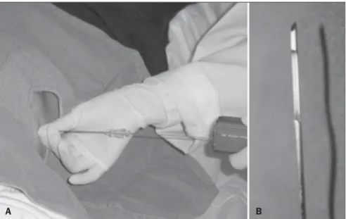
In cases of FNAB, Chiba type needles 22-25-gauge are utilized and the collection is performed by means of the aspiration technique, with back-and-forth movements and negative pressure. The collected mate-rial may be prepared in smears and fixed in alcohol or deposited in a fluid medium (Figure 3).
In the cases of CNB, 18-20-gauge cut-ting needles with an automatic biopsy sys-tem are utilized. Generally, the obtained fragments are preserved in a formaldehyde 10% solution (Figure 4).
The number of collections may vary for both types of needles. The following fac-tors may influence such number: difficul-Figure 2. Tomographic section identifying metal marker attached to patient’s skin and pulmonary lesion allowing the planning of entry point and needle pathway by means of cursors.
ties in the procedure, occurrence of com-plications, specimen’s quality, and required type of pathological analysis. Typically, two or three specimens are collected.
The immediate analysis of the speci-mens by a cytopathologist is recommended by some authors in cases of FNAB. Such a
recommendation is aimed at increasing the biopsy success rate and at reducing the number of collected specimens, as this technique allows the confirmation of the specimen appropriateness from the first puncture. On the other hand, besides re-quiring the availability of such a profes-Figure 3. A: Fine needle aspiration puncture technique utilizing negative pressure and back-and-forth movements B: Chiba type fine needle utilized in fine needle aspiration puncture.
A B
Figure 4. A: Cutting needle (tru-cut) biopsy coupled with automatic spring-activated triggering system. B: Detail of the cutting needle extremity with a lateral cutting section for specimen collection.
Chojniak R et al. Transthoracic needle biopsy of pulmonary nodules
sional, the immediate analysis determines an increase in the total procedure time. With some experience, a higher number of collections can produce comparable re-sults, without an increase in complication indices(8).
Post-procedural care
After the procedure, further images are acquired in order to evaluate the presence of possible complications such as pneu-mothorax or hemorrhage. Patients with no signs of complications are monitored for one hour and are released after confirma-tion of hemodynamic and respiratory sta-bility and chest radiography confirming the absence of pneumothorax. Late complica-tions rarely occur and patients must be in-structed to seek medical assistance in the event of dyspnea or hypotension(3,4,7).
3. Results
Success indices
The literature presents a significant variation in reported accuracy indices in percutaneous biopsy of lung lesions. Many studies report accuracy above 90%.
Success indices in the collection of ap-propriate specimens for analysis and in the achievement of specific diagnosis by means of percutaneous biopsy may vary due to several factors such as patient’s char-acteristics, experience of the service, type of needle and lesion characteristics(2).
Among the main factors considered as being predictive of a higher success index in obtaining a diagnosis by means of per-cutaneous biopsy, one can mention the absence of COPD, large dimensions of the lesion (> 2 cm), peripheral lesion loca-tion and utilizaloca-tion of cutting needles (CNB)(1,2,9).
Needle selection
One of the most studied factors in the analysis of percutaneous biopsy accuracy is the influence of the utilized needle type. In general, cutting needles utilized in CNBs present higher accuracy indices. However, the differences in accuracy should be evaluated in greater detail.
Most studies report high specificity and reliability on results positive for malig-nancy for both types of needles, with rare cases of false-positive results(1,10,11).
False-negative results are most frequent, and for such an occurrence, reported results for different types of needles are clearly different.
Fine Chiba type aspiration needles uti-lized in the collection of specimens for cytological analysis (FNAB) present a higher false-negative index as compared with cutting needles, and depending on the malignancy prevalence in the studied popu-lation, such an index may reach up to 50%(1,10–12).
In cases of benign lesions, more reliable results are obtained by means of cutting needles (tru-cut), since they allow obtain-ing the specimens for histological analysis with false-negative results generally below 10%(1,10,11,13).
Therefore, positive results in cases of lesions highly suspicious for malignancy are reliable with both types of needles; however, a negative result obtained with fine needles (FNAB) must be carefully analyzed, and a new collection should be considered.
For a reliable definition of absence of malignancy, as in the case of low-suspicion solitary nodular lesions, or for the defini-tion of a specific diagnosis of benignity, the utilization of cutting needles should be prioritized(13).
In general, satisfactory success indices in obtaining appropriate specimens for analysis are those above 85%. Sensitivity indices for malignancy are also generally above 85%. And false-positive results are extremely rare and lower than 1%.
In the previous experience reported by the authors, the success indices achieved in obtaining appropriate specimens by FNAB remained in the range between 84% and 88% along the years, and with cutting needles, between 95% and 97%(1,2,10,11,14).
Specific histological results were also more frequently obtained with cutting needles(1,2, 10,11,14). Some factors associated with higher
success indices in the present casuistry in-clude lesions with dimensions > 40 mm, located in the upper lobes and suspicious for malignancy.
Currently, based on the presented results and accumulated experience, the authors perform all the procedures with 18-20-gauge cutting needles. Fine needles are reserved for situations in which the patient
present coagulopathy. And, under ideal conditions, the authors perform three speci-men collections.
Complications
In the literature, the variation of com-plication rates reported for percutaneous biopsy of pulmonary lesions is also consid-erable(4).
Minor complications such as mild focal hemorrhage, pain and vasovagal reaction rarely occur, but are easily resolved. Pneu-mothorax is the most frequent moderate complication, most of times not requiring chest drainage. Several strategies may be adopted in the management of pneumotho-rax identified during or after the procedure, varying with the patient’s clinical status, symptoms, dimensions of the pneumotho-rax and available homecare conditions(4, 7,15) (Figure 5).
Reported rates of pneumothorax occur-rence range from 0% to 61%, and from 3% to 15% for necessity of chest drainage(4).
Factors such as presence of COPD, non-pleural-based lesions, greater depth and small lesion dimensions have been associ-ated with increased risk for development of pneumothorax(4,14,16).
Historically, CNB has been associated with a higher risk for pneumothorax. Much of the data on this subject is based on the utilization of large gauge needles without automatic biopsy systems. Most recent studies have demonstrated rates of occur-rence of pneumothorax similar to those reported for fine needle aspiration (FNAB). Generally, with some experience, pneu-mothorax and drainage indices should be close to 20% and 5% respectively. Lower indices may be expected in cases of pleu-ral-based lesions > 2 cm and absence of COPD.
Some post-procedural actions, such as positioning the patient over the needle en-try point, autologous blood injection into the needle pathway and aspiration of pneumo-thorax, are adopted by some authors to re-duce the pneumothorax index, however the benefit of such actions is still uncertain(17).
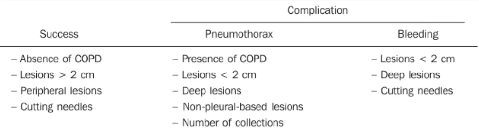
Table 1 Factors identified by different authors as being predictive of success in the acquisition of ap-propriate specimens and of complications occurrence.
Success
– Absence of COPD – Lesions > 2 cm – Peripheral lesions – Cutting needles
Complication
Pneumothorax
– Presence of COPD – Lesions < 2 cm – Deep lesions
– Non-pleural-based lesions – Number of collections
Bleeding
– Lesions < 2 cm – Deep lesions – Cutting needles
COPD, chronic obstructive pulmonary disease.
Figure 5. Pneumothorax management techniques. A: Computed tomography section simultaneously depicting pneumothorax, a relief drain and the biopsy needle. The management of the symptoms of the pneumothorax developed during the procedure allows the continuity of the biopsy. B: Images series acquired during the procedure, demonstrating pneumothorax and after its aspiration by means of a fine needle, which many times avoids drainage.
Table 1 lists the factors identified as predictive of success in the collection of appropriate specimens and the factors most frequently identified as related to relevant complications.
Physicians involved in the performance of percutaneous biopsies must be properly trained to deal with complications, and re-suscitation and chest drainage materials must be easily accessible.
In the authors’ practice, pneumothorax indices ranging between 11% and 16% were identified, with necessity of drainage occurring in 2% to 5% of cases where fine needles were utilized. In cases where cut-ting needles were utilized, such indices were even lower(1,10,14). In spite of the fact
that such data were obtained from differ-ent casuistries, the complication indices for both types of needles are similar, as already
reported by other authors(4,6,12,15). In a
ret-rospective analysis, the authors have iden-tified only the absence of a lesion pleural contact as a predictive factor for the occur-rence of pneumothorax.
In the authors’ practice, as the presence of pneumothorax is identified during the procedure, they go ahead with the
speci-men collection whenever possible and as-piration or a relief drain is utilized in order to stabilize the patient and conclude the collection (Figure 5).
moni-Chojniak R et al. Transthoracic needle biopsy of pulmonary nodules
tored for one hour, and are subsequently released. Patients with more severe or symptomatic pneumothorax may remain under observation or be submitted to aspi-ration or placement of a chest drain.
4. Conclusion
Percutaneous biopsies constitute an in-dispensable procedure in the practice of thoracic oncology. By means of such a pro-cedure, diagnostic problems can be quickly solved at radiology and pathology depart-ments, most of times avoiding more inva-sive procedures. Developments in imaging techniques and biopsy needles, and en-hancement of operators’ experience have led such procedure to become more versa-tile, safe and accurate thus expanding its indications.
REFERENCES
1. Chojniak R, Isberner RK, Viana LM, et al. Com-puted tomography guided needle biopsy: experi-ence from 1,300 procedures. Sao Paulo Med J. 2006;124:10–4.
2. Guimarães MD, Chojniak R, Gross JL, et al. Pre-dictive success factors for CT-guided fine needle aspiration biopsy of pulmonary lesions. Clinics (Sao Paulo). 2009;64:1139–44.
3. Wu CC, Mather MM, Shepard JA. CT-guided per-cutaneous needle biopsy of the chest: preproce-dural evaluation and technique. AJR Am J Roentgenol. 2011;196:W511–4.
4. Manhire A, Charig M, Clelland C, et al. Guide-lines for radiologically guided lung biopsy. Tho-rax. 2003;58:920–36.
5. Baglin TP, Keeling DM, Watson HG; British Committee for Standards in Haematology. Guide-lines on oral anticoagulation (warfarin): third edition – 2005 update. Br J Haematol. 2006;132: 277–85.
6. Topal U, Ediz B. Transthoracic needle biopsy: fac-tors effecting risk of pneumothorax. Eur J Radiol. 2003;48:263–7.
7. Aviram G, Schwartz DS, Meirsdorf S, et al. Tran-sthoracic needle biopsy of lung masses: a survey of techniques. Clin Radiol. 2005;60:370–4. 8. Küçük ÇU, Yilmaz A, Yilmaz A, et al. Computed
tomography-guided transthoracic fine-needle as-piration in diagnosis of lung cancer: a compari-son of single-pass needle and multiple-pass co-axial needle systems and the value of immediate cytological assessment. Respirology. 2004;9: 392–6.
9. Laurent F, Latrabe V, Vergier B, et al. CT-guided transthoracic needle biopsy of pulmonary nodules smaller than 20 mm: results with an automated 20-gauge coaxial cutting needle. Clin Radiol. 2000;55:281–7.
10. Yu LS, Deheinzelin D, Younes RN, et al. Com-puted tomography-guided cutting needle biopsy of pulmonary lesions. Rev Hosp Clin Fac Med Sao Paulo. 2002;57:15–8.
11. Guimaraes MD, de Andrade MQ, da Fonte AC, et al. CT-guided cutting needle biopsy of lung le-sions – an effective procedure for adequate ma-terial and specific diagnose. Eur J Radiol. 2010 Oct 26. [Epub ahead of print].
12. Anderson JM, Murchison J, Patel D. CT-guided lung biopsy: factors influencing diagnostic yield and complication rate. Clin Radiol. 2003;58:791– 7.
13. Gong Y, Sneige N, Guo M, et al. Transthoracic fine-needle aspiration vs concurrent core needle biopsy in diagnosis of intrathoracic lesions: a ret-rospective comparison of diagnostic accuracy. Am J Clin Pathol. 2006;125:438–44. 14. Guimarães MD, Andrade MQ, Fonte AC, et al.
Predictive complication factors for CT-guided fine needle aspiration biopsy of pulmonary le-sions. Clinics (Sao Paulo). 2010;65:847–50. 15. Gohari A, Haramati LB. Complications of CT
scan-guided lung biopsy: lesion size and depth matter. Chest. 2004;126:666–8.
16. Yeow KM, Su IH, Pan KT, et al. Risk factors of pneumothorax and bleeding: multivariate analy-sis of 660 CT-guided coaxial cutting needle lung biopsies. Chest. 2004;126:748–54.
17. Collings CL, Westcott JL, Banson NL, et al. Pneu-mothorax and dependent versus nondependent patient position after needle biopsy of the lung. Radiology. 1999;210:59–64.