346
Radiol Bras. 2017 Set/Out;50(5):338–348 Letters to the Editorhttp://dx.doi.org/10.1590/0100-3984.2015.0031
Luciana Mendes Araújo Borem1, Dimitrius Nikolaos Jaconi Stamoulis2, Ana Flávia Mundim Ramos2
1. Santa Casa de Montes Claros, Montes Claros, MG, Brazil. 2. Universidade Federal do Triângulo Mineiro (UFTM), Uberaba, MG, Brazil. Mailing address: Dr. Dimitrius Nikolaos Jaconi Stamoulis. Departamento de Radiologia e Diag-nóstico por Imagem – UFTM. Avenida Getúlio Guaritá, 130, Nossa Senhora da Abadia. Uberaba, MG, Brazil, 38025-250. E-mail: dimitriusss@hotmail. com.
Known triggers include acute exacerbation of asthma and situa-tions requiring the Valsava maneuver(4).
The combination of spontaneous pneumomediastinum and pneumorrhachis is extremely rare(5,6). Possible causes of pneumorrhachis include use of the drug ecstasy, abscesses, asthma attacks, coughing its, violent vomiting, epidural an -esthesia, lumbar puncture, and thoracic or vertebral surgery or trauma(7,8). In extremely rare cases, meningitis or pneumo-cephalus can occur(7). Pneumorrhachis typically occurs directly, when atmospheric air reaches the epidural space by means of a needle or a penetrating wound from the spine, although it can occur indirectly, as in bronchial asthma. In the case of bronchial asthma, air from the rupture of a peripheral pulmonary alveolus leaks into the pulmonary perivascular interstitium and follows the path of least resistance of the mediastinum to the fascia of the neck. Due to the absence of fascial barriers, air crosses the neural foramen and deposits in the epidural space. In either situation, pneumorrhachis is usually asymptomatic and disap-pears spontaneously within a few weeks.
Whereas CT allows direct visualization of the presence of air in the affected compartment(s), X-rays can reveal signs typi-cal of pneumomediastinum, produced when the air leaving the mediastinum delineates the normal anatomical structures. Such signs include subcutaneous emphysema, the “sail sign” of the thymus, pneumopericardium, the “ring-around-the-artery” sign, the “continuous diaphragm” sign, and the “double bronchial wall” sign(3).
REFERENCES
1. Lopes FPL, Marchiori E, Zanetti G, et al. Pneumomediastino espontâneo após esforço vocal: relato de caso. Radiol Bras. 2010;43:137–9. 2. Bote RP, Moreno AM, Núñez JMF, et al. Enisema epidural associado a
traumatismo torácico grave. Emergencias.2008;20:70–1.
3. Fatureto MC, Santos JPV, Goulart PEN, et al. Pneumomediastino espon -tâneo: asma. Rev Port Pneumol. 2008;14:437–41.
4. van Veelen I, Hogeman PHG, van Elburg A, et al. Pneumomediastinum: a rare complication of anorexia nervosa in children and adolescents. A case study and review of the literature. Eur J Pediatr. 2008;167:171–4. 5. Nascimento J, Gomes M, Moreira C, et al. Caso radiológico. Nascer e
Crescer. 2012;21:153–4.
6. Alves GRT, Silva RVA, Corrêa JRM, et al. Pneumomediastino espontâneo (síndrome de Hamman). J Bras Pneumol. 2012;38:404–7.
7. Aribas OK, Gormus N, Aydogdu Kiresi D. Epidural emphysema associ -ated with primary spontaneous pneumothorax. Eur J Cardiothorac Surg. 2001;20:645–6.
8. Araujo MS, Fernandes FLA, Kay FU, et al. Pneumomediastino, eni-sema subcutâneo e pneumotórax após prova de função pulmonar em paciente com pneumopatia intersticial por bleomicina. J Bras Pneumol. 2013;39:613–9.
Terson’s syndrome: an important differential diagnosis of subarach-noid hemorrhage
Dear Editor,
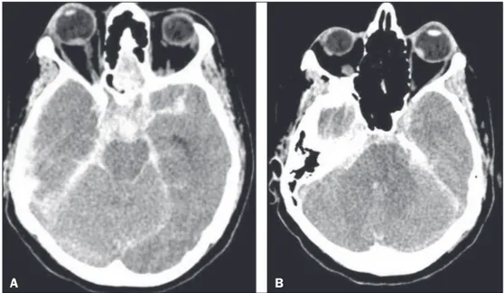
A 42-year-old female patient presented to the emergency room with severe headache and hypertensive urgency (blood pressure, 220/110 mmHg), progressing to left hemiparesis, right anisocoria, and a decreased level of consciousness, with a Glasgow Coma Scale score of 4. Computed tomography (CT) of the brain showed acute subarachnoid hemorrhage (Fisher grade 4), due to rupture of an aneurysm in the anterior circulation,
to-gether with signs of bilateral intraocular hemorrhage (Figure 1). Those indings are consistent with a diagnosis of Terson’s syn -drome.
Terson’s syndrome was initially described as vitreous hem-orrhage secondary to acute subarachnoid hemhem-orrhage, although recent studies have shown that it can also result from traumatic brain injury or even nontraumatic intracerebral hemorrhage(1). Originally described in 1900 by Albert Terson, the syndrome has an incidence of 2.6–27.0% in the context of subarachnoid hemorrhage due to a ruptured aneurysm(2–4). Although the etiol-ogy of the syndrome is controversial, it has been attributed to a
Figure 1. A: CT scan showing signs of subarachnoid hemorrhage and right intraocular hemorrhage, as a sponta-neously hyperattenuating focus in the posterior portion of the right globe. B: CT scan showing left intraocular hemor-rhage.
347
Radiol Bras. 2017 Set/Out;50(5):338–348Letters to the Editor
http://dx.doi.org/10.1590/0100-3984.2016.0053 Ana Paula Alves Fonseca1, Marcos Rosa Júnior1
1. Hospital Universitário Cassiano Antônio Morais da Universidade Federal do Espírito Santo (HUCAM-UFES), Vitória, ES, Brazil. Mailing address: Dra. Ana Paula Alves Fonseca. Rua Major Clarindo Fundão, 110, ap. 604, Praia do Canto. Vitória, ES, Brazil, 29055-655. E-mail: [email protected].
rapid increase in venous or intracranial pressure, which causes rupture of the peripapillary capillaries of the retina or results in compression of the central retinal vein, thus decreasing reti-nal venous drainage, promoting stasis, and provoking hemor-rhage(5).
The diagnosis of intraocular hemorrhage is more accurate-ly conirmed by ophthalmoscopy, although CT can suggest it, with an estimated sensitivity of 66%. The changes seen most frequently are retinal thickening and hyperattenuating nodules overlying the optic disc(6).
Terson’s syndrome most often occurs in patients with se-vere neurological disease, a Glasgow Coma Scale ≤ 8, and an-eurysmal subarachnoid hemorrhage with a Fisher score ≥ 3 at presentation. It is also of note that the rates of morbidity and mortality are high among such patients. In the sample studied by Fountas et al.(7), the mortality was 28.6% among the patients with intraocular hemorrhage, compared with only 2.0% among those without.
Terson’s syndrome is not an uncommon condition, perhaps being underdiagnosed. Given the prognostic implications of this diagnosis for morbidity and mortality, as well as the potential for secondary ocular lesion, it is of extreme relevance to radiolo-gists and other medical professionals, especially in the context of acute subarachnoid hemorrhage(8) but also in other forms of intracranial hemorrhage.
REFERENCES
1. Czorlich P, Skevas C, Knospe V, et al. Terson syndrome in subarachnoid hemorrhage, intracerebral hemorrhage, and traumatic brain injury. Neu-rosurg Rev. 2015;38:129–36.
2. de Vries-Knoppert W. Vitreous indings in a patient with Terson’s syn -drome. Doc Ophthalmol. 1995;90:75–80.
3. Vanderlinden RG, Chisholm LD. Vitreous hemorrhages and sudden in-creased intracranial pressure. J Neurosurg. 1974;41:167–76.
4. Pobereskin LH. Incidence and outcome of subarachnoid haemorrhage: a retrospective population based study. J Neurol Neurosurg Psychiatry. 2001;70:340–3.
5. Ogawa T, Kitaoaka T, Dake Y, et al. Terson syndrome: a case report suggest-ing the mechanism of vitreous hemorrhage. Ophthalmology. 2001;108: 1654–6.
6. Avila M, Cialdini AP, Crivelin M, et al. Vitrectomia na síndrome de Ter -son. Arq Bras Oftalmol. 1997;60:67–71.
7. Fountas KN, Kapsalaki EZ, Lee GP, et al. Terson hemorrhage in patients suffering aneurysmal subarachnoid hemorrhage: predisposing factors and prognostic signiicance. J Neurosurg. 2008;109:439–44.
8. Wajnberg E. Síndrome de takotsubo após hemorragia subaracnóidea. Ra -diol Bras. 2012;45:132–4.
Transient bilateral striatal lesion associated with varicella infection
Dear Editor,
A 5-year-old girl presented to our institution with an 8-day history of dermatological lesions typical of chickenpox, which had evolved to nausea, vomiting, and abdominal pain. During the observation period, she received symptomatic treatment (medication). Because she also experienced somnolence and apathy, she was hospitalized for further diagnostic investigation, evolving to a lack of ine motor coordination, dificulty in walk -ing, tremor, dystonia, generalized tonic-clonic seizures, dysmet-ria, and decomposition of movement. Cerebrospinal luid analy -sis revealed pleocyto-sis with a predominance of lymphocytes
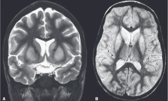
(12 leukocytes with 96% lymphocytes). A computed tomography scan of the head showed no abnormalities. Magnetic resonance imaging (MRI) showed hyperintense lesions in the caudate nu-clei and putamen on T2-weighted and proton density-weighted sequences (Figure 1), without enhancement after contrast ad-ministration. The patient showed gradual improvement and was discharged after 6 days of hospitalization. She was referred to a pediatric neurology clinic. After three months of follow-up, her symptoms had completely disappeared and another MRI of the brain showed regression of the lesions (Figure 2).
Varicella-zoster virus causes chickenpox and is associated with a variety of complications. The most common noncutaneous site of involvement is the central nervous system. Complications
Figure 1. MRI of the brain showing hyperintense lesions in the caudate nuclei and putamen. A: Coronal T2-weighted sequence. B: Axial proton