291 Radiol Bras. 2017 Set/Out;50(5):291–298
Can diffusion-weighted imaging add information
in the evaluation of breast lesions considered suspicious
on magnetic resonance imaging?
A difusão pode acrescentar informações na avaliação de lesões mamárias suspeitas na ressonância magnética?
Camila Souza Guatelli1, Almir Galvão Vieira Bitencourt2, Cynthia Aparecida Bueno de Toledo Osório3, Luciana Graziano1, Alessandra Araújo de Castro4, Juliana Alves de Souza5, Elvira Ferreira Marques6, Rubens Chojniak7
Guatelli CS, Bitencourt AGV, Osório CABT, Graziano L, Castro AA, Souza JA, Marques EF, Chojniak R. Can diffusion-weighted imaging add information in the evaluation of breast lesions considered suspicious on magnetic resonance imaging? Radiol Bras. 2017 Set/Out;50(5):291–298.
Abstract
Resumo
Objective: To assess the role of diffusion-weighted imaging (DWI) in the evaluation of breast lesions classiied as suspicious on magnetic resonance imaging (MRI), correlating the indings with the results of the histological analysis.
Materials and Methods: This was a retrospective, descriptive study based on a review of the medical records of 215 patients who were submitted to MRI with DWI before undergoing biopsy at a cancer center. Apparent diffusion coeficient (ADC) values were cal
-culated for each lesion, and the result of the histological analysis was considered the gold standard.
Results: The mean age was 49 years. We identiied 252 lesions, 161 (63.9%) of which were found to be malignant in the histological analysis. The mean ADC value was higher for the benign lesions than for the malignant lesions (1.50 × 10⁻³ mm2/s vs. 0.97 × 10⁻³
mm2/s), the difference being statistically signiicant (p < 0.001). The ADC cut-off point with the greatest sensitivity and speciicity on
the receiver operating characteristic curve was 1.03 × 10⁻³ mm2/s. When the DWI and conventional MRI indings were combined,
the accuracy reached 95.9%, with a sensitivity of 95.7% and a speciicity of 96.4%.
Conclusion: The use of DWI could facilitate the characterization of breast lesions, especially those classiied as BI-RADS 4, increas
-ing the speciicity and diagnostic accuracy of MRI.
Keywords: Breast neoplasms; Mammography; Ultrasonography, mammary; Magnetic resonance imaging.
Objetivo: Avaliar o papel da sequência em difusão na avaliação de lesões mamárias suspeitas na ressonância magnética (RM), correlacionando seus achados com os resultados histológicos.
Materiais e Métodos: Foi realizado estudo retrospectivo, descritivo, baseado na análise de prontuários médicos de 215 pacientes que realizaram RM com sequência em difusão e que foram submetidas a biópsia em um centro de referência oncológico. Foi
cal-culado o valor do coeiciente de difusão aparente (ADC – apparent diffusion coeficient) para cada lesão e o resultado histológico foi considerado como padrão ouro.
Resultados: A idade média das pacientes foi 49 anos. Foram identiicadas 252 lesões, e destas, 161 (63,9%) eram lesões ma -lignas na avaliação histológica. A média obtida do valor do ADC nas lesões benignas (1,50 × 10⁻³ mm2/s) foi superior à média
das lesões malignas (0,97 × 10⁻³ mm2/s), com signiicância estatística (p < 0,001). O ponto de corte com maior sensibilidade e
especiicidade pela curva receiver operating characteristic foi 1,03 × 10⁻³ mm2/s. Com a combinação da difusão com os achados
da RM, a acurácia chegou a 95,9%, com sensibilidade de 95,7% e especiicidade de 96,4%.
Conclusão: O uso da sequência em difusão pode auxiliar na caracterização das lesões mamárias, principalmente daquelas
classi-icadas como BI-RADS 4, aumentando a especiicidade e a acurácia diagnóstica da RM.
Unitermos: Neoplasias da mama; Mamograia; Ultrassonograia mamária; Ressonância magnética.
Study conducted in the Imaging Department of the A.C.Camargo Cancer Center,
São Paulo, SP, Brazil.
1. MSc, MD, Radiologist at the A.C.Camargo Cancer Center, São Paulo, SP, Brazil. 2. PhD, MD, Radiologist at the A.C.Camargo Cancer Center, São Paulo, SP, Brazil. 3. PhD, MD, Pathologist at the A.C.Camargo Cancer Center, São Paulo, SP, Brazil. 4. MD, Resident in the Imaging Department of the A.C.Camargo Cancer Center, São Paulo, SP, Brazil.
5. MD, Radiologist at the A.C.Camargo Cancer Center, São Paulo, SP, Brazil. 6. MD, Radiologist, Head of the Department of Breast Imaging, A.C.Camargo Cancer Center, São Paulo, SP, Brazil.
7. PhD, MD, Radiologist, Director of the Imaging Department of the A.C.Camargo Cancer Center, São Paulo, SP, Brazil.
INTRODUCTION
Because it provides information regarding the vascu-larization of the breast parenchyma, magnetic resonance imaging (MRI) has greater sensitivity in the detection of breast cancer than do mammography and ultrasound,
Mailing address: Dr. Almir Galvão Vieira Bitencourt. A.C.Camargo Cancer Center – Departamento de Imagem. Rua Professor Antônio Prudente, 211, Liberdade. São
making it an important tool in the screening of high-risk patients with dense breasts(1). MRI also shows greater
accuracy in assessing the extent of the disease and in detecting additional lesions in the contralateral breast during staging, thereby improving surgical and treatment planning(2–4). New MRI techniques have been developed
with the objective of adding functional information to the morphological and kinetic analysis, in order to improve the speciicity of the method. Among such techniques, diffusion-weighted imaging (DWI) is the one that is cur-rently being most widely studied(5).
DWI sequences use gradients that are sensitive to the movement of water molecules(6). Thus, DWI demonstrates differences in the movement of water molecules in tissues. In malignant tumors, increased cell proliferation results in greater cell density, creating more barriers for the diffu-sion of water molecules (restricted diffudiffu-sion), which man-ifests as hypointense signals on DWI. In contrast, benign tumors show lower cell density and a larger extracellular space, thus presenting fewer obstacles to the diffusion of water molecules. Images obtained from apparent diffusion coeficient (ADC) mapping can be analyzed qualitatively and quantitatively. Given these characteristics, DWI ap-pears to be a useful tool for differentiating between benign and malignant lesions, increasing the speciicity of MRI, providing important information for treatment planning and follow-up, as well as allowing the response to neoad-juvant chemotherapy to be evaluated(7–10).
The objective of this study was to evaluate the role of DWI in the evaluation of breast lesions classiied as suspicious on conventional MRI. We also attempted to determine whether DWI indings correlate with those ob -tained in histological and immunohistochemical analyses.
MATERIALS AND METHODS
This was a retrospective descriptive study based on the analysis of medical records and data collected from a group of patients who underwent MRI with DWI between August 2010 and December 2013 at a referral center for cancer. The study was approved by the research ethics committee of the institution.
We initially selected 238 patients in whom MRI scans identiied lesions, who also underwent MRI with DWI, and who were subsequently submitted to percutaneous or surgical biopsy. Of those, 23 were excluded because the DWI evaluation was compromised by movement arti-facts or other technical issues. Therefore, the inal study sample consisted of 215 patients, in whom a total of 252 lesions were identiied.
All MRI scans were performed in a 1.5 T scanner (Signa HDxt; GE Healthcare, Waukesha, WI, USA) with a dedicated breast coil. DWI was performed before the dy-namic phase of contrast enhancement, with array spatial sensitivity encoding technique echo-planar imaging in the axial plane (TR/TE, 4000/94; matrix, 192 × 192; signal
average, 3; slice thickness, 3 mm; distance factor, 20%). The diffusion-sensitizing gradients were applied in two or-thogonal directions, with two b values: 0 and 750 s/mm2.
The MRI scans of the breasts were reviewed on a ded-icated workstation (Advantage Workstation 4; GE Health -care) by a radiologist with experience in breast imaging. The lesions were evaluated and classiied according to the criteria of the Breast Imaging Reporting and Data System (BI-RADS) for MRI, 5th edition, deining the morpho -logical aspects by the type of enhancement (nodular or non-nodular); its distribution, shape, and contours; and the pattern of intravenous contrast uptake in the subse-quent dynamic evaluation.
The DWI sequences were post-processed with com-mercial software (FuncTool 7.4.01d; GE Healthcare). Qualitative and quantitative evaluations were based on the ADC. For the qualitative evaluation, we used gray-scale ADC maps, classifying lesions with restricted diffu -sion as those in which there was high signal intensity on DWI and signal loss on the ADC map. For the quantita -tive evaluation, we calculated the mean ADC, selecting the region of interest (ROI) within the lesion, avoiding ar-eas of necrosis and cystic degeneration. The ADC values were calculated by using the following formula:
ADC = −(1/b) ln (S2/S1)
where S2 and S1 are the intensities of the 0 and 750 s/ mm2b values, respectively(5).
Twenty lesions were excluded from the quantitative analysis because it was not possible to calculate the value of the ADC due to limitations of image recovery on the workstation. Therefore, only the qualitative evaluation of the diffusion was performed in those cases.
Histological data were collected through analysis of the surgical specimen, when available, or of percutaneous biopsy material. Histological types were reported accord-ing to the tumor classiication system of the World Health Organization(11) and the Nottingham (Elston-Ellis) modi-ication(12) of the Scarff-Bloom-Richardson grading
sys-tem.
and diffusion restriction variables. The data collected were compiled in a database created in the program Excel for Windows, and the statistical analysis was carried out with the software Stata, version 11 SE (StataCorp LLC, College Station, TX, USA), the Statistical Package for the Social Sciences, version 16.0 (SPSS Inc., Chicago, IL, USA), and MedCalc, version 15.6.1 (MedCalc Software, Ostend, Belgium).
RESULTS
Of the 215 patients included in the study, only one was male. The mean ± standard deviation for age was 49 ± 12 years (range, 23–88 years). The majority (75.8%) of the patients evaluated were ≥ 40 years of age, 61 (28.4%) had a positive family history of breast cancer, and 19 (8.8%) had a personal history of breast cancer.
Among the 215 patients included, there were a total of 252 lesions, with a mean size of 27 ± 22 mm (range, 4–117 mm). Of the 252 lesions, 210 (83.3%) showed nodular enhancement, 40 (15.8%) showed non-nodular enhancement, and 2 (0.8%) showed no enhancement.
In 106 (42.1%) of the 252 lesions evaluated, per -cutaneous biopsy alone was performed, whereas surgi-cal biopsy alone was performed in 71 (28.2%) and both were performed in 75 (29.8%). The biopsy results indi -cated that 91 (36.1%) of the lesions were benign and 161 (63.9%) were malignant. Benign lesions included but were not limited to ibroadenoma (n = 34), papilloma (n = 12), stromal ibrosis (n = 11), ibrocystic change (n = 8), and atypical lobular hyperplasia (n = 1), the last being the only lesion that showed atypia. Among the malignant lesions, ductal carcinoma in situ (DCIS) accounted for 11 and invasive carcinoma accounted for 150, of which 121 were classiied as invasive carcinoma of no special type (IC-NST).
Table 1 shows the BI-RADS categories, MRI mor -phological features, and MRI dynamic aspects, in relation to the histological results. All of the lesions classiied as BI-RADS 2 or 3 were found to be benign in the histo -pathological analysis. Of the BI-RADS 4 lesions, 74.5% were found to be benign. Among the 30 lesions classi -ied as BI-RADS 5, only one was found to be benign, the histological result in that case being complex sclerosing lesion. We found that, for the distinction between benign and malignant lesions, the BI-RADS MRI classiication had a sensitivity of 100%, a speciicity of 54.9%, a posi -tive predic-tive value (PPV) of 79.7%, a nega-tive predic-tive value (NPV) of 100%, and an accuracy of 83.7%.
In the qualitative analysis, DWI showed restricted diffusion in 216 (85.7%) of the 252 lesions. In the quan -titative analysis, the mean ADC value was 1.13 ± 0.38 × 10⁻³ mm2/s (range 0.38–2.69 10−3 mm2/s). Figures 1 and
2 depict examples of the lesions evaluated.
The comparison between the qualitative DWI anal-ysis and the histopathological indings showed that the
majority (72.7%) of the lesions that presented restricted diffusion were found to be malignant (p < 0.001). For the distinction between benign and malignant lesions, the DWI qualitative analysis showed a sensitivity of 97.5%, a speciicity of 35.2%, a PPV of 72.7%, an NPV of 88.9%, and an accuracy of 75.0%.
The quantitative analysis of diffusion was obtained by calculating the ADC value for each lesion and then comparing it with the histopathological result. The mean ADC value was higher for the benign lesions than for the malignant lesions (1.50 ± 0.35 × 10⁻³ mm2/s vs. 0.97 ±
0.27 × 10⁻³ mm2/s), the difference being statistically
sig-niicant (p < 0.001). The histological results were further consolidated into three groups: invasive carcinomas (n = 149), with a mean ADC of 0.95 × 10⁻³ mm2/s;
precur-sor lesions (n = 12), with a mean ADC of 1.24 × 10⁻³
mm2/s, among which the histopathological diagnosis was DCIS in 11 and atypical lobular hyperplasia in one; and benign lesions (n = 71), with a mean ADC of 1.49 × 10⁻³
mm2/s. The difference among the means was signiicant (p < 0.001).
Table 1—Relationship between the morphological/dynamic characteristics of the lesions and the histopathological indings.
Characteristics of the lesions
BI-RADS 2 3 4 5 6 Nodular lesions Form Irregular Regular Borders Spiculated Irregulars Regulars Internal enhancement Heterogeneous Homogeneous Peripheral Contrast uptake phase
Persistently enhancing Plateau Washout Non-nodular enhancement Distribution Focal Linear or ductal Regional Segmental N 2 48 40 1 0 74 6 68 1 16 58 24 44 6 59 11 6 16 9 2 0 5 Benign (n = 91)
Malignant (n = 161)
(%) (100)* (100) (74.1) (2.9) (0.0) (35.2) (6.9) (55.3) (3.1) (14.7) (85.3) (17.8) (78.6) (26.1) (64.1) (30.6) (6.5) (40.0) (100) (100) (0.0) (21.7) N 0 0 14 33 114 136 81 55 31 93 10 111 12 17 33 25 87 24 0 0 6 18 (%) (0.0) (0.0) (25.9) (97.1) (99.1) (64.8) (93.1) (44.7) (96.9) (85.3) (14.7) (82.2) (21.4) (73.9) (35.9) (69.4) (93.5) (60.0) (0.0) (0.0) (100) (78.3) P NP < 0.001 < 0.001 < 0.001 < 0.001 NP
Among the invasive carcinomas, the mean ADC was not found to be associated with the histological or immu-nohistochemical indings related to tumor aggressiveness (Table 2). Among the lesions classiied as DCIS, there
Table 2—Relationship between the mean ADC values and the histological/im-munohistochemical grade in invasive carcinomas.
Histological and immunohistochemical ind-ings in invasive carcinomas
Histological grade 1
2 3 Nuclear grade
1 2 3
Immunophenotype Her-2
Luminal A Luminal B
Triple negative or basal Estrogen receptor
Negative Positive
Progesterone receptor Negative
Positive HER2
Negative Positive Ki-67 index
1–20% 21–30% > 30%
Mean ADC
1.03 0.90 0.96
0.81 0.96 0.93
0.94 0.95 0.93 0.94
1.00 0.92
0.88 0.96
0.93 0.95
0.94 0.88 0.96
P
0.14
0.83
0.97
0.15
0.15
0.52
0.43
was also no statistical difference in terms of the nuclear grade.
Analysis of the ROC curve (Figure 3) showed an area under the curve of 0.901 (standard error: 0.0199; 95% conidence interval: 0.855–0.936; p < 0.0001). The cut-off ADC value with the highest sensitivity and speciicity, as determined by the ROC curve, was 1.03 × 10⁻³ mm2/s.
The ADC was lower than or equal to that cut-off value in Figure 1. A 64-year-old woman with a nodule in the left breast. Maximum
in-tensity projection reconstructions of the post-contrast subtraction sequence in the sagittal and axial planes (A and B, respectively), showing a circumscribed nodule with heterogeneous enhancement, which presented high signal
inten-sity in the DWI sequence (C) and low signal intensity on the ADC map (D), with
an ADC value of 0.74 × 10⁻³ mm2/s. The histological indings were consistent with a diagnosis of invasive carcinoma of non-special type.
Figure 2. A 35-year-old female with invasive carcinoma in the right breast and presenting with a nodule in the left breast on MRI. Contrast-enhanced T1-weighted images, in the sagittal plane (A) and with subtraction in the axial plane (B), showing a circumscribed nodule with heterogeneous enhancement,
high signal intensity in the DWI sequence (C), low signal intensity on the ADC
map (D), and an ADC value of 1.32 × 10⁻³ mm2/s. The histological indings were consistent with a diagnosis of ibroadenoma.
Figure 3. ROC curve to evaluate the diagnostic accuracy of the ADC value in the diagnosis of breast lesions.
ROC curve
100 - Speciicity
118 lesions, of which 116 (98.3%) were malignant, whereas 7 0 (61.4%) of the 114 in which it was higher than that cut-off value were benign, the difference between the two proportions being statistically signiicant (p < 0.001).
In two cases, the histopathology classiied the le -sions as benign but the ADC values were indicative of malignancy (false-positive results). In both of those cases, the diagnosis was stromal ibrosis without atypia. In ad -dition, there were 44 lesions classiied as malignant in the histopathological analysis but showing ADC values above the cut-off (false-negative results). Among those, the diagnoses were as follows: IC-NST, in 20 patients (45.5%); DCIS, in 9 (20.5%); invasive lobular carcinoma, in 6 (13.6%); carcinoma with intramammary lymph node metastasis, in 1 (2.3%); invasive mucinous carcinoma, in 1 (2.3%); pleomorphic lobular carcinoma, in 1 (2.3%); in -vasive focal tubular carcinoma, in 1 (2.3%); in-vasive pap -illary carcinoma, in 1 (2.3%); mixed ductal and lobular carcinoma, in 1 (2.3%); and metaplastic carcinoma, in 2 (4.5%). When analyzing the histological and immunohis -tochemical features of the invasive false-negative carcino-mas, we found that 91.2% presented histological grade 2 or 3, 69.0% presented nuclear grade 3, and 51.5% were of the luminal B immunophenotype.
Table 3 correlates the BI-RADS classiications, ob -tained from the evaluation of the morphological and dy-namic criteria, with the ADC values, obtained from the DWI, using the cutoff point of 1.03 × 10⁻³ mm2/s. Table 4
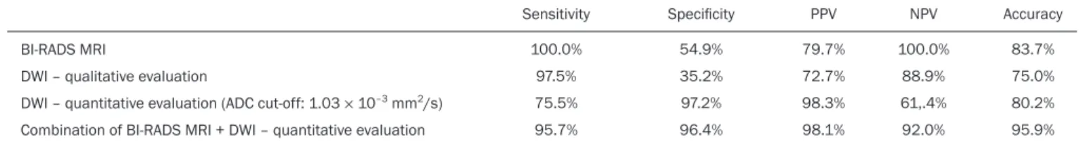
demonstrates the sensitivity, speciicity, PPV, NPV, and accuracy of MRI and DWI, separately and in combina-tion. For the combination of MRI and DWI indings, DWI was considered only in the analysis of the BI-RADS category 4 lesions, as follows:
• BI-RADS 2 or 3, regardless of the DWI indings = probably benign.
• BI-RADS 4, with DWI indicative of benign status = probably benign.
• BI-RADS 4, with DWI indicative of malignant sta -tus = suspected malignancy.
• BI-RADS 5 or 6, regardless of the DWI indings = suspected malignancy.
When MRI and DWI were combined, the accuracy of the tests reached 95.9%, with a sensitivity of 95.7% and a speciicity of 96.4%.
DISCUSSION
The capacity of DWI sequences of MRI to charac-terize the mobility of the water molecules allows indirect evaluation of the microstructure of the tissue by the grad-ing of its cellularity. Based on this principle, it is expected that the use of such sequences will increase the speci-icity of the method and ultimately decrease the number of unnecessary invasive procedures because of the high sensitivity of the contrast-enhanced images(13). Because
DWI sequences are already included in most MRI proto-cols, they do not entail additional costs; they also have an average acquisition time of less than 5 min.
In the present study, the qualitative evaluation based on DWI alone showed high sensitivity (97.5%), although its low speciicity (35.2%) made it incapable of differenti -ating between malignant and benign lesions in the major-ity of cases. In the quantitative analysis, malignant lesions showed signiicantly lower ADC values than did benign lesions. Therefore, the quantitative DWI analysis (ADC measurement) provided a greater contribution to the dif-ferentiation between benign and malignant breast lesions in our study.
Chen et al. conducted a meta-analysis to evaluate the performance of the quantitative DWI analysis. They eval-uated 964 lesions, of which 615 were malignant and 349 benign, and the mean cut-off ADC values for differentia -tion ranged from 0.9 × 10⁻³ mm2/s to 1.76 × 10⁻³ mm2/s,
sensitivity and speciicity ranging from 63% to 100% and from 46% to 97%, respectively. The mean ADC values
Table 4—Sensitivity, speciicity, PPV, NPV, and accuracy of MRI using the BI-RADS criteria alone, of qualitative and quantitative DWI evaluations, and of the combi-nation of the two (BI-RADS MRI + quantitative DWI evaluation), for distinguishing between benign and malignant lesions.
BI-RADS MRI
DWI – qualitative evaluation
DWI – quantitative evaluation (ADC cut-off: 1.03 × 10⁻³ mm2/s)
Combination of BI-RADS MRI + DWI – quantitative evaluation
Sensitivity 100.0% 97.5% 75.5% 95.7% Speciicity 54.9% 35.2% 97.2% 96.4% PPV 79.7% 72.7% 98.3% 98.1% NPV 100.0% 88.9% 61,.4% 92.0% Accuracy 83.7% 75.0% 80.2% 95.9% Table 3—Evaluation of diffusion in the lesions, by BI-RADS category and ADC
cut-off value (1.03 × 10⁻³ mm2/s), in relation to the histological indings.
Category BI-RADS 2 BI-RADS 3 BI-RADS 4 BI-RADS 5 BI-RADS 6 Histological result Benign Malignant N 0 2 0 37 2 31 0 0 0 0 (%) (0.0) (100) (0.0) (100) (22.2) (81.6) (0.0) (0.0) (0.0) (0.0) N 0 0 0 0 7 7 23 9 86 28 (%) (0.0) (0.0) (0.0) (0.0) (77.8) (18.4) (100) (100) (100) (100) P NP NP < 0.001 NP NP
and lobular carcinoma; such lesions can contain areas of normal ibroglandular and adipose tissue (i.e., tissue free of cell hyperproliferation), which can increase the ADC values obtained, thus leading to false-negative re -sults(20–22). High ADC values related to non-nodular en
-hancement also explain the predominance of false-nega-tive results among the cases of DCIS in our sample. De -spite the small number of DCIS cases in our sample (n = 11), these tumors showed a mean ADC value higher than that of invasive carcinomas.
The ADC threshold value for the differentiation of benign and malignant breast lesions should be selected according to the purpose of the examination. If the objec-tive is screening with DWI alone, the use of higher ADC threshold values is recommended, as a means of reduc-ing the risk of false-negative results. However, when DWI is used in conjunction with MRI, the use of lower ADC threshold values is recommended, as a means of reducing the risk of false-positive results(9).
Some studies have compared histological grade and tumor biological markers—expression of estrogen and progesterone hormone receptors; expression of human epidermal growth factor receptor 2 (HER2); and the Ki-67 cell proliferation index—with ADC values, attempting to identify associations, although the results have been inconsistent and occasionally contradictory(23). Belli et
al.(24) studied 289 patients with malignant carcinoma. Comparing ADC values with the histological subtype and grade, the authors found signiicant differences between grade 1 carcinoma and grade 2 or 3 carcinoma, as well as between invasive carcinoma and DCIS. Jeh et al.(25) stud-ied 107 cases of IC-NST in correlation with tumor prog -nostic factors and found ADC values to be signiicantly lower in tumors that were HER2-negative than in those that were HER2-positive. Mori et al.(26) studied 86 cases of IC-NST and demonstrated a signiicant difference in ADC values between tumors with high and low Ki-67 in -dices. Kim et al.(27) studied 67 women with invasive carci -noma and found no signiicant association between ADC values and tumor prognostic factors, including tumor grade and expression of biological markers.
In the present study, the ADC values in malignant tumors were not found to be signiicantly associated with histological or immunohistochemical indings related to aggressiveness. However, the invasive malignant lesions in our sample presented similar characteristics regard-ing histological grade and immunohistochemical proile: 91.9% of the lesions presented histological grade 2 or 3; 74.1% presented nuclear grade 3; and 84.5% were lumi -nal A or lumi-nal B lesions. This relative homogeneity of the histological grade and immunohistochemical proile among the invasive malignant lesions in our sample might have limited the associations with DWI.
When we evaluated the combined use of MRI with DWI compared with MRI alone, we found that their com-ranged from 1.0 × 10⁻³ mm2/s to 1.82 × 10⁻³ mm2/s for
the benign lesions and from 0.87 × 10⁻³ mm2/s to 1.36 ×
10−3 mm2/s for the malignant lesions(9). That considerable
variation is explained by the different protocols used in the studies. The cut-off ADC values obtained in the dif -ferentiation between benign and malignant are dependent upon the respective b values chosen. Therefore, the cut-off value obtained with a b value of 1000 s/mm2 cannot
be used for lesions evaluated with a b value of 500 s/mm2.
The results we obtained with a b value of 750 s/mm2, in
terms of the ADC values, cut-off value, sensitivity, and speciicity, are in agreement with those found in the lit -erature.
Despite the promising capacity of ADC values to differentiate between benign and malignant lesions, the ADC values for malignant and benign lesions can overlap, leading to false-positive and false-negative results. Par-sian et al.(14) studied benign lesions and found that
false-positive results were most often obtained for high-risk le-sions, atypical ductal hyperplasia being the most common subtype. Other studies have often reported false-positive results for intraductal papilloma(15–17). In our study, we
obtained false-positive results for only two lesions, both of which were subsequently diagnosed as stromal ibrosis without atypia, with ADC values of 0.89 × 10⁻³ mm2/s
and 0.90 × 10⁻³ mm2/s, respectively.
It is known that high levels of ADC are frequently as -sociated with benign changes or benign tumors; although some IC-NSTs show ADC values higher than the cut-off established for malignancy, leading to false-negative re-sults(15,17). The malignant histological subtype with the highest ADC values is mucinous carcinoma, which is characterized by low cellularity and a predominance of mucin, therefore often producing false-negative results in DWI(18,19).
bined use resulted in a signiicant increase in speciic -ity (96.4%), without a signiicant reduction in sensitiv-ity (95.7%), corresponding to a signiicant increase in accu -racy (95.9%), conirming our expectations and the data in the literature(28). DWI was particularly useful in cases of lesions categorized as BI-RADS 4, which were respon -sible for the lower speciicity of MRI. Through analysis of the ADC values obtained for the BI-RADS 4 group combined with that of the MRI indings, we were able to propose subdivision of the category BI-RADS 4 into two groups—“probably benign”, comprising lesions with ADC values above the cut-off; and “suspected malignancy”, comprising lesions with ADC values equal to or below the cut-off—with high rates of sensitivity, speciicity, and ac -curacy (95.7%, 96.4%, and 95.9%, respectively).
In the present study, the use of DWI had a greater im-pact on the evaluation of BI-RADS 4 lesions than on that of lesion in other categories, allowing a better, more com-plete assessment of these lesions and leading to tailored practices. These indings are in agreement with those of Almeida et al.(29), who demonstrated that DWI can
im-prove the diagnostic performance of MRI and facilitate the division of BI-RADS 4 lesions into the subcategories 4A, 4B, and 4C. In addition, in comparison with MRI alone, MRI plus DWI can more accurately corroborate benign results in BI-RADS 4 lesions, as well as clarifying the analysis of lesions with a discordant histopathological result from a biopsy fragment, leading to a more accurate surgical evaluation.
The results of the present study should be consid-ered in the context of certain limitations. Because it was a retrospective study, many cases could not be evaluated, because it was not possible to recover the MRI data from our digital archive. We were also forced to exclude ex-aminations in which there were technical dificulties in the acquisition of images due to susceptibility artifacts that resulted in image distortion and impaired the char-acterization of the lesion. It is known that DWI is highly sensitive to such artifacts, and it is hoped that technical innovations currently in development will bring improve-ments in the resolution of DWI of the breast(20). It is also
noteworthy that our patient population, because it com-prised individuals treated at a cancer center, featured a predominance of malignant pathological indings, which could have inluenced the results.
In conclusion, the indings of the present study dem -onstrate that the use of DWI can facilitate the charac-terization of breast lesions, especially those categorized as BI-RADS 4, thus increasing the speciicity and diag -nostic accuracy of MRI. This method provides greater conidence in the management of this patient population and, after further studies involving larger samples have been conducted, might even be used in order to reduce the number of unnecessary biopsies.
REFERENCES
1. Mainiero MB, Lourenco A, Mahoney MC, et al. ACR appropri
-ateness criteria breast cancer screening. J Am Coll Radiol. 2013; 10:11–4.
2. Marques EF, Medeiros MLL, Souza JA, et al. Indications for breast
magnetic resonance imaging in an oncology reference center.
Ra-diol Bras. 2011;44:363–6.
3. American College of Radiology. ACR practice guideline for the per -formance of magnetic resonance imaging (MRI) of the breast.
Res-ton, VA: American College of Radiology; 2014.
4. Mann RM, Kuhl CK, Kinkel K, et al. Breast MRI: guidelines from the European Society of Breast Imaging. Eur Radiol. 2008;18:1307–18. 5. Kul S, Cansu A, Alhan E, et al. Contribution of diffusion-weighted
imaging to dynamic contrast-enhanced MRI in the characterization
of breast tumors. AJR Am J Roentgenol. 2011;196:210–7. 6. Woodhams R, Ramadan S, Stanwell P, et al. Diffusion-weighted im
-aging of the breast: principles and clinical applications.
Radiograph-ics. 2011;31:1059–84.
7. Partridge SC, DeMartini WB, Kurland BF, et al. Quantitative
diffusion-weighted imaging as an adjunct to conventional breast
MRI for improved positive predictive value. AJR Am J Roentgenol. 2009;193:1716–22.
8. Arantes Pereira FP, Martins G, Figueiredo E, et al. The use of
diffusion-weighted magnetic resonance imaging in the differen-tiation between benign and malignant breast lesions. Radiol Bras.
2009;42:283–8.
9. Chen X, Li WL, Zhang YL, et al. Meta-analysis of quantitative dif -fusion-weighted MR imaging in the differential diagnosis of breast
lesions. BMC Cancer. 2010;10:693.
10. Yabuuchi H, Matsuo Y, Sunami S, et al. Detection of non-palpa -ble breast cancer in asymptomatic women by using unenhanced
diffusion-weighted and T2-weighted MR imaging: comparison with
mammography and dynamic contrast-enhanced MR imaging. Eur
Radiol. 2011;21:11–7.
11. Tavassoli FA, Devilee P. World Health Organization Classiication
of Tumours. Pathology and genetics of tumours of the breast and
female genital organs. 5th ed. Lyon, France: IARC Press; 2003. 12. Elston CW, Ellis IO. Pathological prognostic factors in breast
cancer. I. The value of histological grade in breast cancer: experi-ence from a large study with long-term follow-up. Histopathology.
1991;19:403–10.
13. Partridge SC, Rahbar H, Murthy R, et al. Improved diagnostic ac
-curacy of breast MRI through combined apparent diffusion coefi -cients and dynamic contrast-enhanced kinetics. Magn Reson Med.
2011;65:1759–67.
14. Parsian S, Rahbar H, Allison KH, et al. Nonmalignant breast lesions: ADCs of benign and high-risk subtypes assessed as false-positive at dynamic enhanced MR imaging. Radiology. 2012;265:696–706. 15. Woodhams R, Matsunaga K, Kan S, et al. ADC mapping of benign
and malignant breast tumors. Magn Reson Med Sci. 2005;4:35–42. 16. Tozaki M, Fukuma E. 1H MR spectroscopy and diffusion-weighted
imaging of the breast: are they useful tools for characterizing breast
lesions before biopsy? AJR Am J Roentgenol. 2009;193:840–9. 17. Jin G, An N, Jacobs MA, et al. The role of parallel diffusion-weight
-ed imaging and apparent diffusion coeficient (ADC) map values for evaluating breast lesions: preliminary results. Acad Radiol. 2010; 17:456–63.
18. Woodhams R, Kakita S, Hata H, et al. Diffusion-weighted imaging
of mucinous carcinoma of the breast: evaluation of apparent
diffu-sion coeficient and signal intensity in correlation with histologic indings. AJR Am J Roentgenol. 2009;193:260–6.
20. Brandão AC, Lehman CD, Partridge SC. Breast magnetic reso -nance imaging: diffusion-weighted imaging. Magn Reson Imaging
Clin N Am. 2013;21:321–36.
21. Partridge SC, Mullins CD, Kurland BF, et al. Apparent diffusion coeficient values for discriminating benign and malignant breast MRI lesions: effects of lesion type and size. AJR Am J Roentgenol. 2010;194:1664–73.
22. Sahin C, Aribal E. The role of apparent diffusion coeficient values
in the differential diagnosis of breast lesions in diffusion-weighted
MRI. Diagn Interv Radiol. 2013;19:457–62.
23. Costantini M, Belli P, Rinaldi P, et al. Diffusion-weighted imag -ing in breast cancer: relationship between apparent diffusion
coef-icient and tumour aggressiveness. Clin Radiol. 2010;65:1005–12. 24. Belli P, Costantini M, Bui E, et al. Diffusion magnetic resonance
imaging in breast cancer characterisation: correlations between the
apparent diffusion coeficient and major prognostic factors. Radiol Med. 2015;120:268–76.
25. Jeh SK, Kim SH, Kim HS, et al. Correlation of the apparent dif
-fusion coeficient value and dynamic magnetic resonance imaging indings with prognostic factors in invasive ductal carcinoma. J Magn Reson Imaging. 2011;33:102–9.
26. Mori N, Ota H, Mugikura S, et al. Luminal-type breast cancer: cor
-relation of apparent diffusion coeficients with the Ki-67 labeling index. Radiology. 2015;274:66–73.
27. Kim SH, Cha ES, Kim HS, et al. Diffusion-weighted imaging of breast cancer: correlation of the apparent diffusion coeficient value with prognostic factors. J Magn Reson Imaging. 2009;30:615–20. 28. Yabuuchi H, Matsuo Y, Okafuji T, et al. Enhanced mass on
contrast-enhanced breast MR imaging: lesion characterization using combi-nation of dynamic contrast-enhanced and diffusion-weighted MR
images. J Magn Reson Imaging. 2008;28:1157–65.
29. Almeida JRM, Gomes AB, Barros TP, et al. Simple magnetic reso -nance imaging criteria can differentiate ductal carcinomas in situ