389
Radiol Bras. 2017 Nov/Dez;50(6):389–394
Hepatocyte-speciic contrast agent-enhanced magnetic
resonance cholangiography: perioperative evaluation
of the biliary tree
Colangiorressonância com uso de contraste hepatoespecíico: avaliação perioperatória da via biliar
Luciana Carmen Zattar-Ramos1, Regis Otaviano Franca Bezerra2, Luis Tenório de Brito Siqueira3, Marcos Roberto
Menezes4, Claudia da Costa Leite5, Giovanni Guido Cerri6
Zattar-Ramos LC, Bezerra RO, Siqueira LTB, Menezes MR, Leite CC, Cerri GG. Hepatocyte-speciic contrast agent-enhanced magnetic resonance
cholangiography: perioperative evaluation of the biliary tree. Radiol Bras. 2017 Nov/Dez;50(6):389–394.
Abstract
Resumo
A large number of gadolinium chelates have recently been tested in clinical trials. Some of those have already been approved for clinical use in the United States and Europe. Thus, new diagnostic perspectives have been incorporated into magnetic resonance imaging studies. Among such gadolinium chelates are hepatobiliary-speciic contrast agents (HSCAs), which, due to their property of being selectively taken up by hepatocytes and excreted by the biliary ducts, have been widely used for the detection and charac
-terization of focal hepatic lesions. In comparison with conventional magnetic resonance cholangiography (MRC), HSCA-enhanced MRC provides additional information, with higher spatial resolution and better anatomic evaluation of a non-dilated biliary tree. A thorough anatomic assessment of the biliary tree is crucial in various hepatic surgical procedures, such as complex resection in patients with colorectal cancer and living-donor liver transplantation. However, the use of HSCA-enhanced MRC is still limited, because of a lack of data in the literature and the poor familiarity of radiologists regarding its main indications. This pictorial essay aims to demonstrate the use of HSCA-enhanced MRC, with particular emphasis on anatomical analysis of the biliary tree, clinical applications, and the most important imaging indings.
Keywords: Biliary tree; Magnetic resonance imaging; Hepatobiliary-speciic contrast agents.
Recentemente, um grande número de quelantes de gadolínio tem sido testado em ensaios laboratoriais. Alguns deles já foram inclusive aprovados para uso clínico nos Estados Unidos e na Europa. Com isso, novas perspectivas diagnósticas foram incorpora
-das nos estudos de ressonância magnética. Dentre estes quelantes de gadolínio, os contrastes hepatobiliares especíicos (CHBEs) têm sido amplamente utilizados para a caracterização e detecção de lesões focais hepáticas, essencialmente pela propriedade de serem captados pelos hepatócitos e excretados na via biliar. Além disso, os CHBEs trouxeram novas informações na avaliação da árvore biliar quando comparados à colangiorressonância convencional, proporcionando uma maior resolução espacial e melhor avaliação da anatomia da árvore biliar não dilatada. A avaliação da anatomia biliar é de fundamental importância em cirurgias he
-páticas, como ressecções complexas em tumores colorretais ou no transplante hepático com doador vivo, porém, o uso dos CHBEs ainda é restrito para estes propósitos. Em razão da escassa literatura sobre o tema e da pouca familiaridade dos radiologistas com as principais indicações, o presente ensaio iconográico tem por objetivo demonstrar o uso de CHBEs na avaliação perioperatória das vias biliares, ressaltando a avaliação anatômica, as indicações e os principais achados de imagem.
Unitermos: Vias biliares; Ressonância magnética; Contrastes hepatoespecíicos.
Study conducted at the Hospital Sírio-Libanês, São Paulo, SP, Brazil. 1. MD, Radiologist, Hospital Sírio-Libanês, São Paulo, SP, Brazil.
2. MD, Radiologist, Hospital Sírio-Libanês and Instituto do Câncer do Estado de São Paulo (Icesp), São Paulo, SP, Brazil.
3. MD, Radiologist, Hospital Regional de Presidente Prudente and Hospital Nossa Senhora das Graças, Presidente Prudente, SP, Brazil.
4. PhD, MD, Radiologist, Hospital Sírio-Libanês and Instituto do Câncer do Estado de São Paulo (Icesp), São Paulo, SP, Brazil.
5. Associate Professor in the Department of Radiology and Oncology of the Fa-culdade de Medicina da Universidade de São Paulo (FMUSP), Radiologist, Hospital Sírio-Libanês, São Paulo, SP, Brazil.
6. Full Professor in the Department of Radiology and Oncology of the Faculdade de Medicina da Universidade de São Paulo (FMUSP), Radiologist, Hospital Sírio--Libanês, São Paulo, SP, Brazil.
Mailing address: Dr. Luciana Carmen Zattar-Ramos. Hospital Sírio-Libanês. Rua Dona Adma Jafet, 91, Bela Vista. São Paulo, SP, Brazil, 01308-050. E-mail: [email protected].
Received November 8, 2015. Accepted after revision June 7, 2016.
postoperative complications(1–6). Hepatobiliary surgery involves procedures of great technical dificulty. In many
cases, complications related to the biliary tract occur and can be accompanied by unexpected anatomical variations.
In liver transplantation, the rate of such complications
ranges from 10% to 25%, fatal complications occurring in up to 10% of cases(7).
Conventional T2-weighted magnetic resonance chol-angiography (MRC) has the disadvantage of assessing only INTRODUCTION
Gadolinium-based hepatocyte-speciic contrast agents
the anatomy of the biliary tract, providing little informa-tion in cases in which there is no dilatainforma-tion. Therefore, some studies suggest that a thorough evaluation of the biliary system involves a combination of anatomical and functional evaluation techniques(8).
The HSCAs approved for clinical use in Brazil(9),
un-like the contrast agents routinely used in magnetic reso-nance imaging, are selectively captured by functioning hepatocytes and have high rates of elimination (of approx-imately 50%) through the biliary tract(1–5,9). The MRC
ex-amination protocols should be adapted for time optimiza-tion, because it is necessary to obtain late images at 20–30 min after injection of the HSCA(5,6,10–12).
The aim of the present study was to demonstrate the use of HSCA-enhanced MRC in the perioperative evalu-ation of the bile ducts. We highlight the main imaging as-pects that can inform the practice.
METHODS
We selected MRC examinations in which the HSCA gadoxetic acid (Primovist®) was used for the perioperative
assessment of the upper abdomen in patients undergoing hepatobiliary surgery between January 2013 and February 2015. The images were obtained from the digital archives of our institution.
EVALUATION OF THE BILIARY ANATOMY
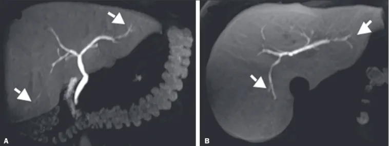
Anatomical variations of the biliary tract occur in ap-proximately 30% of individuals. The correct character-ization of such variations is necessary in order to avoid iatrogenic injuries during hepatobiliary surgery and proce-dures, especially in cases of living donors for liver trans-plantation (Figure 1), in which accurate knowledge of the biliary anatomy is essential(2,4–6,13).
Delineation of the biliary tree occurs in the late hepa-tobiliary phase of acquisition, at 20–30 min after HSCA administration. The MRC sequences acquired with HSCA are T1-weighted, providing images with better spatial resolution than those obtained in the T2-weighted sequences employed in conventional (non-contrast-en-hanced MRC, especially when there is no dilatation of the bile ducts(5,6,10–12), as illustrated in Figure 2.
Figure 1. Preoperative evaluation of a liver donor candidate. The use of HSCA-enhanced MRC allows an excellent evaluation of the normal anatomy of the biliary tree, with delineation of the subsegmental ducts (arrows).
A B
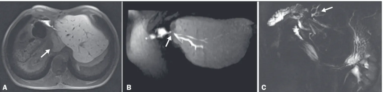
Figure 2. Preoperative evaluation for right hepatectomy in a patient with metastatic colorectal cancer. HSCA-enhanced MRC provides excellent delineation of the biliary tree, demonstrating an accessory bile duct in segment VII (arrow), the recognition of which is important for the surgical planning.
A B C
DIFFERENTIATION BETWEEN BILIARY AND NONBILIARY LESIONS
Making a distinction between cystic lesions near the biliary tree and those actually within the biliary tract is important in clinical management. The presence of the
HSCA within the lumen of a cystic lesion conirms its bili
-ary origin (Figure 3) and allows the differentiation among choledochal cysts, pericholedochal cystic formations, di-verticula, and duodenal duplications(4,5,11).
DIAGNOSIS OF ACUTE CHOLECYSTITIS
Acute cholecystitis is a common cause of acute abdo-men and is initially investigated by ultrasound. However,
when the ultrasound indings are inconclusive,
HSCA-enhanced MRC can be useful, replacing scintigraphy with
diisopropyl iminodiacetic acid (DISIDA scintigraphy).
A lack of HSCA uptake in the gallbladder during MRC
constitutes a speciic sign of acute cholecystitis (similar to
that seen in DISIDA scintigraphy), with the additional ad
-vantage of allowing evaluation of the differential diagno-ses of abdominal pain(4–6,11,13,14). It is noteworthy that the
functional evaluation is considered in combination with the other usual MRC aspects, such as choledocholithia-sis (Figure 4), perfusion disorder (Figure 5), gallbladder
thickening, and peribiliary luid collections(2–4,6,8).
DIAGNOSIS AND GRADING OF BILIARY OBSTRUCTIONS
Changes in the HSCA dynamics in the biliary tract allow the early diagnosis of obstruction, even without evident dilatation (Figure 6), as well as allowing its
clas-siication(2,4,6,10–14), as follows: complete biliary
obstruc-tion—absence of contrast uptake in the distal and proxi-mal portions of a stricture or obstructive lesion (Figure 7); nearly complete biliary obstruction—contrast uptake that
is signiicantly late and only in the proximal portion of a
stricture or obstructive lesion; or partial biliary obstruc-tion—contrast uptake only outside the area of the stric-ture or obstructive lesion. Caution should be exercised when evaluating patients with markedly impaired hepatic function, because the dysfunction changes the way in which the HSCA is metabolized, reducing its uptake in the biliary tract and simulating obstructive processes(6,10,12).
Early, accurate grading and characterization of biliary ob-structions is important in order to guide the therapeutic practice and, occasionally, the surgical planning(2,4,6,10–14).
POSTOPERATIVE EVALUATION OF THE BILIARY
TRACT
Iatrogenic injuries are the most common complications
associated with hepatobiliary surgery, especially performed by laparoscopy. The most common iatrogenic injuries are
istulas, which can occur in the immediate or early post
-operative period, and strictures, which occur later(4–6,11). Figure 3. MRC showing a small
cystic formation (arrows), adja
-cent to the common hepatic duct, in a T2-weighted sequence (A) and a non-contrast-enhanced T1-weighted in-phase sequence (B). An HSCA-enhanced T1-weighted MRC sequence (C) unequivocally demonstrates the diagnosis of bile cyst, after its illing with con
-trast (arrow). A B C
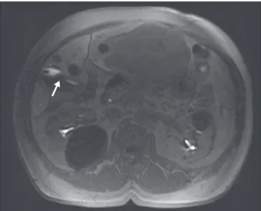
Figure 4. Case of choledocholithiasis. An HSCA-enhanced MRC scan shows a lack of contrast uptake within the gallbladder (arrow).
The use of HSCA-enhanced MRC allows the diag-nosis of extravasations through their direct visualization, as well as demonstrating their origin (Figure 8). Bile duct
injury is graded according to the classiication system de
-vised by Bismuth, as well as that subsequently de-vised by Strasberg(4–6,11).
The combination of conventional (T2-weighted) MRC and HSCA-enhanced MRC has a reported accuracy
of 84% for the detection of biliary istulas(8). Therefore,
the combined use of those two techniques increases the
likelihood of the correct identiication and localization of
the extravasation site (Figure 9)(4–6,11).
Figure 5. Acute cholecystitis. Fast spin-echo axial T2-weighted MRC sequence (A) demonstrating parietal thickening (arrow); non-contrast-enhanced axial T1-weighted MRC sequence (B) showing high-protein biliary content forming a level with a small amount of gas (arrow); and HSCA-enhanced coronal T1-weighted MRC sequence (C) showing no contrast uptake within the gallbladder (arrow). In such cases, the use of morphine can be considered, although it is rarely necessary, given the set of indings suggestive of cholecystitis.
A B C
Figure 7. HSCA-enhanced coro
-nal T1-weighted MRC sequence acquired in the late phase (A) showing that, despite moder
-ate dilation of the biliary tract, there was no elimination of contrast in the hepatobiliary phase, due to obstruction of the distal bile duct (arrow). Non-contrast-enhanced coro
-nal MRC scan (B).
A B
Figure 6. Postoperative evaluation of a hepatectomy patient with biliodigestive anastomosis. An HSCA-enhanced MRC scan shows late contrast uptake in the caudate lobe (A, arrow), suggesting severe obstruction (B, arrow), even before there was signiicant dilation of the biliary tree. An MRC scan acquired three months later showed mild ectasia of the intrahepatic bile ducts of the caudate lobe with no stricture development in the lateral ducts of the left lobe (C, arrow).
A B C
Figure 8. Images acquired in the hepatobiliary phase of HSCA excretion. Note the extravasation of the contrast medium from the main duct of the lateral segment of the left lobe (arrows).
A B C
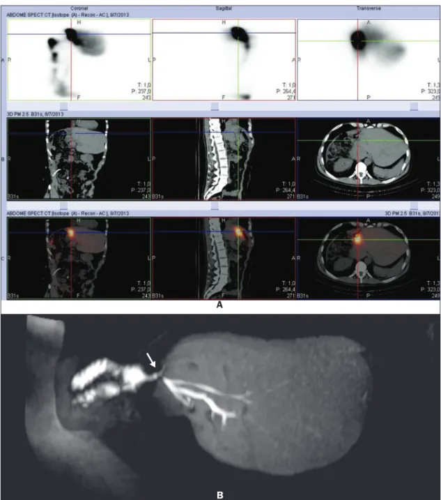
Figure 9. Postoperative evaluation of a hepatectomy patient with biliodigestive anastomosis. DISIDA scintigraphy suggested a liquid collection in the anastomosis plane (A). An HSCA-enhanced MRC scan showed anastomosis integrity and contrast accumulation in the “J” pouch (B, arrow), clearly disproving the hypothesis of a collection or istula.
A
DIFFERENTIAL DIAGNOSIS BETWEEN ABDOMI
-NAL COLLECTIONS AND BILIARY FISTULAS (BI
-LOMAS)
Bilomas are collections of bile outside of the biliary tree. They can be spontaneous, traumatic, or iatrogenic, the majority (70%) occurring in the upper right quadrant of the abdomen. The accurate characterization of a biloma allows it to be distinguished from collections of other na-tures, as well as informing decisions regarding its
manage-ment. A late image after HSCA administration conirms
that the collection is composed of bile and allows the site
of extravasation to be identiied(4,5,11). The use of MRC
with a contrast agent that is speciic for biliary content al
-lows better differentiation among abdominal collections, loculated ascites, and cystic formations adjacent to the bile ducts (Figure 10), providing important additional informa-tion in relainforma-tion to conveninforma-tional T2-weighted MRC(4,5,11).
weighted MRC(3–6). The correct indication and
interpreta-tion of the images can alter the clinical practice, as well as precluding the need for other diagnostic tests, invasive procedures, biopsies, or additional interventions(2,11,12).
REFERENCES
1. Guglielmo FF, Mitchell DG, Gupta S. Gadolinium contrast agent selection and optimal use for body MR imaging. Radiol Clin North Am. 2014;52:637–56.
2. Van Beers BE, Pastor CM, Hussain HK. Primovist, Eovist: what to expect? J Hepatol. 2012;57:421–9.
3. Ringe KI, Husarik DB, Gupta RT, et al. Hepatobiliary transit times of gadoxetate disodium (Primovist®) for protocol optimization of comprehensive MR imaging of the biliary system—what is normal? Eur J Radiol. 2011;79:201–5.
4. Gupta RT. Evaluation of the biliary tree and gallbladder with he-patocellular MR contrast agents. Curr Probl Diagn Radiol. 2013; 42:67–76.
5. Gupta RT, Brady CM, Lotz J, et al. Dynamic MR imaging of the biliary system using hepatocyte-speciic contrast agents. AJR Am J Roentgenol. 2010;195:405–13.
6. Seale MK, Catalano O, Saini S, et al. Hepatobiliary-speciic MR contrast agents: role in imaging the liver and biliary tree. Radio-graphics. 2009;29:1725–48.
7. Hyodo T, Kumano S, Kushihata F, et al. CT and MR cholangiog-raphy: advantages and pitfalls in perioperative evaluation of biliary tree. Br J Radiol. 2012;85:887–96.
8. Kantarci M, Pirimoglu B, Karabulut N, et al. Non-invasive detec-tion of biliary leaks using Gd-EOB-DTPA-enhanced MR cholangi-ography: comparison with T2-weighted MR cholangiography. Eur Radiol. 2013;23:2713–22.
9. Bittencourt LK, Hausmann D, Gasparetto EL, et al. Ressonância magnética do fígado com contraste hepato-especíico: experiência clínica inicial no Brasil. Rev Col Bras Cir. 2012;40:237–40. 10. Goodwin MD, Dobson JE, Sirlin CB, et al. Diagnostic challenges
and pitfalls in MR imaging with hepatocyte-speciic contrast agents. Radiographics. 2011;31:1547–68.
11. Lee NK, Kim S, Lee JW, et al. Biliary MR imaging with Gd-EOB-DTPA and its clinical applications. Radiographics. 2009;29:1707– 24.
12. Reimer P, Schneider G, Schima W. Hepatobiliary contrast agents for contrast-enhanced MRI of the liver: properties, clinical develop -ment and applications. Eur Radiol. 2004;14:559–78.
13. Lee SW, Cha SH, Chung HH, et al. Functional magnetic resonance cholangiography with Gd-EOB-DTPA: a study in healthy volun-teers. Magn Reson Imaging. 2014;32:385–91.
14. Flancbaum L, Choban PS, Sinha R, et al. Morphine cholescintigra-phy in the evaluation of hospitalized patients with suspected acute cholecystitis. Ann Surg. 1994;220:25–31.
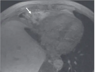
Figure 10. Collection adjacent to the hepatectomy site, which ills with contrast medium in the hepatobiliary phase of HSCA excretion, conirming the diagnosis of biloma (arrow).
CONCLUSION