383 Radiol Bras. 2017 Nov/Dez;50(6):383–388
Breast imaging in patients with nipple discharge
Avaliação imaginológica da paciente com derrame papilarIvie Braga de Paula1, Adriene Moraes Campos2
Paula IB, Campos AM. Breast imaging in patients with nipple discharge. Radiol Bras. 2017 Nov/Dez;50(6):383–388.
Abstract
Resumo
Nipple discharge is a common symptom in clinical practice, representing the third leading breast complaint, after pain and lumps. It is usually limited and has a benign etiology. The risk of malignancy is higher when the discharge is uniductal, unilateral, sponta-neous, persistent, bloody, or serous, as well as when it is accompanied by a breast mass. The most common causes of pathologic nipple discharge are papilloma and ductal ectasia. However, there is a 5% risk of malignancy, mainly ductal carcinoma in situ. The clinical examination is an essential part of the patient evaluation, allowing benign nipple discharge to be distinguished from
suspicious nipple discharge, which calls for imaging. Mammography and ultrasound should be used together as irst-line imaging
methods. However, mammography has low sensitivity in cases of nipple discharge, because, typically, the lesions are small, are
retroareolar, and contain no calciications. Because the reported sensitivity and speciicity of ultrasound, it is important to use the
correct technique to search for intraductal lesions in the retroareolar region. Recent studies recommend the use of magnetic
reso-nance imaging in cases of suspicious nipple discharge in which the mammography and ultrasound indings are normal. The most common magnetic resonance imaging inding is non-mass enhancement. Surgery is no longer the only solution for patients with
suspicious nipple discharge, because short-time follow-up can be safely proposed.
Keywords: Nipple discharge; Mammography; Ultrasonography; Magnetic resonance imaging.
O derrame papilar é um sintoma frequente na prática clínica, correspondendo à terceira queixa mais comum, sendo precedido
apenas por dor e massas palpáveis. A maioria dos derrames papilares é de origem benigna e transitória, sendo deinidos como derrames papilares patológicos os que se apresentam uni ou paucioriiciais, espontâneos, persistentes, serosos ou sanguinolentos
e associados a alteração palpável. Os derrames patológicos são mais frequentemente causados por papiloma ou ectasia ductal, porém, existe risco de malignidade de cerca de 5%, constituído principalmente por carcinoma ductal in situ. O exame clínico é parte essencial na avaliação da paciente, permitindo diferenciar entre derrames papilares tipicamente benignos e derrames papilares
suspeitos, que necessitam de avaliação pelos métodos de imagem. A mamograia e a ultrassonograia devem ser usadas em con
-junto como métodos de imagem de primeira linha, porém, a sensibilidade da mamograia nestes casos é baixa, uma vez que as lesões são comumente retroareolares, pequenas e não calciicadas. A sensibilidade e a especiicidade da ultrassonograia variam
amplamente na literatura, sendo importante o uso de técnicas corretas para a avaliação de lesões intraductais e retroareolares.
Recentemente, a ressonância magnética tem sido indicada nos casos de derrame papilar suspeito com mamograia e ultrassono
-graia normais, sendo o achado mais comum o realce não nodular. A cirurgia não é mais a única solução para as pacientes com
derrame papilar suspeito e todos os exames de imagem normais, tendo em vista que um seguimento em curto prazo pode ser proposto de forma segura.
Unitermos: Derrame papilar; Mamograia; Ultrassonograia; Ressonância magnética.
Study conducted at Conrad Diagnóstico por Imagem, Belo Horizonte, MG, Brazil. 1. MSc, Member of the Colégio Brasileiro de Radiologia e Diagnóstico por Ima
-gem (CBR), MD, Radiologist at Conrad Diagnóstico por Ima-gem, Belo Horizonte, MG,
Brazil.
2. Member of the Colégio Brasileiro de Radiologia e Diagnóstico por Imagem
(CBR), MD, Radiologist at Conrad Diagnóstico por Imagem, Belo Horizonte, MG,
Brazil.
Mailing address: Dr. Ivie Braga de Paula. Conrad Diagnóstico por Imagem. Rua
Rio Grande do Norte, 77, Santa Eigênia. Belo Horizonte, MG, Brazil, 30130-130. E-mail: [email protected].
Received June 15, 2016. Accepted after revision December 14, 2016.
who are not pregnant or breastfeeding. In most cases, sus-picious nipple discharge is caused by benign lesions such as ductal ectasia, in 6–59% of cases, and papilloma, in 35–56%(3). The risk of underlying malignancy is not
negli-gible, ranging from 5% to 23%(2).
Anamnesis and physical examination, with visual in-spection and palpation of the breasts and papillae, play essential roles in the differentiation between physiologi-cal and pathologiphysiologi-cal nipple discharge. The approximate date of onset of the symptom should be investigated, as should its duration, frequency, and quantity, as well as whether it is spontaneous. It is also important to investi-gate the date of the last pregnancy, recent breastfeeding, use of medications (anticoagulants or neuroleptics), trau-ma, and smoking, as well as patient hormonal status and (personal and family) history of breast or ovarian disease.
INTRODUCTION
Nipple discharge is quite common, with a prevalence of 5–10%, representing the third leading breast complaint, after pain and lumps(1,2). It is considered suspicious when
The visual inspection should ideally be made with the aid of a lamp or loupe, which allows nipple discharge to be distinguished from false nipple discharge, which derives from lesions of the nipple-areola complex. The nipple
dis-charge should be deined as uniductal or multiductal and
as unilateral or bilateral. The color of the liquid should be evaluated, which is best done by placing a little of it onto a piece of gauze.
Physiological (i.e., non-suspicious) nipple discharge has the following characteristics: bilateral; non-sponta-neous; previous or intermittent; multiductal; and milky, green or dark in color. In contrast, nipple discharge that is unilateral, spontaneous, persistent, serous, or bloody should be considered pathological and should be investi-gated by imaging.
The color of the secretion determines whether cytol-ogy analysis is necessary. Although cytolcytol-ogy has the ad-vantage of being easy to perform and painless, it has the disadvantage of variable sensitivity, with a > 50% rate of false-negative results for malignant lesions(4). For the
cy-tological examination of the material from the nipple sur-face, the secretion can be placed on a dry slide (if Giemsa
staining is used) or on a slide ixed in ethanol (if Papanico -lau staining is used).
Nipple discharge in men should always be considered
a suspicious inding, because the incidence of carcinoma
in this context is approximately 23%(5). It occurs in 25%
of cases of invasive ductal carcinoma, and axillary lymph node enlargement is common at the time of diagnosis.
Suspicious calciications occur in 13–30% of cases(6).
Imaging methods play a fundamental role in the as-sessment of patients with nipple discharge and make it possible to perform precise imaging-guided biopsies, which provide tissue specimens to be analyzed by the pathologist.
At most facilities, if papilloma is identiied in the biopsy
specimen, surgical excision is performed, because
papil-loma can be associated with carcinoma(7). Recent studies
show that, in cases of papilloma that is single, intraductal, central, and small, diagnosed by vacuum-assisted breast biopsy and presenting no cellular atypia in the pathologi-cal examination, clinipathologi-cal follow-up and imaging can pre-clude the need for surgery(8,9).
IMAGING METHODS FOR THE ASSESSMENT OF NIPPLE DISCHARGE
Mammography
Mammography plays an important role in the diag-nosis of breast diseases(10–15). Although mammography should always be the irst examination requested, it has
low (20–25%) sensitivity in cases of nipple discharge(16),
because the associated lesions are usually retroareolar,
small, intraductal, and noncalciied(17). Therefore,
nega-tive mammography results do not exclude the possibility of underlying disease.
The main mammography inding is calciication. The calciications are typically benign, including eggshell cal
-ciications, which can be associated with papilloma, and rod-shaped calciications, which are usually associated with ductal ectasia. There can also be calciications of sus -picious morphology and distribution, such as pleomorphic
calciications and calciications with a segmental or linear
distribution(1), as depicted in Figure 1. Mammography can
also reveal nodules, focal asymmetry, and ductal ectasia. In cases of nipple discharge, more attention should be paid to the retroareolar region. There are no protocols
in the literature for speciic analysis of that region during
mammography. However, when there is suspicion,
local-ized compression or magniication should be used.
Ultrasound
Ultrasound should always be performed in cases of nipple discharge, even if the alteration has already been
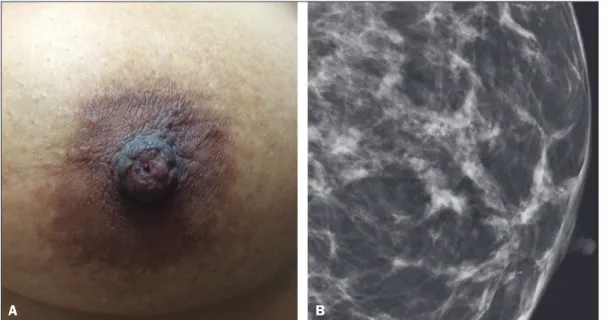
Figure 1. A: Photograph of the nipple-areola complex in a pa-tient with grade II DCIS that is solid, cribriform, and necrotic, with unilateral bloody nipple discharge. B: Magniied me -diolateral oblique view showing ine pleomorphic calciications with segmental distribution in the retroareolar region of the left breast.
noted on mammography(5). Bahl et al.(17) found that, for the detection of ductal carcinoma in situ (DCIS) or in-vasive carcinoma in patients with suspicious nipple
dis-charge, the sensitivity and speciicity of ultrasound were
56% and 75%, respectively.
Appropriate technique includes use of high-frequency transducers, heated gel and ambient temperature con-trol to avoid contraction of the musculature of the nipple and areola. To improve the visualization of the nipple and subareolar regions, certain maneuvers, such as tilting the transducer and observing along the axis of the duct, with discrete peripheral compression, should be used(18).
One of the main ultrasound indings is ductal ectasia, deined as a duct caliber greater than 3 mm. In patients
with suspicious nipple discharge who show focal ductal ectasia with anechoic content, the lesion should be
bi-opsied, because that inding is seen in half of all cases of
papilloma and in 14% of all cases of DCIS(1,19). Focal
duc-tal ectasia in a peripheral location, irregular duct margins, thickening of the duct wall, and hypoechoic adjacent tis-sue are characteristics that can indicate malignancy(20).
In the presence of pathological nipple discharge, sub-areolar nodules and acoustic shadowing should be
clas-siied as BI-RADS 4 or 5 indings. Such indings can be related to DCIS, which is dificult to diagnose by ultra -sound, because false-negative results are obtained in ap-proximately 80% of cases(1).
Doppler ultrasound can facilitate the differentiation between a duct producing viscous secretions and an intra-ductal nodule, because it can reveal vascularization within the latter(17). The most common cause of an intraductal
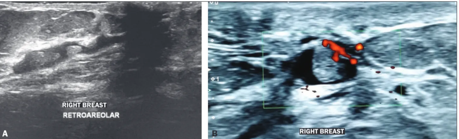
nodule is a single papilloma located a few centimeters from the nipple, usually resulting in ductal obstruction (Figure 2). The characteristics that increase the risk of malignancy are being over 50 years of age, presenting with a nodule larger than 1 cm, and the nodule being located more than 3 cm from the nipple(20).
Ultrasound is important in the second-look evaluation after magnetic resonance imaging (MRI) and can be used to guide biopsies or to facilitate the preoperative
wire-guided localization. Ultrasound is better at detecting nod-ules than non-mass lesions(21), as can be seen in Figure 3.
MRI
There have been few studies on the use of MRI in cases of nipple discharge. According to the European Society of Breast Cancer Specialists, nipple discharge is an emerging indication that has yet to be validated, the evidence produced in the studies warranting only a Grade C recommendation. In clinical practice, MRI can be per-formed in patients with suspicious nipple discharge in
whom mammography and ultrasound indings have been
normal(22). The negative predictive value of MRI is good (approximately 90%), low-grade or very small DCIS le-sions accounting for the false-negative results(22-24). In the
assessment of the location and extent of a lesion, MRI is superior to mammography and ultrasound(1,25). In addi-tion, MRI can identify lesions that initially went unnoticed but could be seen on the second-look ultrasound or mam-mography, especially lesions occurring in the retroareolar region (Figure 4).
The main MRI inding in patients with suspicious
nipple discharge is non-mass enhancement. In a study of 47 patients with suspicious nipple discharge, 59% of the malignant lesions showed non-mass enhancement with segmental distribution, 57% showed heterogeneous en-hancement within the lesion and 40% showed a plateau-type enhancement curve(26). In T1-weighted sequences,
high protein or hemorrhagic content within the duct can appear as an area of high signal intensity, simulating lin-ear or segmental enhancement. In order to differentiate
between the two indings, the pre-contrast and digital sub -traction sequences must be evaluated. In the presence of nipple discharge, a focus of contrast enhancement should be considered suspicious, because it could represent a papilloma.
The main criticisms of MRI are its high cost, the de-tection of additional alterations that can call for other follow-up tests or biopsies unrelated to the initial clinical
complaint, and the dificulty of determining whether the
Figure 2. A: Ultrasound showing intraductal nodules. B: Doppler ultrasound showing vascularity within an intraductal nodule. B
A
RIGHT BREAST
lesion is intraductal or not(25). For that purpose, a second-look ultrasound examination is indispensable.
Galactography
Galactography, also known as ductography, has long been considered the gold standard for the evaluation of nipple discharge. A study by Manganaro et al. evaluated 53 patients with unilateral nipple discharge who under-went galactography and MRI, comparing the two methods in terms of their ability to identify diseases and to distin-guish between benign and malignant lesions. In the
iden-tiication of ductal disease, MRI showed higher sensitivity
than did galactography (98% vs. 49%) and both methods
presented high speciicity. Unlike galactography, MRI was
able to demonstrate not only ductal disease but also le-sions in the adjacent parenchyma(27).
DISCUSSION
Although MRI plays an increasingly greater role in the study of breast cancer(28,29), there have been few studies
on its use in cases of nipple discharge.
Despite the lack of reliable scientiic evidence of the beneit of using MRI in patients with suspicious nipple discharge in whom mammography and ultrasound ind -ings are normal, most authors recommend performing
MRI of the breasts. If the MRI scan identiies a suspicious
lesion, it is now routine practice to use a second-look
ul-trasound to localize the inding. However, if MRI shows
non-mass enhancement with linear or segmental distribu-tion, corresponding to the site of nipple discharge,
second-look mammography with magniication of the region can be useful in the investigation of suspicious calciications,
allowing stereotactic biopsy to be performed. If no abnor-mality is found, an MRI-guided biopsy of the suspicious lesion should be performed(1).
Historically, surgical resection of the terminal breast ducts was the rule for patients with suspicious nipple dis-charge in whom mammography, ultrasound, and MRI all produced normal results. It has recently been shown that the risk of developing a malignant lesion is quite low in such patients, especially if there are no other suspicious clinical signs. In addition, when such patients do develop a malignant lesion, it is a low-grade DCIS or a very small tumor. Therefore, the most recent studies in the literature recommend that patients with suspicious nipple discharge
in whom mammography, ultrasound, and MRI indings
are all normal should be followed for two years, with fol-low-up evaluations every 6 months, until there is sponta-neous resolution of the discharge, which occurs in 81% of the cases(1,16,30). The follow-up protocol can be ultrasound
and clinical examinations every 6 months, together with annual mammography. However, for patients with massive nipple discharge, nipple discharge that causes discomfort, or nipple discharge that persists for more than two years, surgery should be considered(1).
Figure 3. A 64-year-old patient with bloody discharge from the left nipple. A:
Mammography in craniocaudal and mediolateral oblique views, showing focal asymmetry in the retroareolar region. B: T1-weighted MRI sequence with fat suppression, 2 min after intravenous injection of gadolinium, showing a nod-ule with ill-deined margins at the same location. C: Second-look ultrasound showing a hypoechoic intraductal nodule, in correspondence with the mam-mography and MRI indings. Evaluation of a biopsy specimen demonstrated intraductal papilloma without atypia.
A
B
C
FINAL CONSIDERATIONS
The majority of cases of suspicious nipple discharge have a benign cause, the risk of malignancy being approxi-mately 5% and DCIS accounting for most such malignan-cies. After clinical evaluation and physical examination, the imaging investigation begins with mammography and ultrasound, with special attention to the retroareolar re-gion. In such cases, mammography has a sensitivity of 20–25% for the detection of suspicious lesions, compared with 65–85% for ultrasound. When the mammography
and ultrasound indings are normal, MRI can be used,
because it has high sensitivity for lesions of the nipple
and malignant lesions. The most common MRI inding is
non-mass enhancement, being more suspicious for malig-nancy when presenting segmental distribution and
hetero-geneous internal enhancement. When the MRI indings
are suspicious, second-look mammography or ultrasound can facilitate the biopsy process. For patients in whom all imaging examinations produce normal results, a follow-up
protocol involving clinical examination, mammography, and ultrasound can be suggested, given that spontaneous resolution of nipple discharge occurs in a large number of cases.
REFERENCES
1. Lippa N, Hurtevent-Labrot G, Ferron S, et al. Les écoulements mamelonnaires. Journal de Radiologie Diagnostique et Interven-tionnelle. 2015;96:434–50.
2. Chen L, Zhou WB, Zhao Y, et al. Bloody nipple discharge is a pre-dictor of breast cancer risk: a meta-analysis. Breast Cancer Res Treat. 2012;132:9–14.
3. van Gelder L, Bisschops RH, Menke-Pluymers MB, et al. Magnetic resonance imaging in patients with unilateral bloody nipple dis-charge: useful when conventional diagnostic are negative? World J Surg. 2015;39:184–6.
4. Das DK, Al-Ayadhi B, Ajrawi MY, et al. Cytodiagnosis of nipple dis-charge: a study of 602 samples from 484 cases. Diagn Cytopathol. 2001;25:25–37.
5. Muñoz Carrasco R, Álvarez Benito M, Rivin del Campo E. Value of mammography and breast ultrasound in male patients with nipple discharge. Eur J Radiol. 2013;82:478–84.
Figure 4. A 44-year-old patient with suspicious nipple discharge. Mam-mography showing dense breasts with focal asymmetry in the supero-lateral quadrant of the left breast. Ultrasound, obtained at another facil-ity, showing no alterations. A: Sagittal T1-weighted MRI sequence with fat suppression, showing ductal ectasia with hemorrhagic and high protein content in the superolateral quadrant of the left breast. B: MRI with digital subtraction 2 min after intravenous administration of contrast medium, showing non-mass enhancement with segmental distribution and heteroge-neous enhancement in the supero-lateral quadrant of the left breast. C:
Contrast-enhanced MRI with digital subtraction, showing a nodule with ill-deined margins and ring enhance -ment in the superolateral quadrant of the left breast, in correspondence with the mammography inding. D: Second-look ultrasound showing a nodule with ill-deined margins in the superolateral quadrant of the left breast, in correspondence with the MRI indings. Evaluation of a biopsy specimen demonstrated grade II in-vasive mucinous carcinoma.
A B
6. Lattin GE Jr, Jesinger RA, Mattu R, et al. From the radiologic pa-thology archives: diseases of the male breast: radiologic-pathologic correlation. Radiographics. 2013;33:461–89.
7. Glenn ME, Throckmorton AD, Thomison JB 3rd, et al. Papillomas of the breast 15 mm or smaller: 4-year experience in a community- based dedicated breast imaging clinic. Ann Surg Oncol. 2015;22: 1133–9.
8. The American Society of Breast Surgeons. Consensus guideline on concordance assessment of image-guided breast biopsies and man-agement of borderline or high-risk lesions. [cited 2017 Aug 25]. Available from: https://www.breastsurgeons.org/new_layout/about/ statements/PDF_Statements/Concordance_and_High%20RiskLe-sions.pdf.
9. Kibil W, Hodorowicz-Zaniewska D, Popiela TJ, et al. Vacuum-assist-ed core biopsy in diagnosis and treatment of intraductal papillomas. Clin Breast Cancer. 2013;13:129–32.
10. Freitas-Junior R, Rodrigues DCN, Corrêa RS, et al. Contribution
of the Uniied Health Care System to mammography screening in
Brazil, 2013. Radiol Bras. 2016;49:305–10.
11. Schwingel R, Almeida O, Ferreira TS. Fat necrosis associated with
the use of oral anticoagulant therapy: atypical mammographic ind -ings. Radiol Bras. 2016;49:269–70.
12. Koch H. Mammography as a method for diagnosing breast cancer. Radiol Bras. 2016;49(6):vii.
13. Villar VCFL, De Seta MH, Andrade CLT, et al. Evolution of mam-mographic image quality in the state of Rio de Janeiro. Radiol Bras. 2015;48:86–92.
14. Avelar MS, Almeida O, Alvares BR. Mammographic artifact leading to false-positive result. Radiol Bras. 2015;48:198–9.
15. Paixão L, Oliveira BB, Viloria C, et al. Monte Carlo derivation of il -tered tungsten anode X-ray spectra for dose computation in digital mammography. Radiol Bras. 2015;48:363–7.
16. Ashfaq A, Senior D, Pockaj BA, et al. Validation study of a modern treatment algorithm for nipple discharge. Am J Surg. 2014;208;222– 7.
17. Bahl M, Baker JA, Greenup RA, et al. Diagnostic value of ultra-sound in female patients with nipple discharge. AJR Am J Roent-genol. 2015;205:203–8.
18. Da Costa D, Taddese A, Cure ML, et al. Common and unusual dis-eases of the nipple-areolar complex. Radiographics. 2007;27 Suppl 1:S65–77.
19. Yang WT, Tse GMK. Sonographic, mammographic, and histopatho-logic correlation of symptomatic ductal carcinoma in situ. AJR Am J Roentgenol. 2004;182:101–10.
20. Ferris-James DM, Iuanow E, Mehta TS, et al. Imaging approaches to diagnosis and management of common ductal abnormalities. Radiographics. 2012;32:1009–30.
21. Candelaria R, Fornage BD. Second-look US examination of MR-detected breast lesions. J Clin Ultrasound. 2011;39:115–21. 22. Lorenzon M, Zuiani C, Linda A, et al. Magnetic resonance imaging
in patients with nipple discharge: should we recommend it? Eur Radiol. 2011;21:899–907.
23. Boisserie-Lacroix M, Adenet C, Trillaud H. Evaluation of suspi-cious nipple discharge with MRI: review of 50 cases. J Radiol. 2011;92:412–20.
24. Morrogh M, Morris EA, Liberman L, et al. The predictive value of ductography and magnetic resonance imaging in the management of nipple discharge. Ann Surg Oncol. 2007;14:3369–77.
25. Eiada R, Chong J, Kulkarni S, et al. Papillary lesions of the breast: MRI, ultrasound, and mammographic appearances. AJR Am J Roentgenol. 2012;198:264–71.
26. Tokuda Y, Kuriyama K, Nakamoto A, et al. Evaluation of suspicious nipple discharge by magnetic resonance mammography based on breast imaging reporting and data system magnetic resonance imag-ing descriptors. J Comput Assist Tomogr. 2009;33:58–62.
27. Manganaro L, D’Ambrosio I, Gigli S, et al. Breast MRI in patients with unilateral bloody and serous-bloody nipple discharge: a com-parison with galactography. Biomed Res Int. 2015;2015:806368. 28. Almeida JRM, Gomes AB, Barros TP, et al. Predictive performance
of BI-RADS magnetic resonance imaging descriptors in the context
of suspicious (category 4) indings. Radiol Bras. 2016;49:137–43.
29. Bitencourt AGV. Subdividing BI-RADS category 4 breast lesions ob-served on magnetic resonance imaging: is it feasible? Radiol Bras. 2016;49(3):v.