Estrogen and progesterone receptor testing in breast
carcinoma: concordance of results between local and
reference laboratories in Brazil
Teste de receptores de estrógeno e progesterona em carcinoma de mama:
concordância dos resultados entre laboratórios locais e de referência no Brasil
Sheila Cristina Lordelo Wludarski
I, Lisandro Ferreira Lopes
I, Ívison Xavier Duarte
I, Filomena Marino Carvalho
II, Lawrence
Weiss
III, Carlos Eduardo Bacchi
IVPathology Consultancy, Botucatu, São Paulo, Brazil
ABSTRACT
CONTEXT AND OBJECTIVE: Breast cancer accounts for approximately one quarter of all cancers in fe-males. Estrogen and progesterone receptor testing has become an essential part of the clinical evaluation of breast carcinoma patients, and accurate results are critical in identifying patients who may benefit from hormone therapy. The present study had the aim of investigating the concordance of the results from hormone receptor tests between a reference laboratory and local (or community) laboratories in Brazil.
DESIGN AND SETTING: Retrospective study at a reference pathology laboratory.
METHODS: The concordance in the results from hormone receptor tests between a reference laboratory and 146 local laboratories in Brazil was compared in relation to 500 invasive breast carcinoma cases, using immunohistochemistry.
RESULTS: There was concordance in 89.4% (447/500 cases) and 85.0% (425/500 cases) of the results from estrogen ( = 0.744, P < 0.001) and progesterone ( = 0.688, P < 0.001) receptor tests, respectively, be-tween local and reference laboratories. This was similar to findings in other countries. The false negative rates from estrogen and progesterone receptor tests in local laboratories were 8.7% and 14.4%, respec-tively. The false positive rates from estrogen and progesterone receptor tests in local laboratories were 15.5% and 16.0%, respectively.
CONCLUSION: Technical and result interpretation issues may explain most of the discordances in hor-mone receptor testing in local laboratories. Validation of estrogen and progesterone receptor tests at local laboratories, with rigorous quality control measures, is strongly recommended in order to avoid erroneous treatment of breast cancer patients.
RESUMO
CONTEXTO E OBJETIVO: O carcinoma de mama é responsável por cerca de um quarto de todos os cân-ceres em mulheres. O teste de receptores de estrógeno e progesterona se tornou parte essencial da ava-liação clínica de pacientes com carcinoma de mama; assim, resultados precisos são fundamentais para identificação de pacientes que podem se beneficiar da terapia hormonal. O presente estudo tem por objetivo investigar a concordância nos resultados do teste de receptores hormonais entre um laboratório referência e laboratórios locais (ou comunitários) do Brasil.
TIPO DE ESTUDO E LOCAL: Estudo retrospectivo em laboratório referência em patologia no Brasil.
MÉTODOS: A concordância nos resultados dos testes de receptores hormonais entre um laboratório re-ferência e 146 diferentes laboratórios locais brasileiros foi comparada em 500 casos de carcinoma invasivo de mama através da imunoistoquímica.
RESULTADOS: Houve concordância de 89,4% (447/500 casos) e 85,0% (425/500 casos) nos resultados dos testes de receptores de estrógeno ( = 0,744, P < 0,001) e progesterona ( = 0,688, P < 0,001), respecti-vamente, entre laboratórios locais e referência, similar à descrita em outros países. A taxa de resultados falso-negativos nos testes de receptores de estrógeno e progesterona em laboratórios locais foi de 8,7% e 14,4%, respectivamente. A taxa de resultados falso-positivos nos testes de receptores de estrógeno e progesterona em laboratórios locais foi de 15,5% e 16,0%, respectivamente.
CONCLUSÃO: Questões técnicas e de interpretação dos resultados podem explicar a maior parte das discordâncias nos testes de receptores hormonais em laboratórios locais. A validação dos testes de re-ceptores de estrógeno e progesterona pelos laboratórios locais com medidas de controle de qualidade rigorosas é fortemente recomendada de modo a evitar o tratamento errôneo de pacientes com carcino-ma de carcino-macarcino-ma.
IMD. Associate Pathologist, Pathology
Consultancy, Botucatu, São Paulo, Brazil.
IIMD, PhD. Associate Professor, Department
of Pathology, Faculdade de Medicina da Universidade de São Paulo (FMUSP), São Paulo, Brazil.
IIIMD, PhD. Chair, Division of Pathology, City
of Hope National Medical Center, Duarte, California, United States.
IVMD, PhD. Director and Chief Pathologist,
Pathology Consultancy, Botucatu, São Paulo, Brazil.
KEY WORDS: Breast neoplasms. Receptors, estrogen. Receptors, progesterone. Immunohistochemistry. Brazil.
INTRODUCTION
Breast cancer is one of the most common human neoplasms, accounting for one quarter of all cancers in females.1 Hormone
therapy is frequently used in breast carcinoma treatment because it reduces the relative risk of recurrence by more than 50% in patients with hormone-sensitive tumors, thus leading to signii-cant improvements in survival. For these reasons, determination of estrogen receptor (ER) and progesterone receptor (PgR) status has become an essential part of the clinical evaluation of all breast carcinoma patients, and accurate results are critical in identify-ing patients who may beneit from hormone therapy.2-8
Immu-nohistochemistry (IHC) is currently the most common method used for determining ER and PgR status. Low cost and applica-bility to routinely processed and archived tissue samples are the main advantages of IHC. However, discordances in ER and PgR testing have been reported in the literature, and they have been mostly correlated with technical issues, including ixative and ix-ation issues, immunohistochemical methodology and diversity of interpretation of results.9-29
OBJECTIVE
Considering that the accuracy of ER and PgR testing in breast carcinoma is extremely important in selecting the hormone ther-apy, the present study had the aim of investigating the concor-dance of the results from ER and PgR tests using IHC between a reference laboratory (Pathology Consultancy, Botucatu, São Paulo, Brazil) and local (or community) laboratories in Brazil.
METHODS
Institutional certiications
his study was approved by the Scientiic Committee of the Department of Pathology, Faculdade de Medicina da Univer-sidade de São Paulo (FMUSP), and by the Ethics Committee for Research Projects of Hospital das Clínicas (HC), FMUSP (CAPPesq, protocol no 1238/09).
Validation at the reference laboratory
For any clinical assay to be validated, the results need to be com-pared with a standard. For ER and PgR testing, the recommended approach towards validation is based on comparison of the assay results with results obtained by another laboratory using a test-ing method that has been previously validated against the clini-cal outcome or using proiciency-testing material that has been validated by showing a consensus of results among multiple lab-oratories in a peer group (which must include lablab-oratories with validated assays). ER and PgR IHC assays that are not subjected to direct clinical validation may be validated by showing at least 90% agreement for positive results and at least 95% agreement for negative results (a minimum of 20 positive and 20 negative speci-mens are required) with testing performed on the same material
in another laboratory that provides written attestation that it is in conformity with the testing requirements of the American Society of Clinical Oncology (ASCO) and College of American Patholo-gists (CAP) and that ofers fully validated ER and PgR assays.6,7,30
Pathology Consultancy, a reference laboratory located in Botucatu, São Paulo State, Brazil, performs approximately 7,000 ER and PgR assays by means of IHC annually. In order to fur-ther validate the ER and PgR testing by Pathology Consultancy, we compared the results from ER and PgR IHC assays between a CAP-accredited laboratory that performs fully validated ER and PgR testing (PhenoPath Laboratories, Seattle, Washington, United States) and Pathology Consultancy. For validation pur-poses, 255 invasive breast carcinoma samples (a 2-mm tissue core for each sample) were distributed over nine tissue microarray (TMA) blocks. Unstained 3-μm-thick histological sections from each TMA block were obtained and used to determine the ER and PgR status using IHC, both in PhenoPath Laboratories and in Pathology Consultancy.
In Pathology Consultancy, the sections were deparainized in xylene and rehydrated in graded alcohols and phosphate-buf-ered saline (PBS). he sections were then subjected to antigen retrieval in a pressure pan using citric acid (0.21%, pH 6.0) for a total of eight minutes, followed by a 20-minute cool-down period at room temperature. Subsequently, the slides were incubated overnight with the primary antibody. he SP1 rabbit monoclo-nal antibody (code RM-9101-S, hermo Fisher Scientiic, Fre-mont, California, United States; dilution 1:1000) was used for ER testing. he PgR 636 mouse monoclonal antibody (code M3569, Dako, Carpinteria, California, United States; dilution 1:1600) was used for PgR testing. Ater incubation with the primary anti-body, the slides were washed with PBS, and incubated with Nov-olink polymer (code 7161, Novocastra Laboratories, Newcastle upon Tyne, United Kingdom) for 30 minutes. Diaminobenzidine was used as the chromogen, and the sections were then counter-stained with hematoxylin and coverslipped. he same antibodies (SP1 for ER and PgR 636 for PgR) were also used at PhenoPath Laboratories, using fully validated technical procedures.
he results from the ER and PgR assays were compared between PhenoPath Laboratories and Pathology Consultancy, in line with the most recent consensus of ASCO and CAP: the ER and PgR tests were considered positive if at least 1% of the tumor nuclei in the samples tested were positive, irrespective of the intensity of staining.6,7,30
Case selection for comparison between reference and local laboratories
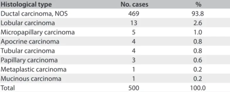
Histological type No. cases %
Ductal carcinoma, NOS 469 93.8
Lobular carcinoma 13 2.6
Micropapillary carcinoma 5 1.0
Apocrine carcinoma 4 0.8
Tubular carcinoma 4 0.8
Papillary carcinoma 3 0.6
Metaplastic carcinoma 1 0.2
Mucinous carcinoma 1 0.2
Total 500 100.0
Table 1. Histological classification of invasive breast carcinomas according to World Health Organization (WHO), 2003
NOS = not otherwise specified.
hese local laboratories consisted of community-based laborato-ries with low volumes of ER and PgR testing. he cases included in the study were sent to the reference laboratory by oncologists for conirmation of breast cancer diagnosis and for ER and PgR retesting.
All the cases were morphologically reviewed in the reference laboratory by at least two diferent pathologists in order to con-irm the diagnosis of invasive breast carcinoma and to classify the tumors in accordance with the World Health Organization (WHO) classiication.1
Information regarding the ER and PgR testing performed by the local laboratories was retrieved from the immunohistochem-ical reports, including the geographic location of the laboratory, details about ixation of the specimen (ixative used and ixa-tion time), type of breast carcinoma tested (in situ or invasive carcinoma), use of tissue controls, IHC methodology (antibod-ies, antigen retrieval technique and detection system speciica-tions), ER and PgR results and interpretation criteria for report-ing the results. Parain-embedded breast carcinoma tissue from all cases was used for ER and PgR testing in the reference labo-ratory. he same parain blocks from the local laboratories were used. IHC was used to determine ER and PgR status, as previ-ously described. In accordance with the most recent consensus of ASCO and CAP,6,7 the ER and PgR results were considered
positive if at least 1% of the tumor nuclei in the samples tested were positive, irrespective of the intensity of staining. he ER and PgR results were interpreted blindly by experienced patholo-gists, without knowledge of the previous results deined by local laboratories.
Statistical analysis
he reference and local laboratories results for ER and PgR were compared using Pearson’s chi-square (χ2) test. Kappa statistics
were used as a concordance measurement. Sensitivity, speci-icity, false negative rate, false positive rate, positive predic-tive value, negapredic-tive predicpredic-tive value, overall accuracy and the Youden index were calculated. he signiicance level used in all tests was 5%.31-34
RESULTS
Validation by the reference laboratory
As previously presented, 255 invasive breast carcinomas sam-ples were tested for ER and PgR for validation purposes. In the CAP-accredited laboratory (PhenoPath Laboratories), 172 sam-ples were considered positive and 83 were negative for ER; 164 samples were considered positive and 91 were negative for PgR. he overall concordance between PhenoPath Laboratories and Pathology Consultancy was 98.8% (252/255 samples) and 98.4% (251/255 samples) for ER and PgR, respectively. For positive results, the concordance was 100.0% for both ER and PgR. For negative results, the concordance was 96.4% for ER and 95.6% for PgR. hese results fully validate the ER and PgR testing per-formed at Pathology Consultancy, according to recent recom-mendations for validating ER and PgR IHC assays.6,7,30
Comparison of results between reference and local laboratories
he 500 cases of invasive breast carcinoma included in the study were classiied as invasive ductal carcinoma that was not other-wise speciied (93.8%) and invasive lobular carcinoma (2.6%). Micropapillary, apocrine, tubular, papillary, metaplastic and mucinous carcinomas were rarely found (3.6%), as shown in
Table 1. Cases from all geographic regions of Brazil were
rep-resented. Most of the cases were from the southeastern region (70.0%), followed by the southern (12.8%), northeastern (8.0%), central-western (6.6%) and northern (2.6%) regions. he total number of cases from each local laboratory ranged from 1 to 41 cases.
Information about specimen ixation (including the ixative used and ixation time) was found in only 32 reports (6.4%) from local laboratories, and all of them reported that formalin was the ixative. Fixation time was not found in any of the reports. Most reports (67.6%) did not provide any information about the type of breast carcinoma (in situ or invasive carcinoma) that was
tested in IHC assays. Only 209 reports (41.8%) provided infor-mation on the use of tissue controls for IHC assays.
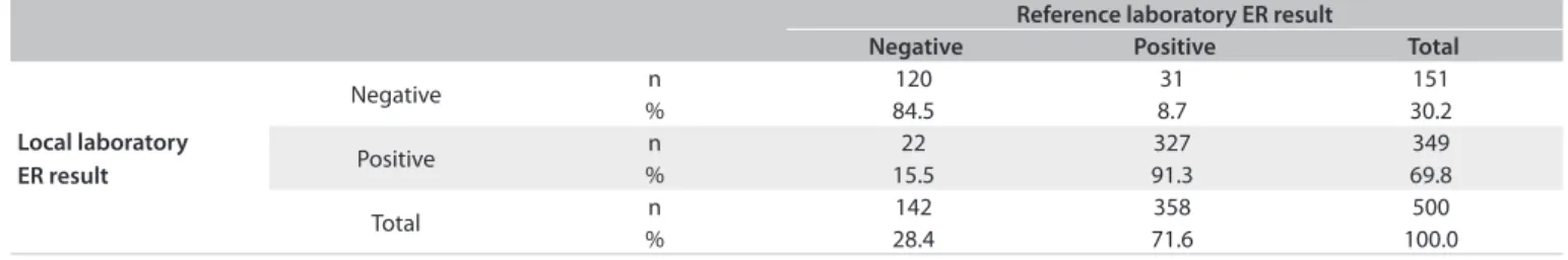
Reference laboratory ER result
Negative Positive Total
Local laboratory ER result
Negative n 120 31 151
% 84.5 8.7 30.2
Positive %n 15.522 91.3327 69.8349
Total n 142 358 500
% 28.4 71.6 100.0
Table 2. Distribution of estrogen receptor (ER) results between reference and local laboratories (kappa statistic k = 0.744, P < 0.001;
Pearson’s chi-square association test χ2 = 273.889, P < 0.0001)
Table 3. Distribution of progesterone receptor (PgR) results between reference and local laboratories (kappa statistic k = 0.688, P < 0.001;
Pearson’s chi-square association test χ2 = 234.490, P < 0.0001)
Reference laboratory PgR result
Negative Positive Total
Local laboratory PgR result
Negative n 163 44 207
% 84.0 14.4 41.4
Positive n 31 262 293
% 16.0 85.6 58.6
Total n 194 306 500
% 38.8 61.2 100.0
Authors, country and year of publication ER concordance PgR concordance
Viale et al.,22 25 countries, 2007 6058/6205 (97.6%) 4202/5237 (80.2%)
Badve et al.,23 USA, 2008 694/769 (90.2%) 649/769 (84.4%)
Gelber et al.,27 several countries, 2009 4323/4931 (87.6%)
-Wludarski et al., Brazil, 2011 (present study) 447/500 (89.4%) 425/500 (85.0%)
Table 4. Comparison of the concordance results for estrogen (ER) and progesterone (PgR) receptors between reference/central and local laboratories reported in the literature and by the present study
he antigen retrieval method was not speciied in 198 reports (39.6%). When available, heat-induced antigen retrieval was used. he detection system was not speciied in 128 reports (25.6%). When available, LSAB (labeled streptavidin biotin) was used most frequently (60.5%), followed by ABC (avidin bio-tin complex) (15.3%), “polymer” (15.3%), EnVision™ (5.9%), ADVANCE™ HRP (2.4%) and PAP (peroxidase anti-peroxidase) (0.6%).he comparison of ER and PgR results between the refer-ence and local laboratories is shown in Tables 2 and 3.
Using kappa statistics as a concordance measurement (κ = 0.744; P < 0.001; 95% conidence interval 0.679-0.809), the over-all accuracy of ER testing in local laboratories was 89.4% (447/500 cases). ER-positive and negative results were concordant between the reference and local laboratories in 91.3% (327/358) and 84.5% of the cases (120/142), respectively. Pearson’s chi-square (χ2) association test for ER results between the reference and
local laboratories revealed χ2 = 273.889 (P < 0.0001), which
indi-cates a signiicant association in the results. he sensitivity, spec-iicity, false negative rate, false positive rate, positive predictive value, negative predictive value and Youden index of ER test-ing in local laboratories were 91.3%, 84.5%, 8.7%, 15.5%, 93.7%, 79.5% and 75.8%, respectively. Using kappa statistics as a con-cordance measurement (κ = 0.688; P < 0.001; 95% conidence interval 0.623-0.753), the overall accuracy of PgR testing in local
laboratories was 85.0% (425/500 cases). PgR-positive and nega-tive results were concordant between the reference and local lab-oratories in 85.6% (262/306) and 84.0% of the cases (163/194), respectively. Pearson’s chi-square (χ2) association test for PgR
results between the reference and local laboratories revealed χ2
= 234.490 (P < 0.0001), which indicates a signiicant association in the results. he sensitivity, speciicity, false negative rate, false positive rate, positive predictive value, negative predictive value and Youden index of PgR testing in local laboratories were 85.6%, 84.0%, 14.4%, 16.0%, 89.4%, 78.7% and 69.6%, respectively.
DISCUSSION
Brazil has a population of approximately 190 million people.35
he incidence of breast cancer in Brazil is about 50,000 new cases per year,36 and it is considered to be an important public health
problem.
Hormone receptor status should be deined in all newly diag-nosed, invasive breast carcinomas as well as in recurrences, in order to determine patient eligibility for hormone therapy, which provides substantial survival beneit for patients with hormone-positive tumors. Accurate determination of ER and PgR status is, therefore, critical for ensuring that patients receive appropri-ate therapy.2-8,30 However, discordances in hormone receptor
test-ing ustest-ing IHC have been reported in diferent laboratories from several countries, and these probably relate to technical issues, including delayed or inadequate ixation, non-optimized anti-gen retrieval and diversity of interpretation and reporting of results.9-29
In 2000, Rhodes et al.10 demonstrated that there was
consid-erable variability between laboratories (200 laboratories in 26 countries) regarding ER results, especially in relation to detec-tion of breast cancers with low ER positivity, with a false-negative rate ranging from 30% to 60%. his variability between laborato-ries probably related to diferences in IHC methodology, accord-ing to these authors.
In 2001, in a study that involved 105 laboratories, the same authors showed that the eiciency of the antigen retrieval step was the single most important contributory factor inluencing the overall reproducibility of the hormone assays. Reliable assays were found in the majority of centers known to have clinically validated results. Inadequate assay sensitivity, with subsequent weak staining, was the main cause of poor and variable results at laboratories that used microwave antigen retrieval; heating times that were too short were identiied as the principal contrib-utory factor. Extension of the heating time resulted in signiicant improvement regardless of all other variables in the immunohis-tochemical protocol. hey also stated that continual participation in external quality assessment programs was an efective way to identify and improve variables that inluence the reliability of immunohistochemical assays for ER and PgR determination, thereby assisting in technical validation and standardization.14
Regitnig et al.15 investigated the variability in the results from
ER and PgR testing performed by diferent laboratories in Aus-tria. hey found that the variability in the results was greater when participants used their own IHC staining method. In 2007, Viale et al.22 evaluated locally versus centrally assessed ER and
PgR status among a signiicant number of breast cancer patients. Out of 105 tumors that were considered to be ER-negative in local laboratories, 81 were found to have positive cell rates of at least 1% at the central laboratories. Out of 6,100 tumors that were found locally to be ER-positive, 66 were found to have no
staining centrally. he discordance was more marked for PgR than for ER. Because of these results, Regitnig et al.15
recom-mended that the ER and PgR status should be reviewed in central laboratories whenever possible. Badve et al.23 showed a
concor-dance rate of 90% in ER testing between local and central labora-tories. For PgR, the concordance between local and central lab-oratories was 84%. A recent inquiry into ER testing practices in Canada revealed that approximately one third of 1,023 ER assays were scored falsely negative when retested in a central laboratory. he possible causes of such discrepancies related to turnover and lack of relevant training of pathologists and technologists, lack of appropriate quality assurance methods, and inadequate quality control policies and practices. he false negative ER assays were found to have poor ixation, negative internal controls, and/or absent internal controls.26 In 2009, Gelber et al.27 reported that
4.3% of the tumors that tested ER-positive in local laboratories were found to be negative (false positive) on central review. More than 20% of the tumors that tested locally as ER-negative were shown to exhibit at least some expression of ER (false negative) on central reference laboratory review. Table 4 presents a
com-parison of the concordance results for ER and PgR between labo-ratories reported in the literature and by the present study.
Pathology Consultancy is considered to be a reference labo-ratory in Brazil because of its high volume of ER and PgR test-ing (approximately 7,000 assays every year). In the present study, we compared the results from ER and PgR testing on 500 invasive breast carcinomas between a reference laboratory and 146 diferent local laboratories in Brazil. Local, low-volume laboratories from all geographic regions of Brazil were represented. Even though the overall concordance of the results from ER and PgR assays between the reference and local laboratories was high (89.4% for ER and 85.0% for PgR) and the ER-positive assays were concordant in 91.3% of the cases, the ER-negative assays were concordant in only 84.5%, a result that is lower than the minimum recommended. he same was true for the PgR-positive and negative results. hey were concordant in 85.6% and 84.0% of the cases, respectively. As pre-viously stated, at least 90% and 95% agreement for positive and negative results is recommended.6,7,30 he false negative rates for
ER and PgR testing in the local laboratories were 8.7% and 14.4%, respectively. his means that patients who are misclassiied as hav-ing ER-negative tumors are denied the potential beneit of hor-mone treatment. he false positive rates for ER and PgR testing in the local laboratories were 15.5% and 16.0%, respectively. his means that patients who were misclassiied as having ER-positive tumors will be exposed unnecessarily to the risks and costs of inef-fectual treatment. he risks include a decrease in bone density with an increased fracture risk, an increased risk of thromboem-bolic events and an increased risk of endometrial cancer.37 Fixation
that were probably not validated as recommended by ASCO and CAP, and heterogeneity in interpretation criteria for reporting the results could explain most of the ER and PgR testing discordances between the local and reference laboratories in Brazil.
CONCLUSION
his study presents relatively high concordance of the results from ER and PgR testing between local laboratories and a ref-erence laboratory in Brazil, similar to indings from other coun-tries. However, false-negative and false-positive results occur, which may relate to technical and interpretation issues. hese results may be associated with erroneous treatment of breast can-cer patients. We believe that validation of ER and PgR testing and standardization of the interpretation of the results, with rigorous quality control measures at local laboratories, are crucial. More-over, reference laboratories could assist in validating local labo-ratories’ ER and PgR assays.
REFERENCES
1. Tavassoli FA, Devilee P. World Health Organization Classification of Tumours. Pathology and Genetics of Tumours of the Breast and Female Genital Organs. Lyon: IARC Press; 2003.
2. Knight WA, Livingston RB, Gregory EJ, McGuire WL. Estrogen receptor as an independent prognostic factor for early recurrence in breast cancer. Cancer Res. 1977;37(12):4669-71.
3. Early Breast Cancer Trialists’ Collaborative Group (EBCTCG). Effects of chemotherapy and hormonal therapy for early breast cancer on recurrence and 15-year survival: an overview of the randomised trials. Lancet. 2005;365(9472):1687-717.
4. Fisher B, Redmond C, Brown A, et al. Treatment of primary breast cancer with chemotherapy and tamoxifen. N Engl J Med. 1981;305(1):1-6. 5. Fisher B, Redmond C, Brown A, et al. Influence of tumor estrogen
and progesterone receptor levels on the response to tamoxifen and chemotherapy in primary breast cancer. J Clin Oncol. 1983;1(4):227-41.
6. Hammond ME, Hayes DF, Dowsett M, et al. American Society of Clinical Oncology/College of American Pathologists guideline recommendations for immunohistochemical testing of estrogen and progesterone receptors in breast cancer (unabridged version). Arch Pathol Lab Med. 2010;134(7):e48-72.
7. Hammond ME, Hayes DF, Dowsett M, et al. American Society of Clinical Oncology/College Of American Pathologists guideline recommendations for immunohistochemical testing of estrogen and progesterone receptors in breast cancer. J Clin Oncol. 2010;28(16):2784-95.
8. Gown AM. Current issues in ER and HER2 testing by IHC in breast cancer. Mod Pathol. 2008;21 Suppl 2:S8-S15.
9. Allred DC, Harvey JM, Berardo M, Clark GM. Prognostic and predictive factors in breast cancer by immunohistochemical analysis. Mod Pathol. 1998;11(2):155-68.
10. Rhodes A, Jasani B, Barnes DM, Bobrow LG, Miller KD. Reliability of immunohistochemical demonstration of oestrogen receptors in routine practice: interlaboratory variance in the sensitivity of detection and evaluation of scoring systems. J Clin Pathol. 2000;53(2):125-30. 11. Rhodes A, Jasani B, Balaton AJ, Miller KD. Immunohistochemical
demonstration of oestrogen and progesterone receptors: correlation of standards achieved on in house tumours with that achieved on external quality assessment material in over 150 laboratories from 26 countries. J Clin Pathol. 2000;53(4):292-301.
12. Rhodes A, Jasani B, Balaton AJ, Barnes DM, Miller KD. Frequency of oestrogen and progesterone receptor positivity by immunohistochemical analysis in 7016 breast carcinomas: correlation with patient age, assay sensitivity, threshold value, and mammographic screening. J Clin Pathol. 2000;53(9):688-96. 13. Layfield LJ, Gupta D, Mooney EE. Assessment of Tissue Estrogen
and Progesterone Receptor Levels: A Survey of Current Practice, Techniques, and Quantitation Methods. Breast J. 2000;6(3):189-96. 14. Rhodes A, Jasani B, Balaton AJ, et al. Study of interlaboratory reliability
and reproducibility of estrogen and progesterone receptor assays in Europe. Documentation of poor reliability and identification of insufficient microwave antigen retrieval time as a major contributory element of unreliable assays. Am J Clin Pathol. 2001;115(1):44-58. 15. Regitnig P, Reiner A, Dinges HP, et al. Quality assurance for detection
of estrogen and progesterone receptors by immunohistochemistry in Austrian pathology laboratories. Virchows Arch. 2002;441(4):328-34.
16. Parker RL, Huntsman DG, Lesack DW, et al. Assessment of interlaboratory variation in the immunohistochemical determination of estrogen receptor status using a breast cancer tissue microarray. Am J Clin Pathol. 2002;117(5):723-8.
17. Layfield LJ, Goldstein N, Perkinson KR, Proia AD. Interlaboratory variation in results from immunohistochemical assessment of estrogen receptor status. Breast J. 2003;9(3):257-9.
18. Wells CA, Sloane JP, Coleman D, et al. Consistency of staining and reporting of oestrogen receptor immunocytochemistry within the European Union--an inter-laboratory study. Virchows Arch. 2004;445(2):119-28.
19. Diaz LK, Sneige N. Estrogen receptor analysis for breast cancer: current issues and keys to increasing testing accuracy. Adv Anat Pathol. 2005;12(1):10-9.
20. Phillips T, Murray G, Wakamiya K, et al. Development of standard estrogen and progesterone receptor immunohistochemical assays for selection of patients for antihormonal therapy. Appl Immunohistochem Mol Morphol. 2007;15(3):325-31.
21. Francis GD, Dimech M, Giles L, Hopkins A. Frequency and reliability of oestrogen receptor, progesterone receptor and HER2 in breast carcinoma determined by immunohistochemistry in Australasia: results of the RCPA Quality Assurance Program. J Clin Pathol. 2007;60(11):1277-83.
value of centrally reviewed expression of estrogen and progesterone receptors in a randomized trial comparing letrozole and tamoxifen adjuvant therapy for postmenopausal early breast cancer: BIG 1-98. J Clin Oncol. 2007;25(25):3846-52.
23. Badve SS, Baehner FL, Gray RP, et al. Estrogen- and progesterone-receptor status in ECOG 2197: comparison of immunohistochemistry by local and central laboratories and quantitative reverse transcription polymerase chain reaction by central laboratory. J Clin Oncol. 2008;26(15):2473-81.
24. Hede K. Breast cancer testing scandal shines spotlight on black box of clinical laboratory testing. J Natl Cancer Inst. 2008;100(12):836-7, 844.
25. Mathews AW. Bad cancer tests drawing scrutiny. The Wall Street Journal. Available from: http://online.wsj.com/article/ SB119941325367266813.html. Accessed in 2011 (Mar 14).
26. Cameron MA. Commission of inquiry on hormone receptor testing. Canada, Government of Newfoundland and Labrador; 2009. Available from: http://www.cihrt.nl.ca/final%20report/index.pdf. Accessed in 2011 (Mar 21).
27. Gelber RD, Gelber S; International Breast Cancer Study Group; Breast International Group. Facilitating consensus by examining patterns of treatment effects. Breast. 2009;18 Suppl 3:S2-8.
28. Allred DC. Problems and solutions in the evaluation of hormone receptors in breast cancer. J Clin Oncol. 2008;26(15):2433-5. 29. Allred DC. Commentary: hormone receptor testing in breast cancer:
a distress signal from Canada. Oncologist. 2008;13(11):1134-6. 30. Fitzgibbons PL, Murphy DA, Hammond ME, Allred DC, Valenstein
PN. Recommendations for validating estrogen and progesterone receptor immunohistochemistry assays. Arch Pathol Lab Med. 2010;134(6):930-5.
31. McCall RB. Fundamental statistics for psychology. New York: Harcourt, Brace & World Inc.; 1970.
32. Cohen J. A coefficient of agreement for nominal scales. Educational and Psychological Measurement. 1960;20(1):37-46. Available from: http://epm.sagepub.com/content/20/1/37.extract. Accessed in 2011 (Mar 14).
33. Hilden J, Glasziou P. Regret graphs, diagnostic uncertainty and Youden’s Index. Stat Med. 1996;15(10):969-86.
34. Youden WJ. Index for rating diagnostic tests. Cancer. 1950;3(1):32-5. 35. Brasil. Ministério do Planejamento, Orçamento e Gestão. Instituto
Brasileiro de Geografia e Estatística. IBGE divulga os eesultados da coleta do Censo 2010. Available from: http://www.ibge.gov.br/home/ presidencia/noticias/noticia_visualiza.php?id_noticia=1744&id_ pagina=1. Accessed in 2011 (Mar 14).
36. Brasil. Ministério da Saúde. Instituto Nacional de Câncer. Estimativa 2010. Incidência de câncer no Brasil. Síntese de Resultados e Comentários. Câncer de mama. Available from: http://www1.inca. gov.br/estimativa/2010/index.asp?link=conteudo_view.asp&ID=5. Accessed in 2011 (Mar 14).
37. Visvanathan K, Chlebowski RT, Hurley P, et al. American society of clinical oncology clinical practice guideline update on the use of pharmacologic interventions including tamoxifen, raloxifene, and aromatase inhibition for breast cancer risk reduction. J Clin Oncol. 2009;27(19):3235-58.
Acknowledgements: The authors thank the staff at Pathology Consultancy for their excellent technical assistance, and Mr. Antonio Bruni for his help with statistical procedures
Sources of funding: None
Conflict of interest: None
Date of first submission: November 15, 2010
Last received: March 23, 2011
Accepted: March 28, 2011
Address for correspondence: Sheila Cristina Lordelo Wludarski Carlos Eduardo Bacchi Consultoria em Patologia Rua Major Leônidas Cardoso, 739 Botucatu — São Paulo (SP) — Brasil CEP 18602-010