○ ○ ○ ○ ○ ○ ○ ○ ○ ○ ○ ○ABSTRACT○ ○ ○ ○ ○ ○ ○ ○ ○ ○ ○ ○ ○ ○ ○ ○ ○ ○ ○ ○ ○ ○ ○ ○ ○ ○
Introduction
The introduction of new drugs and treat-ment methods during the neonatal period has improved the prognosis for critically ill babies. The survival rate is now more than 70% for babies with birth weight between 700 and 900 g, and more than 90% for babies with birth weight greater than 1000 g.1,2 Nevertheless, this improvement may be accompanied by higher morbidity due to prematurity and/or intensive care sequelae in most organs and systems.
The lung is a particularly sensitive or-gan and serious lung injuries during the first few weeks of life can affect lung develop-ment.3,4 Koumbourlis et al.5 noted that post-natal lung injury in premature infants is as-sociated with decreases in pulmonary vol-umes, changes in lung mechanics, airway obstruction and hyperinflation.
Very little research has been carried out for characterizing lung function5,6,7,8 and mor-phology9,10 in premature infants at the time of discharge. It is well known that chest x-rays are not sensitive enough to reveal lung parenchymal abnormalities and there is fre-quently a discrepancy between how the lung appears on an x-ray and the given respiratory symptoms.11 Although lung mechanics stud-ies are reliable, they are not available on a clini-cal basis in most centers.
It has recently been shown that high reso-lution computed tomography can yield bet-ter results than conventional chest x-rays, thus suggesting that this may be an additional imaging technique to be used on infants with pulmonary disease.12
Up to the present day, we are unaware of
• José Roberto Ramos
• Pedro Daltro
• Márcia Boechat
• José Maria de Andrade Lopes
high-resolution computed
tomography of the chest in
very low birth weight
premature infants
Instituto Fernandes Figueira, Fundação Oswaldo Cruz, Rio de Janeiro, Brazil
CONTEXT: Premature infant lung development may be affected by lung injuries during the first few weeks of life. Lung injuries have been associated with changes in lung mechanics.
OBJECTIVE: To evaluate an association between lung mechanics and lung structural alterations in very low birth weight infants (birth weight less than 1500 g).
DESIGN: A cross-sectional evaluation of pulmonary mechanics (lung compliance and lung resistance) and high resolution computed tomography of the chest at the time of discharge, in 86 very low birth weight infants born at Instituto Fernandes Figueira, a tertiary public healthcare institution in Rio de Janeiro, Brazil. Lung compliance and re-sistance were measured during quiet sleep. High resolution computed tomography was performed using Pro Speed-S equipment.
MAIN MEASUREMENTS: Statistical analysis was per-formed by means of variance analysis (ANOVA/ Kruskal Wallis). The significance level was set at 0.05.
RESULTS: Abnormal values for both lung compliance and lung resistance were found in 34 babies (43%), whereas 20 (23.3%) had normal values for both lung compliance and lung resistance. The mean lung compliance and lung resistance for the group were respectively 1.30 ml/cm H2O/kg and
63.7 cm H2O/l/s. Lung alterations were found
via high-resolution computed tomography in 62 (72%) infants. Most infants showed more than one abnormality, and these were described as ground glass opacity, parenchymal bands, atelectasis and bubble/cyst. The mean compliance values for in-fants with normal (1.49 ml/cm H2O/kg) high
reso-lution computed tomography, 1 or 2 abnormali-ties (1.31 ml/cm H2O/kg) and 3 or more
abnor-malities (1.16 ml/cm H2O/kg) were significantly
different (p = 0.015). Our data were insufficient to find any association between lung resistance and the number of alterations via high-resolution computed tomography.
CONCLUSION: The results show high prevalence of lung functional and tomographic abnormalities in asymptomatic very low birth weight infants at the time of discharge. They also show an association between lung morphological and functional ab-normalities.
KEY WORDS: Premature infant. Pulmonary function test. Lung compliance. Lung resistance. High reso-lution computed tomography.
any study showing an association between lung mechanical abnormalities and structural alterations of the lung parenchyma or airways in very low birth weight infants. The objec-tive of this study was to conduct a pulmonary evaluation of very low birth weight infants by means of a pulmonary function test and high resolution computed tomography of the chest. We also aimed to demonstrate an association between lung mechanics and structural altera-tions of lung parenchyma in very low birth weight infants before hospital discharge.
○ ○ ○ ○ ○ ○ ○ ○ ○ ○ ○ ○ ○ ○ ○ ○ ○ ○ ○ ○
Methods
We studied newborn infants of adequate development for their gestational age13 with birth weights of less than 1,500 g and gesta-tional ages of less than 34 weeks, born between January 1998 and August 2000 at Instituto Fernandes Figueira (Fundação Oswaldo Cruz, Rio de Janeiro, Brazil), a tertiary public healthcare institution. Gestational age was calculated from the mother’s last menstrua-tion and confirmed by an obstetric ultrasound examination prior to the 20th week of preg-nancy. Babies with congenital malformations, infections and genetic syndromes, and those that were small for the gestational age, were excluded from this study.
The study was approved by the Research Ethics Committee of Instituto Fernandes Figueira and informed consent for the infants’ inclusion in the study was obtained from ei-ther one or both parents.
Evaluation of lung function
Shortly before discharge from the
nurs-Original A
ery, a pulmonary function test was performed by a neonatologist who was unaware of the babies’ clinical history. The children were clini-cally stable and breathing room air. The pul-monary function test was carried out without sedation, with the baby in an incubator, in the supine position in quiet sleep (during non-REM sleep).
The following physiological signs were measured: flow, esophageal pressure and air-way pressure. Airflow was measured using a face mask connected to a pneumotachograph (Fleisch 00) and an ultrasensitive pressure transducer (Validyne MP 45). Volume was given through the electronic integration of the flow signals collected. The flow signal was cali-brated using a precision flowmeter. Esophageal pressure was measured using a water-filled catheter (Argyle 8) positioned in the lower third of the esophagus. The catheter was in-troduced through the oral cavity and an oc-clusion test was carried out to verify whether its positioning was adequate. This consisted of obstructing the airway at the end of the inspiration and checking whether the esophageal pressure was similar to the pres-sure in the mouth generated at the obstruc-tion. The test is considered satisfactory when the alteration between occlusion and esophageal pressure is lower than 5%. When the pressures have equal results it means that there is no leak on the mask and the position-ing of the catheter is correct. The catheter was connected to a Validyne DP 45 pressure trans-ducer. Airway pressure was also measured
us-ing a Validyne DP 45 pressure transducer at-tached to the face mask.
All data were digitally input to a micro-computer through a 1280-A Data Translation Systems analog-digital board. The analog read-ings were interpreted by PCLAB software (Data Translations). The analysis and data processing were carried out using the Anadat/ Labdat software program (RHT Infodat, Montreal, Canada). The data collected were stored on a floppy disk in binary format for processing and analysis. Between 20 and 50 breathing cycles were selected per file. Lung resistance and lung compliance were calculated using linear regression analysis.14 In the linear regression technique (computer-based calcu-lation) the description of the lung mechani-cal properties are derived from the general motion equation for the lung: P = (1/C x V) + (R x F) + (I x d2V/dt2), where P is the total pressure measured at any point of the respira-tory cycle, i.e. the pressure developed by the respiratory muscles, C stands for compliance, V for volume, R for resistance, F for flow, I for inertance and d2V/dt2 for acceleration. The values for lung compliance were calculated and standardized for body weight.
Lung compliance values of less than 1.2 ml/ cm H20/kg,15 and lung resistance greater than 50 cm H2O/l/s16 were defined as abnormal.
Pulmonary structural evaluation via high resolution computed tomography of the chest
High resolution computed tomography was performed using Pro Speed-S (General
Electric) equipment using high resolution slices of 90 mAs (60 mA for 1.5 seconds) and 120 kV. Each test consisted of 6 to 9 axial slices of 1 mm in thickness, at intervals of 10-15 mm. The tests were carried out without sedation, using the technique devel-oped by Lucaya et al.17 (1996), which allows low doses of radiation without the loss of image quality. The tomographic images were analyzed independently by two radiologists who were unaware of any of the children’s clinical histories. The inter-observer reliabil-ity for evaluating normal and abnormal to-mographic images was assessed via Kappa statistics.18 Cases of disagreement between test results were solved by reaching a con-sensus between the specialists. Consequently, the data presented in this work is the result of agreement between two radiologists.
The parenchymal alterations visible via high resolution computed tomography were described as follows, according to the type and extent of the abnormality: a) aeration distur-bance, b) parenchymal bands, c) atelectasis, d) consolidation, e) bronchial wall thickness, f ) “ground-glass” opacity, g) interlobular sep-tal thickening, h) subpleural opacity, or i) bubble/cyst.19
We defined bronchopulmonary dysplasia (BPD) as oxygen dependence beyond the 28th day of life, in association with chest x-ray ab-normalities.20
○ ○ ○ ○ ○ ○ ○ ○ ○ ○ ○ ○ ○ ○ ○ ○ ○ ○ ○ ○
Data analysis
The compliance and resistance values for each group of tomographic alterations were considered to be the outcome variable and the comparisons were performed via variance analysis. ANOVA, the variance analysis for comparisons between more than two means, was used in cases of homogeneous variance and normal distribution of samples, and Kruskal/Wallis variance analysis was used in cases of unequal variance. p values of less than 0.05 were taken to indicate significant differ-ences between groups.
Data were analyzed by using the STATA Intercooled program (Stata Corporation, Col-lege Station, TX) .
○ ○ ○ ○ ○ ○ ○ ○ ○ ○ ○ ○ ○ ○ ○ ○ ○ ○ ○ ○
Results
A total of 179 babies with birth weight of less than 1,500 g and gestational age of less than 34 weeks were admitted to the Neonatology Department between January 1998 and August 2000. Of these, 20 (11.2%) died during the hospitalization period and, in 4 cases, the parents refused to participate in
Table 1. Characteristics of the study group, formed by babies born in Instituto Fernandes Figueira, from 1998 to 2000
Birth weight (g) 1,101 (235.8)* Gestational age (weeks) 28 (2.3)*
Apgar score at 5 min 8 ‡
Duration of neonatal unit stay (days) 59 (26.6)*
Male sex – n (%) 43 (50.0)
Cesarean section delivery – n (%) 45 (52.3) Birth weight < 1,000 g – n (%) 27 (31.3) Gestational age < 32 weeks – n (%) 67 (89.3) Oxygen dependency (hours) 149 ‡
*mean ± (standard deviation); ‡ median.
Table 2. Neonatal morbidity of very low birth weight infants who had adequate weight for their gestational age, born in Instituto Fernandes Figueira, from 1998 to 2000
Neonatal morbidity n (%)
Apnea 60 (69.8)
the study. A total of 58 babies were excluded from the study, for the following reasons: 41 (22.9%) were small for the gestational age, 7 (3.9%) had genetic syndromes, 7 (3.9%) con-genital malformations and 3 concon-genital infec-tions (1.6%). Ninety-seven children satisfied the inclusion criteria for the study. Of these, 11 did not undergo computed tomography of the chest and were therefore not included in the study. Hence, the study was carried out using a sample of 86 children. Sixty-three mothers (73.3%) received antenatal steroids. The characteristics of the subjects included are described in Table 1. The neonatal morbidity encountered is shown in Table 2.
At the time of the pulmonary function test, the mean weight was 1846 ± 451 g and the corrected gestational age, 36.0 ± 2 weeks. The mean value for lung compliance was 1.30 ± 0.44 ml/cm H2O/kg and for resistance, 63.7 ± 24.87 cm H2O/l/s. Only 20 of all the tests undertaken (n = 86) showed normal values for compliance and resistance: 39 (45.3%) had compliance/kg of less than 1.2 ml/cm H2O/ Kg and 57 (69.5%) had resistance of greater than 50 cm H2O/l/s. In 34 babies (43%), the values for compliance and resistance were ab-normal according to our criteria.
A total of 24 children (28%) were classi-fied as having bronchopulmonary dysplasia, and the mean lung compliance value in this group was 1.0 ± 0.35 ml/cm H2O/kg (95% confidence interval, CI of 0.88-1.12). For the infants without bronchopulmonary dysplasia, the mean value was 1.41 ± 0.42 ml/cm H2O/ kg (95% CI of 1.35-1.47; p < 0.05). The lung resistance was 70.04 ± 28.26 cm H2O/l/s (95% CI of 60.38-79.70) in bronchopulmo-nary dysplasia infants and 61.20 ± 23.20 cm H2O/ml/s (95% CI of 58.13-64.27) in those without bronchopulmonary dysplasia.
The inter-observer reliability for evaluat-ing normal and abnormal high resolution com-puted tomography was 0.71 (measured via Kappa statistics). Among the 86 high resolu-tion computed tomography scans, only 24 (27.9%) were considered normal. Aeration dis-turbance occurred in 33 cases (38.4%), parenchymal band in 30 (34.9%), atelectasis in 27 (31.4%), “ground-glass” opacity in 37 (43.0%), thickening of interlobular septa in 16 (18.6%), subpleural opacity in 18 (20.9%) and bubbles/cyst in 8 (9.3%). Most infants pre-sented more than one abnormality (Table 3).
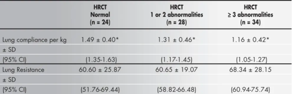
The mean values of lung compliance for the groups with normal tomography, 1 or 2 alterations and 3 or more alterations were re-spectively 1.49 ± 0.40 ml/cm H2O/kg (95% CI of 1.35-1.63), 1.31 ± 0.46 (95% CI of
1.17-1.45) and 1.16 ± 0.42 (95% CI of 1.05-1.27). The variance analysis performed on the lung compliance values had a statistically sig-nificant result (p = 0.015).
The mean lung resistance values for the groups with normal tomography, 1 or 2 altera-tions and 3 or more alteraaltera-tions were respec-tively 60.60 ± 25.87 cm H2O/l/s (95% CI of 51.76-69.44), 60.65 ± 19.07 cm H2O/l/s (95% CI of 58.82-66.48) and 68.34 ± 28.15 cm H2O/l/s (95% CI of 60.94-75.74). The vari-ance analyses for lung resistvari-ance values among the three groups of tomographic alterations were not statistically significant (Table 4).
We were able to verify an association be-tween lung compliance and several tomo-graphic abnormalities.
○ ○ ○ ○ ○ ○ ○ ○ ○ ○ ○ ○ ○ ○ ○ ○ ○ ○ ○ ○
Discussion
This was a prospective study undertaken in a neonatal intensive care unit with prema-ture very low birth weight infants. The clini-cal data from these babies showed moderate severity among this group of infants, with 31% of the babies having birth weights of less than 1,000 g and about 45% requiring mechanical ventilation. The incidence of bronchopulmo-nary dysplasia was 28%, defined as the use of oxygen beyond 28 days of life.
Various techniques can be used for meas-uring lung compliance and lung resistance in newborns. Among the commonly used tech-niques are multiple occlusion, the
conven-1
2
tional (Mead-Whittenberger) method and lin-ear regression. In this study, the latter was used. The conventional and linear regression tech-niques measure lung compliance and resist-ance, whereas the multiple occlusion tech-nique measures compliance and resistance of the respiratory system, which includes meas-urements of lung and thoracic wall mechan-ics. Both the conventional and linear regres-sion techniques have the advantage of mak-ing the measurement durmak-ing spontaneous breathing, without the need for the respira-tory muscles to relax.21 The multiple occlu-sion and conventional techniques have the limitation of needing to assume a linear re-lation between pressure and volume. How-ever, several authors have already described a non-linear relationship between pressure and volume in newborns with pulmonary diseases.22 Moreover, the high compliance of the thoracic wall observed in extremely pre-mature infants (birth weight less than 1,000 g) may also alter the linearity of the pres-sure/volume relationship, since it can cause uneven distribution of pleural pressure and inadequate air filling of the lungs.
In the linear regression technique, the analysis is done by computer and calculation of the compliance and resistance is done at several points in the respiratory cycle (not only two or three, as in the conventional tech-nique), which gives rise to high precision in the results and diminishes the influence of possible artifacts. Computer-based techniques
Table 3. High-resolution computerized tomography: number of abnormalities among very low birth weight infants who had adequate weight for their gestational
age before discharge, born in Instituto Fernandes Figueira, from 1998 to 2000
number of abnormalities number of patients (%)
None 24 (27.9)
1 or 2 28 (32.6)
≥ 3 34 (39.5)
Table 4. High-resolution chest tomography versus. lung compliance and resistance, for very low birth weight infants who had adequate weight for their gestational age, born in Instituto Fernandes Figueira, from 1998 to 2000
HRCT HRCT HRCT
Normal 1 or 2 abnormalities ≥≥≥≥≥ 3 abnormalities (n = 24) (n = 28) (n = 34)
Lung compliance per kg 1.49 ± 0.40* 1.31 ± 0.46* 1.16 ± 0.42* ± SD
(95% CI) (1.35-1.63) (1.17-1.45) (1.05-1.27) Lung Resistance 60.60 ± 25.87 60.65 ± 19.07 68.34 ± 28.15 ± SD
(95% CI) (51.76-69.44) (58.82-66.48) (60.94-75.74)
allow data to be collected for extended peri-ods of time and the distribution of various points on the regression curve to be observed. Thus, identification of non-linear points al-lows their exclusion and respiratory mechan-ics can then be calculated. The conventional and multiple occlusion techniques require many manual calculations for the assessment of pulmonary mechanics of the newborn, and they are slow and arduous. Authors who have compared pulmonary compliance and resist-ance measured by the conventional and lin-ear regression techniques have found excellent correlation coefficients.22,23
We found a high prevalence of abnormali-ties in the pulmonary function test and high resolution computed tomography, which probably reflects lung injury caused by expo-sure of the immature lung to oxygen and me-chanical ventilation. About 72% of the ba-bies had some tomographic alteration and 45% displayed altered lung compliance.
The mean values of lung compliance (1.30 ± 0.44 ml/cm H2O/kg) and lung resistance (63.7 ± 24.87 cm H2O/l/s) in our sample are in agreement with the literature. Zaballos et al.24 studied the lung function of 18 very low birth weight premature infants (average weight of 1,288 ± 196 g and gestational age of 30.1 ± 2.7 weeks) and found that, for children of more than 7 days old, the mean compliance and resistance values were respectively 1.31 ± 0.3 ml/cm H2O/kg and 61.3 ± 36.4 cm H2O/ l/s, which are comparable with our results. Abbasi and Bhutani16 studied the lung me-chanics of very low birth weight infants with gestational ages of less than 34 weeks, who did not need mechanical ventilation, and found values of 1.4 ± 0.1 for compliance and 49.0 ± 6.0 for resistance. In our laboratory, Viviani21 studied the lung function of 13 pre-mature infants without respiratory disease in the neonatal period, with postnatal ages rang-ing from 28 to 47 days and gestational ages of between 32 and 34 weeks, and found mean values of lung compliance and resistance of respectively 1.55 ± 0.6 ml/cm H2O/kg and 53.6 ± 16.7 cm H2O/l/s.
Our data also show a significant associa-tion between high resoluassocia-tion computed to-mography alterations and decreased lung
compliance (Table 4). In most cases, the ba-bies with more than three abnormalities seen via computed tomography used more me-chanical ventilation and oxygen, and devel-oped chronic lung disease. Studies in new-born animals have shown that hyperoxia and barotrauma can cause morphological and functional alterations in the lung. Warner et al.25 showed that prolonged neonatal hype-roxia in mice caused a decrease in alveolar septation, an increase in the size of terminal airways, an increase in lung fibrosis and an increase in the number of inflammatory cells in the lung. Hyperoxia also caused functional alterations with a decrease in lung volume and compliance. Davis et al.26 concluded that, 48 hours after being submitted to hyperoxia and hyperventilation, animals pre-sented decreased dynamic lung compliance and increased lung resistance. Moderate-to-serious atelectasis, exsudate fibrosis, edema and inflammation were all identified in these animals via microscopy.
Our tomographic findings seem to reflect these microscopic alterations described in ani-mals. Compromised interstices can therefore affect lung distensibility and, consequently, the compliance. Decreased lung compliance can also be explained by peribronchial fibrosis, substitution of normal lung tissue by fibrotic parenchymal bands27 and decreased numbers of alveoli due to lung injury.4 The relation-ship between high resolution computed tom-ography and compliance in our data indicates primarily parenchymal disease.
The lack of an association between pul-monary resistance and lung morphology can be due to the following factors:
a) The disease in these premature infants mainly involves the pulmonary paren-chyma and not so much the airways. Re-cent articles have described altered lung anatomy in newborns who died of bron-chopulmonary dysplasia due to factors that were present in the pre-surfactant era such as severe fibrosis, airway injury, hy-perplasia and lung inflammation, which mainly showed as decreases in alveolar septation and vascular development.28 b) There may have been minor alterations in
the airways that were not evident from the
high-resolution computed tomography. c) The test method used consisted of a series
of images during spontaneous respiration, given that the patients in this study had very low weight and were breathing room air, thus not justifying the use of a more aggressive method such as tracheal intuba-tion, which would have allowed the test to be done in two phases: inspiratory and ex-piratory apnea. This method of a series of inspirations and expirations with apnea is especially known as a means for detecting the presence of injuries that suggest that the small airways are compromised.29
Very few studies have been published using high resolution computed chest tomography on babies, but none has attempted to correlate these findings with functional lung tests in very low birth weight infants. Papers published previously involving high resolution computed tomogra-phy on premature infants basically focused on babies who developed bronchopulmonary dys-plasia and were evaluated when they were close to or over one year old, and therefore utilized methodologies quite different from ours. Oppenheim et al.12 analyzed high resolution computed tomography from 23 children with bronchopulmonary dysplasia and an average age of 4 years, and found tomographic abnor-malities in all the children. The alterations in-cluded multifocal areas of hyperaeration and well-defined linear and subpleural opacities. In 20 of the 23 children the three types of tomo-graphic alterations were concurrent. Lee et al.30 evaluated the tomographic results from 6 chil-dren with bronchopulmonary dysplasia and an average age of 4 months, and found bubbles and focal emphysema in three patients and at-electasis in three others. Although these stud-ies were carried out on older children, the ab-normalities described, including bubbles, opaci-ties, and hyperaeration, were similar to those shown in our study.
1. Hack M, Wright LL, Shankaran S, et al. Very-low-birth-weight outcomes of the National Institute of Child Health and Human Development Neonatal Network, November 1989 to October 1990. Am J Obstet Gynecol 1995;172(2 Pt 1):457-64. 2. Cooper TR, Berseth CL, Adams JM, Weisman LE. Actuarial
survival in the premature infant less than 30 weeks’ gestation. Pediatrics 1998;101(6):975-8.
3. Hakulinen AL, Heinonen K, Lansimies E, Kiekara O. Pulmo-nary function and respiratory morbidity in school-age children born prematurely and ventilated for neonatal respiratory insuf-ficiency. Pediatr Pulmonol 1990;8(4):226-32.
4. Jobe AH, Ikegami M. Mechanisms initiating lung injury in the preterm. Early Hum Dev 1998;53(1):81-94.
5. Koumbourlis AC, Motoyama EK, Mutich RL, Mallory GB, Walczak SA, Fertal K. Longitudinal follow-up of lung function from childhood to adolescence in prematurely born patients with neonatal chronic lung disease. Pediatr Pulmonol 1996;21(1):28-34.
6. Gerhard T, Reifenberg L, Duara S, Bancalari E. Comparison of dynamic and static measurements of respiratory mechanics in infants. J Pediatr 1989;114(1):120-5.
7. Chan KN, Wong YC, Silverman M. Relationship between in-fant lung mechanics and childhood lung function in children of very low birthweight. Pediatr Pulmonol 1990;8(2):74-81. 8. Fitzgerald DA, Mesiano G, Brosseau L, Davis GM. Pulmonary
outcome in extremely low birth weight infants. Pediatrics 2000;105(6):1209-15.
9. Moya MP, Bisset GS, Auten RL, Miller C, Hollingworth C, Frush DP. Reliability of CXR for the diagnosis of bronchopul-monary dysplasia. Pediatr Radiol 2001;31(5):339-42. 10. Yuksel B, Greenough A, Karani J, Page A. Chest radiograph
scoring system for use in pre-term infants. Br J Radiol 1991;64(767):1015-8.
○ ○ ○ ○ ○ ○ ○ ○ ○ ○ ○ ○ ○ ○ ○ ○ ○ ○ ○ ○ ○ ○ ○ ○ ○ ○ ○ ○ ○ ○ ○ ○ ○ ○ ○ ○ ○ ○ ○ ○ ○ ○ ○ ○ ○ ○ ○ ○ ○ ○ ○ ○ ○ ○ ○ ○ ○ ○ ○ ○ ○ ○ ○ ○
REFERENCES
11. Northway WH, Moss RB, Carlisle KB, et al. Late pulmonary sequelae of bronchopulmonary dysplasia. N Engl J Med 1990;323(26):1793-9.
12. Oppenheim C, Mamou-Mani T, Sayegh N, de Blic J, Scheinmann P, Lallemand D. Bronchopulmonary dysplasia: value of CT in identifying pulmonary sequelae. AJR Am J Roentgenol 1994;163(1):169-72.
13. Lubchenco LO, Hansman C, Dressler M, Boyd E. Intrauterine growth as estimated from liveborn birth-weight data at 24 to 42 weeks gestation. Pediatrics 1963;32:793-6.
14. Greenspan JS, Abbasi S, Bhutani VK. Sequential changes in pulmonary mechanics in the very low birth weight (less than or equal to 1000 grams) infant. J Pediatr 1988;113(4):732-7. 15. Respiratory mechanics in infants: physiological evaluation in
health and disease. American Thoracic Society/European Res-piratory Society. Am Rev Respir Dis 1993;147(2):474-96. 16. Abbasi S, Bhutani VK. Pulmonary mechanics and energetics of
normal, non-ventilated low birthweight infants. Pediatr Pulmonol 1990;8(2):89-95.
17. Lucaya J, Garcia-Pena P, Souza P, Sotil J. Low-dose and special chest CT techniques. Pediatr Radiol 1996;65-8. 18. Fleiss JL. Statistical methods for rates and proportions. 2nd ed.
New York: Wiley; 1981.
19. Webb WR, Müller NL, Naidich DP. HRCT findings of lung disease. In: Webb WR, Muller NL, Naidich DP, eds. High-Resolution CT of the lungPhiladelphia: Lippincott-Raven; 1996. p.41-109.
20. Bancalari E, Abdenour GE, Feller R, Gannon J. Bronchopul-monary dysplasia: clinical presentation. J Pediatr 1979;95(5 Pt 2):819-23.
21. Viviani AR. Avaliação do desenvolvimento funcional pulmonar dos recém-nascidos pretermo. [thesis]. Rio de Janeiro: Instituto Fernandes Figueira, Fundação Oswaldo Cruz; 1997.
22. Ramos JRM. Estudo da complacência e respiratório do recém-nascido a termo. [thesis]. Rio de Janeiro: Instituto Fernandes Figueira, Fundação Oswaldo Cruz, 1994.
23. Silva Neto G, Gerhardt T, Silberg A, Gerhardt T, Claure N, Duara S, Bancalari E. Nonlinear pressure/volume relationship and measurement of lung mechanics in infants. Pediatr Pulmonol 1992;12(3):146-52.
24. Benito Zaballos MF, Pedraz García C, Salazar A-Villalobos V. Función pulmonar en recién nacidos pretérmino y a término du-rante el periodo neonatal: I. patrón respiratorio. [Pulmonary func-tion in preterm and full term infants during the neonatal period: 1. respiratory pattern]. An Esp Pediatr 1991;35(4):243-7. 25. Warner BB, Stuart LA, Papes RA, Wispé JR. Functional and
pathological effects of prolonged hyperoxia in neonatal mice. Am J Physiol 1998;275(1 Pt 1):L110-7.
26. Davis JM, Dickerson B, Metlay L, Penney DP. Differential ef-fects of oxygen and barotrauma on lung injury in the neonatal piglet. Pediatr Pulmonol 1991;10(3):157-63.
27. Van Lierde S, Smith J, Devlieger H, Eggermont E. Pulmo-nary mechanics during respiratory distress syndrome in the prediction of outcome and differentiation of mild and severe bronchopulmonar y dysplasia. Pediatr Pulmonol 1994;17(4):218-24.
28. Coalson JJ. Pathology of chronic lung disease of early infancy. In: Bland RD, Coalson JJ, eds. Chronic lung disease in early infancy. New York: Marcel Dekker; 2000. p.85-124. 29. Siegel MJ. Lung, pleura and chest wall. In: Siegel MJ, editor.
Pediatric body CT. Philadelphia: Lippincott-William & Wilkins; 1999.p.101-40.
Mecânica pulmonar e tomografia computa-dorizada de tórax de alta resolução em recém-nascidos prematuros de muito baixo peso
CONTEXTO: O desenvolvimento pulmonar do
bebê prematuro pode ser afetado por lesão ao pulmão nas primeiras semanas de vida, podendo ocasionar alterações na mecânica pulmonar.
OBJETIVO: Avaliar a relação entre a mecânica
pulmonar e as alterações estruturais pulmo-nares em prematuros de muito baixo peso (peso de nascimento inferior a 1.500 g).
TIPO DE ESTUDO: Estudo de corte
transver-sal avaliando a mecânica pulmonar (medidas a complacência e a resistência) e estrutura pulmonar (através da tomografia de tórax de alta resolução) próximo à alta hospitalar de 86 prematuros de muito baixo peso, nasci-dos no Instituto Fernandes Figueira, uma instituição pública, terciária, no Rio de Ja-neiro, Brasil. A complacência e resistência pulmonares foram medidas durante o sono tranqüilo. A tomografia de tórax foi realiza-da com o equipamento Pro Speed-S.
MAIN MEASUREMENTS: Na análise
estatís-tica, utilizamos análise de variância para múl-tiplas comparações (ANOVA, Kruskal Wallis). O nível de significância estatística empregado foi 0,05.
○ ○ ○ ○ ○ ○ ○ ○ ○ ○ ○ ○ ○ ○ ○ ○ ○ ○ ○ ○ ○ ○ ○ ○ ○ ○ ○ ○ ○ ○ ○ ○ ○ ○ ○ ○ ○ ○ ○ ○ ○ ○
RESUMO
The paper (partial data) was presented at the Annual Meet-ing of the Pediatric Academic Societies, USA, 1999.
Rosane Reis de Mello, MD,PhD. Follow-up clinic, De-partment of Neonatology, Instituto Fernandes Figueira, Fundação Oswaldo Cruz, Rio de Janeiro, Brazil.
Maria Virgínia Peixoto Dutra, MD, MSc. Epidemiol-ogy Center, Instituto Fernandes Figueira, Fundação Oswaldo Cruz, Rio de Janeiro, Brazil.
José Roberto Ramos, MD, MSc. Department of
Neonatology, Instituto Fernandes Figueira, Fundação Oswaldo, Rio de Janeiro, Brazil.
Pedro Daltro, MD. Department of Radiology, Instituto Fernandes Figueira, Fundação Oswaldo Cruz, Rio de Ja-neiro, Brazil.
Márcia Boechat, MD. Department of Radiology, Instituto Fernandes Figueira, Fundação Oswaldo Cruz, Rio de Ja-neiro, Brazil.
José Maria de Andrade Lopes, MD, PhD. Department
of Neonatology, Instituto Fernandes Figueira, Fundação Oswaldo Cruz, Rio de Janeiro, Brazil.
Sources of funding: Not declared
Conflict of interest: Not declared
Date of first submission: July 11, 2002
Last of received: December 17, 2002
Accepted: May 19, 2003
Address for correspondence
Rosane Reis de Mello
Avenida Rui Barbosa 716 — Flamengo. Rio de Janeiro/RJ — Brasil — CEP 22250-020 Tel. (+55 21) 2553-3384 Ramal 5224/5235 Fax (+55 21) 2553-3384 Ramal 5319/5320 E-mail: [email protected] COPYRIGHT©2003, Associação Paulista de Medicina
○ ○ ○ ○ ○ ○ ○ ○ ○ ○ ○ ○ ○ ○ ○ ○ ○ ○ ○ ○
Publishing information
RESULTADOS: Em 34 bebês (43%), foram
en-contrados resultados alterados tanto para a complacência quanto para a resistência pul-monares e, em 20 crianças (23,3%), ambos os resultados foram normais. As médias da complacência e resistência pulmonares da população estudada foram respectivamente 1,30 ml/cm H2O/Kg e 63,7 cm H2O/l/s. Em 62 bebês (72%), os exames tomográficos apresentavam alterações pulmonares. A mai-oria das crianças mostrou mais de uma alte-ração, descritas como opacidade em vidro fosco, banda parenquimatosa, atelectasia, bolha/cisto. As médias dos valores de com-placência para as crianças com tomografia de tórax normal, 1 ou 2 alterações e 3 ou mais alterações, foram significativamente difertes: 1,49, 1,31, 1,16 (p = 0,015). Não en-contramos relação entre resistência pulmo-nar e o número de alterações tomográficas.
CONCLUSÃO: Os resultados mostram alta
prevalência de anormalidades funcionais e tomográficas em bebês prematuros, assin-tomáticos, próximo à alta hospitalar. Mostram também uma relação entre anormalidades pulmonares morfológicas e funcionais.
PALAVRAS CHAVE: Prematuro. Prova de