* Study carried out in the Department of General Surgery of the Universidade Federal de Pernambuco – UFPE, Federal University of Pernambuco – Hospital das Clínicas and in the Department and Clinical Surgery of the São José Memorial Hospital, Recife (PE) Brazil.
1. PhD in Surgery. Universidade Federal de Pernambuco – UFPE, Federal University of Pernambuco – Recife (PE) Brazil.
2. Professor of Basic Clinical Practice and Surgical Technique. Universidade Federal de Pernambuco – UFPE, Federal University of Pernambuco – Recife (PE) Brazil.
3. Masters in Surgery. Universidade Federal de Pernambuco – UFPE, Federal University of Pernambuco – Recife (PE) Brazil. 4. Adjunct Professor of Surgery. Universidade Federal de Pernambuco – UFPE, Federal University of Pernambuco – Recife (PE) Brazil.
5. Full Professor of Abdominal Surgery and Basic Surgical Technique. Universidade Federal de Pernambuco – UFPE, Federal University of Pernambuco – Recife (PE) Brazil.
6. Professor of Pulmonology at the School of Medical Sciences. Universidade Federal de Pernambuco – UFPE, Federal University of Pernambuco – Recife (PE) Brazil.
Correspondence to: Josemberg Marins Campos. Rua Joaquim Nabuco, 92, 1101, Graças, CEP 51011-000, Recife, PE, Brasil. Tel/Fax 55 81 3441-4460. E-mail: berg@elogica.com.br
Submitted: 20 March 2006. Accepted, after review: 8 August 2006.
Gastrobronchial fistula as a rare complication of gastroplasty for obesity.
A report of two cases*
Josemberg Marins Campos1, Luciana Teixeira de Siqueira2, Marconi Roberto de Lemos Meira3,
Álvaro Antônio Bandeira Ferraz4, Edmundo Machado Ferraz5, Murilo José de Barros Guimarães6
Abstract
Gastrobronchial fistula is a rare condition as a complication following bariatric surgery. The management of this condition requires the active participation of a pulmonologist, who should be familiar with aspects of the main types of bariatric surgery. Herein, we report the cases of two patients who presented recurrent subphrenic and lung abscess secondary to fistula at the angle of His for an average of 19.5 months. After relaparotomy was unsuccessful, cure was achieved by antibiotic therapy and, more importantly, by stenostomy and endoscopic dilatation, together with the use of clips and fibrin glue in the fistula. These pulmonary complications should not be treated in isolation without a gastrointestinal evaluation since this can result in worsening of the respiratory condition, thus making anesthetic management difficult during endoscopic procedures.
Keywords: Fistula; Lung abscess; Subphrenic abscess; Obesity, surgery; Endoscopy.
Introduction
Obesity, which is a chronic disease associated with comorbidities, should be controlled by physical exercise and changes in dietary habits, and surgical treatment is indi-cated when clinical measures are inefficient.(1)
There are various surgical techniques to control obesity. However, the procedure most often employed has been gastroplasty involving the Fobi-Capella technique. Recently, another technique, known as sleeve gastrectomy, has been applied. It involves partial vertical resection of the stomach, which is transformed into a tube in order to reduce its storage capacity.
Although the two surgical techniques present good results in terms of weight loss and control of comorbidi-ties, both can lead to early postoperative complications, of which gastric fistula is one of the most severe due to the occurrence of abdominal sepsis and respiratory altera-tions, especially in the left lung, resulting from the common occurrence of subphrenic abscess.(2) These consequences for
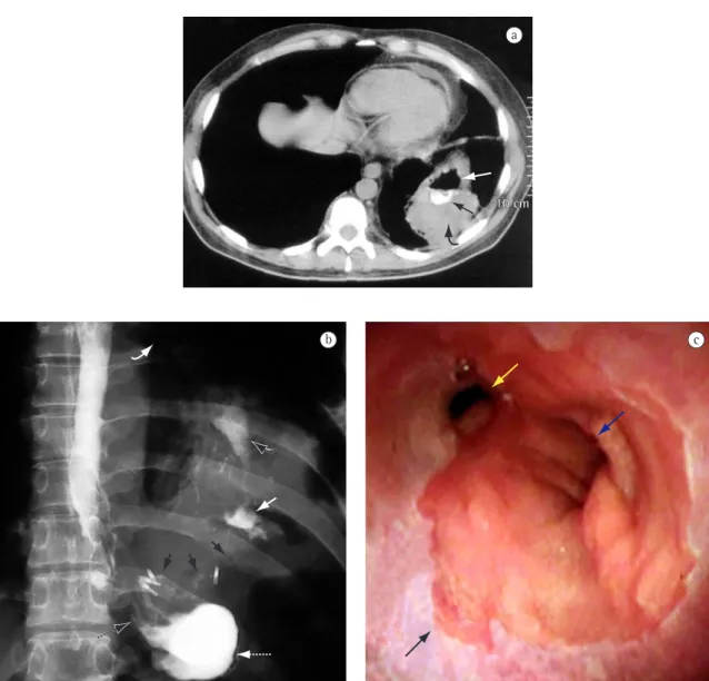
the definitive closure of the fistula, which resulted in the resolution of the lung abscess.
Case 2
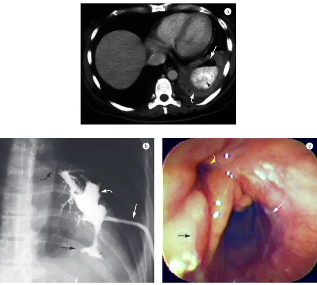
A 44-year-old female patient who underwent vertical banded gastroplasty (sleeve gastrectomy) by laparoscopy developed gastric fistula and presented five episodes of subphrenic abscess over an 8-month period. Her clinical symptoms began with pain in the left shoulder and worsened to include fever, cough, leukocytosis, worsening of overall health status, and excessive weight loss. The last two episodes were accompanied by cough with purulent expectoration, and computed tomography of the chest diag-nosed an abscess at the left lung base (Figure 2a), secondary to subphrenic abscess at the angle of His, and gastric stenosis at the level of the band and the incisura angularis. The lung abscess resulting from the gastrobronchial fistula was drained through a catheter seen on a contrast X-ray (Figure 2b), and endoscopic imaging showed the internal opening of the fistula at the angle of His (Figure 2c).
The patient was monitored by a pulmonologist, an endoscopist, and a surgeon. She was treated with antibiotic therapy, respiratory therapy, and nutritional support provided through a nasogastric tube. In addition, the patient underwent explora-tory laparoscopy for removal of the band, at which point a severe inflammatory obstruction was seen in the left subphrenic region and in the esophagogas-tric junction, making it impossible to gain surgical access to the fistula.
Therefore, we carried out six endoscopic stenos-tomy sessions using an electric scalpel and dilatation of the stenotic area, thereby allowing satisfactory food intake, nutritional recovery, and cicatrization of the lung abscess, after the permanent closure of the fistula.
Discussion
Gastric fistula following bariatric surgery occurs in 0.9 to 2.6% of the cases, reaching 8% in second operations, and is most often located at the level of the angle of His, probably as a result of ischemia at the apex of the lateral stapling.(1)
The fistula secretion moves to the subphrenic region and can cause abdominal sepsis, complica-tions in the left lung, and gastrocutaneous fistula. Less frequently, drainage into the excluded stomach Typically, the subphrenic infectious process
causes gastrocutaneous fistula, pleural effusion, and respiratory infection.(3) However, there have
been few reports of the occurrence of gastrobron-chial fistula following bariatric surgery. Therefore, we report herein the satisfactory evolution of two patients with lung abscess secondary to fistula at the angle of His following gastroplasty.
Case report
Case 1
bronchial fistula, and left lung abscess.(4) Certainly,
this also occurred in our study.
The patients analyzed presented aspects in common, which facilitated the communication of secretion between the stomach and the respi-ratory system. Stenosis of the gastric pouch or of the gastrojejunal anastomosis increased the pres-sure and directed the food to the fistula at the angle of His, leading to the appearance of recurrent (gastrogastric fistulas) occurs.(3) Drainage into the
bronchial tree (gastrobronchial fistula) has been reported as a complication following trauma, esoph-agogastric surgery (transhiatal esophagectomy due to neoplasias or megaesophagus), splenectomy, and perforated gastric ulcer.(4,5) In these abdominal
surgeries, thereis the development of subphrenic abscesses develop and cross the diaphragm via the lymphatic route, causing pleural effusion,
gastro-10 cm 10 cm a
b c
bronchoscopy has been presented as a diagnostic and therapeutic method for cases of gastrobron-chial fistula, especially following esophagectomy, and this facilitates the evaluation and adequate clearance of the airways.(4,5) In the present study,
this procedure was not necessary given the rapid improvement of the respiratory condition after anti-biotic therapy, discontinuation of the oral diet, and initiation of the diet provided through a gastros-tomy or a nasogastric tube.
The classical treatment of digestive fistula includes nutritional support and surgical resection subphrenic infection, in the absence of an exit route
to the skin, since the abdominal tube had already been removed.
These two patients belong to a group of 27 patients with gastric fistula treated endoscopi-cally. The specific characteristic of the two cases was the recurrent character of the fistula for a prolonged period, the stenosis of the gastric pouch or of the gastrojejunal anastomosis, and the absence of a drainage route for the abdominal infection.
In conjunction with digestive endoscopy, imaging of the chest and imaging of the abdomen,
Figure 2 - Case 2: a) Computed tomography of the chest: abscess at the left lung base (curved white arrow) containing air (white arrow) and contrast coming from the gastric pouch (black arrow); b) Contrast X-ray: catheter (white arrow) draining lung abscess (curved white arrow), subphrenic abscess (black arrow), and bronchial tree (curved black arrow); and c) Endoscopic imaging: esophagogastric junction (black arrow), gastric pouch (white arrow), and fistula at the angle of His (yellow arrow).
a
condition, thus making anesthetic management difficult during the performance of surgical and endoscopic procedures aimed at curing the fistula and the respiratory problems.
Therefore, in cases of persistent respiratory infection in the early or late postoperative period following bariatric surgery, a gastrointestinal evalua-tion by imaging and endoscopic examinaevalua-tion should be promptly carried out with the aim of diagnosing and designing a plan for treating a possible fistula involving the operated stomach.
References
1. Rocha LCM, Lima Jr. GF. Papel da endoscopia na obesidade mórbida. In: Savasi-Rocha PR, Coelho LGV, Diniz MTC, Nunes TA, editores. Tópicos em Gastroenterologia - 13. Rio de Janeiro: Editora Medsi; 2003. p.53-75.
2. Merkle EM, Hallowell PT, Crouse C, Nakamoto DA, Stellato TA. Roux-en-Y gastric bypass for clinically severe obesity: normal appearance and spectrum of complications at imaging. Radiology. 2005;234(3):674-83.
3. Stanczyk M, Deveney CW, Traxler SA, McConnell DB, Jobe BA, O’Rourke RW.Gastro-gastric fistula in the era of divided Roux-en-Y gastric bypass: strategies for prevention, diagnosis and management. Obes Surg. 2006;16(3):359-64. 4. Jeganathan R, Pore N, Clements WD. Gastrobronchial
fistula - a complication of splenectomy. Ulster Med J. 2004;73(2):126-8.
5. Purucker EA, Sudfeld S, Matern S. Gastrobronchial fistula after caustic injury due to lye ingestion. Endoscopy. 2003;35(3):252.
6. Bennie MJ, Sabharwal T, Dussek J, Adam A. Bronchogastric fistula successfully treated with the insertion of a covered bronchial stent. Eur Radiol. 2003;13(9):2222-25.
7. Maluf-Filho F, Moura EGH. Prótese plástica auto-expansível no tratamento da fístula complexa pós-cirurgia bariátrica. GED. 2005;24(4):203.
of the fistulous pathway, the latter being associ-ated with considerable morbidity due to the intense inflammatory process in the upper abdomen, in addition to the risk of injury of the adjacent struc-tures, and becoming a laborious procedure due to the frequent need to re-operate,(1) thereby leading to
infection of the incision site and its consequences. Therefore, endoscopic treatment is a minimal inva-sive and efficient means of resolving gastric stenosis, which is considered the leading cause of perpetua-tion of the fistula, as demonstrated in this study.
In addition to the endoscopic treatment methods used in the study cited, the use of esophageal or tracheobronchial stent, which is applied in gastro-bronchial fistula followingesophagectomy, has been implemented.(6) In the two patients analyzed, the use
of a self-expanding plastic stent was not indicated due to the scarcity of available data on the subject at the time. Recently, we successfully used this method in two patients with gastric fistulas larger than 10 mm in diameter, since it has the advantage of reducing the number of endoscopy sessions, in addition to allowing early food intake, which is in accordance with the current literature.(7)
Considering the complexity of the obese patient and of the gastrobronchial infectious process, it is essential that a joint evaluation be carried out by specialists, including pulmonologists, surgeons, radiologists, and endoscopists.

