Rev Odontol UNESP. 2017 Sept-Oct; 46(5): 255-260 © 2017 - ISSN 1807-2577
ORIGINAL ARTICLE
Doi: http://dx.doi.org/10.1590/1807-2577.02517
Radiographic analysis of 1000 cast posts in Sergipe state, Brazil
Avaliação radiográica de 1000 retentores intrarradiculares metálicos fundidos no Estado de Sergipe
Caroline Garcez MENDONÇA
a*, José Rogério Vieira de ALMEIDA
a, Wilton Mitsunari TAKESHITA
a,
Fábio MARTINS
a, Monica Silveira PAIXÃO
aaFaculdade de Odontologia, UFS – Universidade Federal de Sergipe, Aracaju, SE, Brasil
Resumo
Introdução: Dentes com ampla destruição coronária e tratados endodonticamente necessitam de retentores intrarradiculares para viabilizar a restauração protética. Apesar do crescente uso de pinos intrarradiculares pré-fabricados, os retentores intrarradiculares metálicos fundidos ainda são muito utilizados. A qualidade desses é importante para a longevidade do tratamento restaurador, pois podem levar ao insucesso quando seus princípios fundamentais não são seguidos.
Objetivo: Avaliar radiograficamente os princípios protéticos de 1000 retentores intrarradiculares metálicos fundidos em dentes unirradiculares e a coerência dos mesmos com os princípios para sua confecção. Material e método: Foram selecionadas radiografias periapicais digitais de 1000 dentes unirradiculares que receberam tratamento endodôntico e retentores intrarradiculares metálicos fundidos, através do acervo de uma clínica radiológica (Centro de Imagem, Aracaju/SE, Brasil). As imagens foram analisadas através de um software de mensuração (ImageJ, EUA) de acordo com os princípios fundamentais protéticos: comprimento e diâmetro do pino, relação do pino com a crista óssea, contiguidade ao canal, espaço entre o remanescente obturador e o pino, quantidade de material obturador e ausência de lesão periapical. Os dados foram tabulados, analisados qualitativamente em ideais e não ideais e submetidos ao teste do Qui-Quadrado (α=0,05). Resultado: Apenas 6,7% dos retentores intrarradiculares fundidos analisados foi confeccionado de forma satisfatória. Conclusão: Muitos critérios protéticos são negligenciados durante a confecção de retentores intrarradiculares metálicos fundidos, resultando em trabalhos inadequados que podem comprometer a longevidade do tratamento restaurador.
Descritores: Técnica para retentor intrarradicular; pinos dentários; prótese dentária; radiografia.
Abstract
Introduction: Endodontically treated teeth with extensive coronary destruction require posts and cores to enable prosthetic restoration. Despite the increasing use of prefabricated posts, cast metal posts and cores are still widely used. The quality of the latter is important for the longevity of restorative treatment, and failure can occur if the fundamental principles are not followed. Objective: To radiographically evaluate the prosthetic principles of 1000 cast metal post-and-core restorations performed in single-rooted teeth and their coherence with the principles used for their confection. Material and method: Digital periapical radiographs of 1000 endodontically treated, single-rooted teeth with cast metal posts and cores were selected from the collection of a radiology clinic (Centro de Imagem, Aracaju/SE, Brazil). The images were analyzed using a measurement software (ImageJ, USA) in accordance with the fundamental prosthetic principles: length and diameter of the post, ratio between post and bone crest, contiguity of post to the root canal, gap between post and the remaining root canal filling, amount of remaining root canal filling, and absence of periapical lesion. The data were qualitatively analyzed, classified into ideal and not ideal, and submitted to the chi-square test (α=0.05). Result: Only 6.7% of the cast metal posts analyzed were satisfactorily fabricated. Conclusion: Many prosthetic criteria are neglected during the manufacturing of cast metal post and cores, resulting in inadequate work that may compromise the longevity of restorative treatments.
Descriptors: Post-and-core technique; dental posts; dental prosthesis; radiography.
INTRODUCTION
Endodontically treated teeth require special care when they are prosthetically restored because of the substantial tooth structure loss caused by removal of carious lesion, crown fracture, and access for endodontic treatment. Restoration of this lost dental structure, so that the tooth can again develop its functions in the oral cavity,
dental remnant conditions; it should consider the amount of dental remnant let ater removal of carious tissue and existing restorations, as well as the root bony implantation and periapex of the tooth3.
Cast metal posts are versatile because they can copy the root canal anatomy, ensuring better adaptation, correcting the position of the crown of badly positioned teeth, and improving the distribution of masticatory loads on the root2,4. hey can be fabricated using both indirect and direct techniques: in the irst case, a plaster impression is sent to a dental laboratory for post construction; in the latter case, an acrylic resin post is built up in the patient’s mouth, providing better retention2.
Studies on the radiographic evaluation of cast metal posts and cores and their coherence with the basic preparation principles for acceptable treatment show that only a low percentage of posts obey ideal conditions3,5-11. he most unfavorable failures associated with this type of system are loss of retention due to displacement12,13, root fractures14, and risk of corrosion2,15. In search for the origin of these failures, authors have suggested that the problem lies in the way cast posts and cores are being manufactured3,5-7,13. Aiming to reduce these failures, prefabricated posts have appeared as new option of simpler and faster confection; however, care must be taken when performing additional denture wear maneuvers to adapt the root canal space to the selected post shape in order to avoid weakening of the dental remnant and/or root perforations16.
Although the cast metal post-and-core system can present failures, it is still widely used with indication for elements with little dental remnant and for support of prosthetic restorations and posterior teeth3,5,6,12,17.
In view of the need for root retention, much is discussed about endodontic treatment being compromised ater restoration using this system17, as well as with respect to maintenance, or emergence of periapical lesion5,7,17-20. It is believed that a well-performed endodontic treatment leads to greater care in the execution of prosthesis, reinforcing that this is a basic criterion for the beginning of the prosthetic stage preparation, in such a way that these two stages are inseparable and must be conducted properly to obtain success5,6.
herefore, the objective of this study was to radiographically assess endodontically treated, single-rooted teeth with cast metal posts and cores, analyzing whether the prosthetic criteria fundamental to a restorative treatment are adequate or not.
MATERIAL AND METHOD
his study was approved by the Human Research Ethics Committee (CEPSH) of Universidade Federal de Sergipe under protocol no. 107999 (CAAE: 50369015.4.0000.5546). Ater approval, the study was conducted through the selection of digital periapical radiographs of 1000 endodontically treated, single-rooted teeth with cast metal posts and cores. hese periapical radiographs of patients who sought this service for complementary radiographic examination were obtained from the digital collection of a radiology clinic (Centro de Imagem, Aracaju/SE, Brasil). he radiographic images were saved in a ile and identiied with numbers to maintain the conidentiality of the patients’ identity.
he radiographic images were assessed using the same previously calibrated viewer. he images were enlarged and measured according to the evaluated criteria using a graphic analysis program (ImageJ, 1.44p, National Institutes of Health, USA). According to the program, the digital measures obtained in pixels were converted to millimeters with precision of up to three decimals. To this end, the sotware was calibrated from a known size of the image (in this case, the actual dimensions of the radiographic ilm). One of the long axes of the ilm was gauged and this measure was inserted in millimeters for the program calibration. No distinction between age and gender of patients was considered.
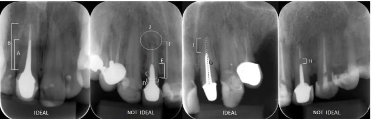
All radiographs containing endodontically treated teeth with cast metal posts and cores were expanded, and the measures were taken observing the absence or presence of failures according to the following prosthetic principles: length of post, diameter of post, ratio between post and bone crest, contiguity of post to root canal, gap between post and the remaining root canal illing, amount of remaining root canal illing, and absence of periapical lesion. he following prosthetic data were considered satisfactory: cast metal posts and cores with post length equal to 2/3 of root length (1mm error margin); post diameter equal to 1/3 of root diameter (0.5mm error margin); post:bone crest ratio with the post reaching 1/2 of root bony implantation; post contiguous to root canal with no deviations; absence of gap between post and the remaining root canal illing (0.2mm error margin); remaining root canal illing in the apical third of the tooth, with a minimum of 4mm of guta percha (below 4mm was considered inadequate and above 4mm was considered adequate, as long as not combined with short posts); and absence of periapical lesion (Figure 1).
he radiography viewer was calibrated according to the criteria evaluated, which were selected based on the methodology of previous studies and according to the standards described in the speciic scientiic literature2,3,5-9,11,17-21.
Each image obtained was opened in the program, enlarged, gauged, evaluated according to the prosthetic criteria, and included in an Excel spreadsheet. A new spreadsheet was prepared with the results of the 1000 images in which all the data were grouped, without the numerical values and only classiied as ideal and not ideal, for statistical analysis. he chi-square hypothesis test, which aimed to ind a dispersion value for two nominal variables and evaluate the association between the qualitative variables, was applied for statistical assessment.
RESULT
Based on the data obtained from the 1000 cast metal posts and cores, frequency was veriied for all evaluated prosthetic criteria (Figure 2).
he following levels of adequacy were veriied for the assessed prosthetic criteria: post length (13.5%), post diameter (80.1%), post:bone crest ratio (19.4%), post contiguity to root canal (92.9%), absence of gap between post and the remaining root canal illing (51.9%), amount of remaining root canal illing not associated with short post (8.3%), and absence of periapical lesion (85.8%). Only 6.7% of all cast metal posts were satisfactorily fabricated, meeting all the prosthetic criteria evaluated, and considered adequate.
he results of this study were analyzed using the chi-square test (α=0.05) with the aim of verifying the existence of signiicant association between the prosthetic criteria found in the assessed radiographs and the success of prosthetic rehabilitation with cast metal posts and cores.
Regarding the relationship between the prosthetic criteria found in the radiographs and the success of prosthetic restorations using cast metal posts and cores, Table 1 presents the signiicant association between all the prosthetic criteria analyzed (p<0.05) and shows the strength between them (Phi coeicient of association).
Among the prosthetic criteria observed, the amount of root canal illing presented the highest positive correlation with the success of endodontic treatment (89%), followed by post length (68%). Correlation between contiguity of post to root canal and success of endodontic treatment was signiicantly low (7%).
Reliability tests showed that assessments of the position of cast metal posts were considered excellent in the intra-examiner evaluation, with Kappa values ranging from 0.918 to 1.00.
DISCUSSION
With the evolution of dental materials and the implementation of adhesive systems, many changes have occurred in dental specialties, and despite the growing use of prefabricated posts1, cast metal posts and cores are still widely used3,5,6,12,17, and their situation requires assessment.
According to the radiographic sample evaluated, only 6.7% of the cast metal posts properly followed all the principles evaluated. Durighetto et al.5 reported a similar result - 10.6% of the cast metal posts analyzed were satisfactorily fabricated.
he present study observed that the vast majority (86.5%) of cast metal posts and cores were manufactured disrespecting the basic 2/3 rule (post length must be 2/3 of root canal length) (Figure 2). his fact was also corroborated in the studies by Bonfante et al.3 (85%); Pinzetta et al.10 (96.9%); Nimigean et al.9 (70.09%); Minguini et al.8 (80%); and Vital et al.11 (73.75%), which reported unacceptable standards for length of cast metal posts. Shorter-than-ideal post length may cause displacement by improper retention and generation
of forces of the tooth fulcrum, thereby increasing the chances of root fracture due to second-degree lever behavior22. his situation is further aggravated when cast metal posts do not follow the fulcrum rule, that is, they do not reach a minimum of 1/2 of the root bony implantation. No major drawbacks are associated with this procedure for situations in which posts are longer than 2/3 of the root length - long posts, as long as the excessive length does not compromise the amount of remaining root canal illing and does not result in excessive dentin removal3.
With respect to post diameter, 80.1% of the radiographic images analyzed in this study presented ideal results, i.e., 1/3 of root diameter (Figure 2), whereas Nimigean et al.9, Minguini et al.8, and Vital et al.11 reported ideal results below 42.41, 40, and 45%, respectively. Such diferences may have occurred because those authors used smaller samples. Very extensive preparations, with large posts, may increase resistance and retention, but present greater probability of root fracture23,24, whereas insuicient preparation, with thin posts, may be interesting to maintain the remaining dentin, but hinder preparation impression during confection, and are likely to produce short posts3,6,18. he data provided in relation to the diameter were limited to the mesiodistal direction.
Regarding the post:bone crest ratio (fulcrum rule), only 19.4% of the cases were within the ideal standards (Figure 2), which was corroborated by Hilgert et al.6, who reported 13.65%. Bonfante et al.3, Minguini et al.8, and Vital et al.11 found 31.8, 45, and 43.75% of ideal root bony implantation, respectively. When a minimum of half of the bone crest is not reached by the post, concentration of forces acting of the dental fulcrum may occur, thus increasing the
probability of root fracture. It was observed that, because the great majority of the analyzed posts were short, they did not reach half of the root bony implantation. Conditions of considerable bone loss were also observed, which would contraindicate the use of posts, and would thus condemn the dental element.
Absence of contiguity between post and root canal may cause root fracture25. In this study, 92.9% of the analyzed cast metal posts and cores presented satisfactory results, with no deviations observed (Figure 2). However, no studies evaluating this criterion were found in the literature; thus we proposed that further studies on such assessment be conducted.
As for the gap between post and the remaining root canal illing, ideally no gap should exist, with tolerance of 0.2mm; in this study, 51.9% of the radiographic results were considered acceptable (Figure 2). he remaining 48.1% of cases presented gaps ranging from 0.2mm to 6.225mm, which was the largest space observed. Bonfante et al.3 found 70.9% of cases with gaps >0.2mm, and similarly, Hilgert et al.6 observed 69.35%. Removal of excess root canal illing is usually not accompanied by lengthening of the post, leaving an empty space inside the root6. his gap may house microorganisms, thus compromising the result of endodontic treatment, potentiating the emergence of periapical lesion3,20,21. he presence of this gap means some degree of failure or negligence in the process of preparing, obtaining, and cementing cast metal posts and cores.
he amount of remaining root canal illing presented the highest negative correlation with treatment success, 91.7% (Figure 2). A large number of treatments with amount of root canal illing Table 1. Association between prosthetic criteria and treatment success (n=1000)
Prosthetic criteria
Treatment success
TOTAL
(n=1000) X2 Phi Φ p value Yes
(n=67)
No (n=933)
Post length (2/3 of root size):
Yes 67 68 135 460.125 0.68 <0.001*
No 0 865 865
Post diameter (1/3 of root size):
Yes 67 734 801 17.841 0.13 <0.001*
No 0 199 199
Post:bone crest ratio (1/2 of post inserted in the bone crest):
Yes 67 127 194 298.350 0.54 <0.001*
No 0 806 806
Contiguity to root canal:
Yes 67 862 929 5.488 0.07 0.019*
No 0 71 71
Gap between post and the remaining root canal illing:
Yes 67 452 519 66.553 0.25 <0.001*
No 0 481 481
Amount of root canal illing (4 mm):
Yes 67 16 83 793.386 0.89 <0.001*
No 0 917 917
Absence of periapical lesion:
Yes 67 791 858 11.885 0.11 <0.001*
No 0 142 142
below ideal were observed, thus being a risk factor for apical sealing and restoration longevity. here are no disadvantages for cases in which the illing extends beyond 4mm without compromising the ideal length of the cast metal post; however, when the amount of illing extended beyond the ideal, the posts were oten short. Bonfante et al.3 and Hilgert et al.6 found results diferent from those of the present study, which can be justiied by the fact that they adopted 3mm of remaining root canal illing as ideal.
Presence of periapical lesion is a criterion that generates doubts7,17,18,20,21, considering that without the initial control of treatment, it is impossible to know whether the existing lesion has regressed, developed, or remained stationary. Hommez et al.18 and Durighetto et al.5 concluded that the presence of a post in the root canal has no inluence on apical health. Periapical lesions were present in 14.2% of the radiographic images investigated in this study (Figure 2), conirming the idea advocated by the authors.
When properly executed, prosthetic rehabilitation using cast metal posts and cores follow fabrication and cementation principles that allow treatment success rates, providing maximum retention and resistance without causing root damage, as well as favoring the dissipation and dispersion of masticatory load by the remaining
root6. However, when the prosthetic criteria are neglected, longevity of the restorative treatment may be compromised.
his is a cross-sectional study; therefore, it was not possible to verify the longevity of the cast metal posts and cores whose criteria were considered inadequate. Although periapical radiographs are an elective examination for the assessment of cast metal posts, their images are two-dimensional and may present distortions.
CONCLUSION
Within the limitations of this study, the authors conclude that, according to the prosthetic criteria evaluated, most of the cast metal posts and cores radiographically assessed were inadequately fabricated.
ACKNOWLEDGEMENTS
he authors are grateful to the radiologic clinic Centro de Imagem (Aracaju/SE) for their contribution with this research by providing space and access to their collection of digital periapical radiographs.
REFERENCES
1. Pilo R, Cardash HS, Levin E, Assif D. Effect of core stiffness on the in vitro fracture of crowned, endodontically treated teeth. J Prosthet Dent. 2002 Sep;88(3):302-6. PMid:12426501. http://dx.doi.org/10.1067/mpr.2002.127909.
2. Prado MAA, Kohla JCM, Nogueira RD, Martins VRG. Retentores intrarradiculares: revisão da literatura. UNOPAR Cient Ciênc Biol Saúde. 2014;16(1):51-5.
3. Bonfante G, Fagnani CM, Miraglia SS, Silva W Jr. Avaliação radiográfica de núcleos metálicos fundidos intrarradiculares. RGO (Porto Alegre). 2000 Jul/Set;48(3):170-4.
4. Rennó DG, Contin I. Avaliação da adaptação de retentores intra-radiculares fundidos obtidos por modelagem direta com RAAQ – estudo in vitro. RPG Rev Pós Grad. 2006 Jan/Mar;13(1):25-30.
5. Durighetto IL, Biffi JCG, Dirighetto AF Jr, Caram CM. Avaliação das características da contenção intra-radicular e tratamentos endodôntico em radiografias periapicais de 1000 dentes. Ciênc Odontol Bras. 2007 Abr/Jun;10(2):31-9.
6. Hilgert E, Buso L, Mello EB, Valera MC, Araújo MAM. Avaliação radiográfica de retentores intra-radiculares metálicos fundidos. Ciênc Odontol Bras. 2004;7(4):52-9. http://dx.doi.org/10.14295/bds.2004.v7i4.240.
7. Klautau EB, Souza PS, Barros CMTM, Garcia V, Maranhão KM. Radiographic evaluation of endodontic treatment and radicular retainer quality. Salusvita. 2009;28(1):21-9.
8. Minguini ME, Mantovani MB, Lolli LF, Silva CO, Progiante P, Marson FC. Estudo clínico de pinos intrarradiculares diretos e indiretos em região anterior. Uningá Review. 2014 Out/Dez;20(1):15-20.
9. Nimigean VR, Buţincu L, Nimigean V. A radiographic study regarding post retained restorations. Rom J Morphol Embryol. 2012;53(3 Suppl):775-9. PMid:23188439.
10. Pinzetta AL, Inoue RT, Feltrin PP. Avaliação radiográfica da proporção comprimento de pinos intra-radiculares em relação ao comprimento radicular em dentes suporte de próteses parciais fixas unitárias e compostas. RGO (Porto Alegre). 2006 Out/Dez;54(4):302-7.
11. Vital RA, Ribeiro JS, Rocha SS. Evaluation of the pattern dimensions of cast-metal posts in uniradicular teeth. Rev Odontol UNESP. 2015 Mar/Apr;44(2):99-102. http://dx.doi.org/10.1590/1807-2577.1066.
12. Balkenhol M, Wöstmann B, Rein C, Ferger P. Survival time of cast post and cores: a 10-year retrospective study. J Dent. 2007 Jan;35(1):50-8.; published online Jun 5, 2006. PMid:16750593. http://dx.doi.org/10.1016/j.jdent.2006.04.004.
13. Gómez-Polo M, Llidó B, Rivero A, Del Río J, Celemin A. A 10-year retrospective study of the survival rate of teeth restored with metal prefabricated posts versus cast metal posts and cores. J Dent. 2010 Nov;38(11):916-20.; published online Aug 14, 2010. PMid:20713117. http://dx.doi.org/10.1016/j.jdent.2010.08.006.
14. Jung RE, Kalkstein O, Sailer I, Roos M, Hämmerle CH. A comparison of composite post buildups and cast gold post-and core buildups for the restoration of nonvital teeth after 5 to 10 years. Int J Prosthodont. 2007 Jan/Feb;20(1):63-9. PMid:17319366.
16. Souza EM, Pappen FG, Leonardi DP, Flores VO, Berbert FLCV. O papel da anatomia radicular na colocação de pinos pré-fabricados: uma visão endodôntica. RGO (Porto Alegre). 2007 Jan/Mar;55(1):77-82.
17. Rosalem CGC, Mattos CMA, Guerra SMG. Association between intra-radicular posts and periapical lesions in endodontically treated teeth. J Appl Oral Sci. 2007 Jun;15(3):225-9. PMid:19089134. http://dx.doi.org/10.1590/S1678-77572007000300013.
18. Hommez GMG, Coppens CRM, De Moor RJG. Periapical health related to the quality of coronal restauration and root fillings. Int Endod J. 2002;35(8):680-689. Int Endod J. 2002 Aug;35(8):680-9. PMid:12196221. http://dx.doi.org/10.1046/j.1365-2591.2002.00546.x.
19. Kirkevang LL, Orstavik D, Horsted-Bindslev P, Wenzel A. Periapical status and quality of root fillings and coronal restorations in a Danish population. Int Endod J. 2000 Nov;33(6):509-15. PMid:11307254. http://dx.doi.org/10.1046/j.1365-2591.2000.00381.x.
20. Ozkurt Z, Kayahan MB, Sunay H, Kazazoğlu E, Bayirli G. The effect of the gap between the post restoration and the remaining root canal filling on the periradicular status in a Turkish subpopulation. Oral Surg Oral Med Oral Pathol Oral Radiol Endod. 2010 Jul;110(1):131-5. PMid:20610303. http://dx.doi.org/10.1016/j.tripleo.2010.02.036.
21. Moshonov J, Slutzky-Goldberg I, Gottlieb A, Peretz B. The effect of the distance between post and residual gutta-percha on the clinical outcome of endodontic treatment. J Endod. 2005 Mar;31(3):177-9. PMid:15735463. http://dx.doi.org/10.1097/01.don.0000137646.07662.8e. 22. Russi S, Leonardi P. Verificação radiográfica de alguns princípios relacionados às coroas com pino. Rev Fac Farm Odontol Araraquara.
1968;2(2):161-8. PMid:5258572.
23. Fokkinga WA, Kreulen CM, Bronkhorst EM, Creugers NH. Up to 17-year controlled clinical study on post-and-cores and covering crowns. J Dent. 2007 Oct;35(10):778-86.; published online Aug 22, 2007. PMid:17716800. http://dx.doi.org/10.1016/j.jdent.2007.07.006.
24. Trope M, Maltz DO, Tronstad L. Resistance to fracture of restored endodontically treated teeth. Endod Dent Traumatol. 1985 Jun;1(3):108-11. PMid:3893998. http://dx.doi.org/10.1111/j.1600-9657.1985.tb00571.x.
25. Dekon SFC, Zavanelli AC, Rezende CA, Martins LRM. Falhas e soluções na confecção dos núcleos metálicos fundidos. JBC J Bras Clin Odontol Integr. 2004 Jul/Set;8(46):347-51.
CONFLICTS OF INTERESTS
he authors declare no conlicts of interest.
*CORRESPONDING AUTHOR
Caroline Garcez Mendonça, Departamento de Odontologia, Campus da Saúde, UFS – Universidade Federal de Sergipe, Rua Cláudio Batista, s/n, Sanatório, 49060-100 Aracaju - SE, Brasil, e-mail: [email protected]