Rev Odontol UNESP. 2017 Sept-Oct; 46(5): 249-254 © 2017 - ISSN 1807-2577
ORIGINAL ARTICLE
Doi: http://dx.doi.org/10.1590/1807-2577.04917
Ultramorphology of pre-treated adhesive interfaces between
self-adhesive resin cement and tooth structures
Ultramorfologia de interfaces adesivas entre cimento resinoso auto-adesivo e
estruturas dentárias pré-tratadas
Carolina Nemesio de Barros PEREIRA
a*, Bruno DALEPRANE
b, Giovani Lana Peixoto de MIRANDA
c,
Cláudia Silami de MAGALHÃES
a, Allyson Nogueira MOREIRA
aaFaculdade de Odontologia, UFMG – Universidade Federal de Minas Gerais, Belo Horizonte, MG, Brasil bClínica privada, Vitória, ES, Brasil
cClínica privada, Belo Horizonte, MG, Brasil
Resumo
Introdução: Restaurações estéticas indiretas são preferencialmente cimentadas utilizando-se cimentos resinosos convencionais e sistema adesivo de condicionamento total ou cimentos resinosos autoadesivos. Estes últimos são tecnicamente menos sensíveis e aderem aos tecidos dentários sem tratamento prévio ou aplicação de adesivo, com um único passo para sua aplicação aos tecidos dentários. Objetivo: Comparar qualitativamente as interfaces adesivas de dois cimentos resinosos autoadesivos e um cimento resinoso convencional, sob microscopia eletrônica de varredura.
Material e método: 42 coroas de incisivos bovinos foram seccionadas e as faces vestibulares planificadas expondo esmalte (E) ou dentina (D). Os subgrupos foram definidos de acordo com o tipo e tempo de condicionamento: E1-sem tratamento, E2-37% de ácido fosfórico por 15 segundos, E3-37% de ácido fosfórico por 30 segundos; D1-sem tratamento, D2-37% de ácido fosfórico durante 5 segundos; D3-11,5% de ácido poliacrílico durante 15 segundos. Um bloco de resina foi unido a cada substrato usando os cimentos resinosos autoadesivos RelyX U100 e RelyX U200 (3M ESPE) (n=3). Como referência de camada híbrida, foram cimentados seis blocos de resina com RelyX ARC e o sistema adesivo Scotchbond Multi-Purpose(esmalte-EA, dentina-DA). Após armazenamento (7 dias, umidade, 37±1°C), as amostras foram preparadas para análise microscópica. Resultado: Nos espécimes ARC, houve formação de camada híbrida em EA e DA. U100 E1 mostrou lacunas na interface adesiva, enquanto E2 e E3 apresentaram boa interação para ambos os cimentos autoadesivos. Houve interação superficial com U100 e U200 em D1, enquanto em D2 e D3, foram observadas tags de resina apenas para U100. Conclusão: Concluiu-se que o condicionamento do substrato pode aumentar a interação entre cimentos resinosos autoadesivos e os tecidos dentários, embora este não seja o caso do RelyX U200 e da dentina.
Descritores: Dentina; condicionamento dentário; cimento resinoso; microscopia eletrônica de varredura.
Abstract
E2 and E3 showed interaction for both self-adhesive cements. There was superficial interaction with bothU100 and U200 in D1, while in D2 and D3, resin tags were only observed in the case of U100. Conclusion: It was concluded that substrate conditioning may enhance the interaction between self-adhesive resin cements and dental tissues, although this is not the case for RelyX U200 and dentine.
Descriptors: Dentin; dental etching; resin cement; scanning electron microscopy.
INTRODUCTION
Resin-based cements are widely used for luting inlays, onlays, and veneer restorations. Conventional resin cements are based on etch-and-rinse technique, which require treatment of the dental structure before application of the low-viscosity composite resin1.
hus, clinicians must be competent in the highly sensitive technique of luting, as well as in the use of diferent materials and procedures, which vary depending on the adhesive system chosen. To minimize these problems and reduce the sensitivity of the technique, self-adhesive luting material, which involves only one step, has been introduced. he manufacturers of self-adhesive resin cements claim that they are suitable for all restorative materials2. However, despite signiicant
improvements in adhesive dental materials, the bonding interface remains the main weakness of dental restorations3.
Self-adhesive cements undergo a micromechanical bonding with dental substrate and chemical reaction with the calcium ions in hydroxyapatite4. Additionally, their simpliied application have shown
bond strength to dentine, but not to enamel, that is similar to those of conventional resin cements1,2. However, six self-adhesive resin
cements had lower dentine bond strength values than conventional resin cements with etch-and-rinse adhesives5. Notwithstanding the
technical simplicity of self-adhesive resin cement application, the absence of conditioning may create a limited decalcifying substrate, harming the difusion of resin monomers into the dentine6. It was
suggested dental substrate treatment that can enhance both the hybrid layer and bond strength results. Furthermore, it is not yet clear whether enamel conditioning with phosphoric acid is clinically required before luting with self-adhesive cements3,6-12.
Previous formulations of self-adhesive resin cements (RelyX Unicem and RelyX U100, 3M ESPE) have a high viscosity and therefore require a greater cementation pressure1,13. To compensate
this important limitation, rheological properties were changed in the new cement (RelyX U200, 3M ESPE) maintaining the original chemical properties while decreasing the viscosity.
he aim of this study was to carry out an ultramorphological characterization of the diferent resin cements adhesive interfaces with enamel and dentine. he adhesive interfaces of two self-adhesive resin cements and a conventional resin cement ater diferent surface treatments were evaluated under scanning electron microscopy (SEM). he hypothesis suggested was that substrate treatments afect the dentine and enamel hybrid layer.
MATERIAL AND METHOD
Crowns of 42 bovine incisors were sectioned using a diamond disc under air–water cooling. he incisors were then split into two groups: the enamel group (E), in which the buccal faces of the
incisors were lattened and wet polished with 200, 320, 400, and 600-grit SiC paper (Norton S.A., São Paulo, SP, Brazil), and in the dentine group (D) the buccal surrounding enamel was removed using diamond burs (#2214; KG Sorensen, Cotia, SP, Brasil) and the dentine surfaces were lattened and polished as described above. Within the enamel group, three subgroups were created for each cement (n=3): E1—no treatment; E2—etched with 37% phosphoric acid (Condac, FGM) for 15 seconds; E3—etched with 37% phosphoric acid for 30seconds. For the dentine group, three subgroups (n=3) were created for each cement: D1—no treatment; D2—etched with 37% phosphoric acid for 5 seconds; D3—conditioned with 11.5% polyacrylic acid solution (dentine conditioner; Vidrion, SS White, RJ, Brazil) for 15 seconds under friction. As a reference, one enamel subgroup (E/ARC) and one dentine subgroup (D/ARC) (n=3) were treated using a conventional resin cement RelyX ARC/Scotchbond Multi-Purpose Plus (ARC/SBMP; 3M ESPE; St. Paul, MN, USA). Forty-two composite resin blocks (5 × 5 × 2 mm) were prepared with a microhybrid composite resin layered into a silicon mold. Photoactivation was performed using a light-emitting diode (1300mW/cm2; Bluephase, IvoclarVivadent) for 60 seconds. One side
of the resin blocks was abraded with 600-grit SiC paper under water cooling to create a lat surface with standardized roughness. he blocks were then thoroughly rinsed with distilled water and dried at room temperature. A thin SBMP adhesive layer was applied.
Two self-adhesive resin luting agents—RelyX U100 (U100) (3M ESPE, St, Paul, MN, USA) and RelyX U200 (U200) (3M ESPE, St. Paul, MN, USA) were mixed according to the manufacturer’s instructions and applied to the dental surface. he pre-cured resin block was positioned and pressed onto the cement using5N load and excess cement was removed with microbrush. he resin cement was light cured for 20 seconds on each block side, and then for 120 seconds through the resin block, (1300mW/cm2; Bluephase,
times for 10 minutes each. Ater drying at room temperature for 10 minutes, the samples were placed into a hermetically sealed container with silica gel for at least 24 hours before carbon-sputtering under a low-vacuum (Balzers SCD 050). Analysis was performed using a low-vacuum scanning electron microscope (FEG–FEI; FEG Quanta 200F) at an accelerating voltage between 15 and 30 kV. he images were obtained in increasing magniications to highlight the morphological characteristics of the surfaces.
RESULT
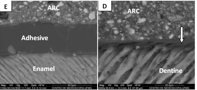
RelyX ARC resin cement samples showed an interaction between the adhesive system and the enamel–dentine hybrid layer (Figure 1). Analysis of RelyX U100 specimens showed gaps in the adhesive
interface between the resin cement and the unetched enamel (Figure 2: U100-E1). here was interaction between the cement and the enamel etched for 15 seconds (Figure 2: U100-E2) and 30 seconds (Figure 2: U100-E3). Analysis of RelyX U200 specimens showed that the unetched enamel lacked a full adhesive interface (Figure 2:U200-E1). he interaction was more efective when the phosphoric acid was applied for 30 seconds compared with 15 seconds (Figure 2: U200-E2 and U200-E3). he unconditioned dentine surface bonded with RelyX U100 resin cement had a supericial interaction (Figure 3: U100-D1). Furthermore, the images of the etched dentine showed that both treatments yielded cement tags in the dentine tubules (Figure 3: U100-D2 and U100-D3). he interface analysis showed a supericial interaction between RelyX U200 resin cement and unetcheddentine (Figure 3: U200-D1). RelyX
Figure 1. RelyX ARC interface ater total-etch (phosphoric acid 37%) and Scotchbond Multipurpose Plus: (E) enamel, (D) dentin. Arrow shows adhesive tag in hybrid layer area.
U200 interacted more with dentine treated with polyacrylic acid (Figure 3: U200-D3) than phosphoric acid (Figure 3: U200-D2).
DISCUSSION
Self-adhesive cements were introduced to simplify the cementation technique without any pre-treatment of the enamel or dentine, reducing chairside, clinical steps, and operator errors. However, it has been questioned whether treatment of the tooth surface would improve the interaction between these cements and dental tissues. Several studies have reported that self-adhesive cements have limited interaction with enamel, as well as low bond strength to dentine1,2,7,9,14,15. In the present study, there were gaps at
the interface between RelyXU100 and unetched enamel, suggesting that the interaction was not efective. In contrast, when the enamel was conditioned with phosphoric acid for 15 seconds or 30 seconds, no gaps were found. his result reinforces previous indings suggesting prior enamel conditioning to improve self-adhesive cement interaction with substrate.
A clinical trial concluded that the self-adhesive resin cement RelyX Unicem can be used for ceramic inlay luting in conjunction with selective enamel etching. In fact, they found that survival rates were even better than those without enamel etching16. A similar
pattern was found in the present analysis of U200 cement, whereby cement interacted more with etched enamel than with unconditioned enamel. herefore, it is likely that, in the case of unconditioned enamel, there is an irregular interaction between the cement and the substrate12. Besides that, cracks between the substrates
in the adhesive interface, observed by Cantoro et al.15 as well as
in the present study, maybe exacerbated by sample dehydration
procedures during SEM preparation, as discussed in a previous study17. hus, the hypothesis suggested could be accepted once
dentine or enamel hybrid layer ultramorphology was afected, depending on substrate treatment.
he resin cement RelyX Unicem has particles of glass ionomer cement, which bonds to dentine when polyacrylic acid modiies the smear layer1. his acid has numerous carbonyl ions that form
hydrogen bonds, which promote substrate wettability. Ater a microtensile test, RelyX Unicem was found to confer greater bond strength ater dentine treatment with polyacrylic acid, suggesting that bonding is enhanced ater substrate conditioning18. Furthermore, the
interaction between self-adhesive cement and dentine is only supericial, showing that it has a reduced capacity to completely dissolve the smear layer and interact with the underlying dentine1,13,
as observed in D1 subgroup in the present study.
he present study showed RelyX U100 tags in etched dentine samples. However, demineralization by phosphoric acid etching for 5 seconds produced a greater degradation of peritubular dentine and the presence of wider and shorter tags. However, polyacrylic acid showed a selective dissolution pattern, preserving peritubular dentine and allowing the formation of longer and narrower tags; this suggests that the cement interacts with the dentine. In contrast, Hikita et al.14 demonstrated that the bond strength of RelyX Unicem
to phosphoric acid-etched dentine was signiicantly lower due to inadequate iniltration of the viscous cement on the thick and compact collagen mesh, which was exposed by the phosphoric acid for 15 seconds.
In the present study, it was not possible to obtain images from whole interface between dentine etched by phosphoric acid and RelyX U200, suggesting that the dentine matrix had degraded
further in this experimental subgroup, leading to disruption of the total interface. Polyacrylic-acid etched dentine generated a full interface, with the modiied smear layer interacting less with U200 than withU100, and without tag formation.
he adhesive potential of RelyX Unicem to enamel and dentine may be due to diferent interfacial microstructures and regional dental tissues1,2,19. In particular, the smear layer and underlying dentine
are regarded as solid structures that probably rapidly counteract the acidity of viscous solutions, thereby limiting the etching ability of acidic monomers in creating an evident hybrid layer19. In this
regard, Al-Assaf et al.20 reported that RelyX Unicem has the lowest
bond strength values of all conventional resin cements, and that it provides no visible hybrid layer with the methodology used. Conversely, another study demonstrated that RelyX U100 confers less nanoleakage than conventional resin cement, and that it produces adhesive interfaces that are better sealed3. Furthermore, dentine
pre-treatment with polyacrylic acid improves the microtensile bond strength of RelyX Unicem18,21, decreasing the surface energy
and enhancing adhesion to dentine22. Selective etching of dentine
with phosphoric acid prior to luting results in the most efective bonding of all self-adhesive resin cements, suggesting that bonding
can be achieved with self-adhesive resin cements without any pre-treatment steps, such as etching, priming, or bonding, which, according to the manufacturers, can compromise bonding ability10.
Although the bonding strength of RelyX Unicem to dentine was lower than that of conventional resin cements it was more reliable less sensitive to variations in handling and aging23.
Bond strength and restoration adaptation to the dental structure using self-etching and self-adhesive dual-curing cements are enhanced if a seating force greater than inger pressure is maintained throughout the initial self-curing period; such a force decreases the porosity of the cement13. According to the manufacturers, low
viscosity is an advantage of RelyX U200 over U100, because it means that less pressure during cementation, and that the product can be in “automix” form. Since that cementing pressure was equal for the cements used, our results suggest that increasing the lowability did not ensure greater interaction of U200 with dentine as tags were not formed.
It was concluded that enamel etched with phosphoric acid and dentine etched with polyacrylic acid improved RelyX U100 and U200 cement interaction. Polyacrylic acid etching was more efective in the interaction between dentine and U200.
REFERENCES
1. De Munck J, Vargas M, Van Landuyt K, Hikita K, Lambrechts P, Van Meerbeek B. Bonding of an auto-adhesive luting material to enamel and dentin. Dent Mater. 2004 Dec;20(10):963-71. PMid:15501325. http://dx.doi.org/10.1016/j.dental.2004.03.002.
2. Abo-Hamar SE, Hiller KA, Jung H, Federlin M, Friedl KH, Schmalz G. Bond strength of a new universal self-adhesive resin luting cement to dentin and enamel. Clin Oral Investig. 2005 Sep;9(3):161-7.; published online Apr 27, 2005. PMid:15856343. http://dx.doi.org/10.1007/ s00784-005-0308-5.
3. Stape TH, Menezes MS, Barreto BC, Naves LZ, Aguiar FH, Quagliatto PS, et al. Influence of chlorhexidine on dentin adhesive interface micromorphology and nanoleakage expression of resin cements. Microsc Res Tech. 2013 Aug;76(8):788-94.; published online Jun 5, 2013. PMid:23737406. http://dx.doi.org/10.1002/jemt.22230.
4. Gerth HU, Dammaschke T, Züchner H, Schäfer E. Chemical analysis and bonding reaction of RelyX Unicem and Bifix composites--a comparative study. Dent Mater. 2006 Oct;22(10):934-41.; published online Dec 20, 2005. PMid:16364427. http://dx.doi.org/10.1016/j. dental.2005.10.004.
5. Viotti RG, Kasaz A, Pena CE, Alexandre RS, Arrais CA, Reis AF. Microtensile bond strength of new self-adhesive luting agents and conventional multistep systems. J Prosthet Dent. 2009 Nov;102(5):306-12. PMid:19853172. http://dx.doi.org/10.1016/S0022-3913(09)60180-3. 6. Monticelli F, Osorio R, Mazzitelli C, Ferrari M, Toledano M. Limited decalcification/diffusion of self-adhesive cements into dentin. J Dent
Res. 2008 Oct;87(10):974-9. PMid:18809754. http://dx.doi.org/10.1177/154405910808701012.
7. Duarte S Jr, Botta AC, Meire M, Sadan A. Microtensile bond strengths and scanning electron microscopic evaluation of self-adhesive and self-etch resin cements to intact and etched enamel. J Prosthet Dent. 2008 Sep;100(3):203-10. PMid:18762032. http://dx.doi.org/10.1016/ S0022-3913(08)60179-1.
8. D’Arcangelo C, De Angelis F, D’Amario M, Zazzeroni S, Ciampoli C, Caputi S. The influence of luting systems on the microtensile bond strength of dentin to indirect resin-based composite and ceramic restorations. Oper Dent. 2009 May-Jun;34(3):328-36. PMid:19544823. http://dx.doi.org/10.2341/08-101.
9. Lin J, Shinya A, Gomi H, Shinya A. Bonding of self-adhesive resin cements to enamel using different surface treatments: bond strength and etching pattern evaluations. Dent Mater J. 2010 Aug;29(4):425-32.; published online Jul 23, 2010. PMid:20668359. http://dx.doi.org/10.4012/ dmj.2009-140.
10. Pisani-Proença J, Erhardt MC, Amaral R, Valandro LF, Bottino MA, Del Castillo-Salmeron R. Influence of different surface conditioning protocols on microtensile bond strength of self-adhesive resin cements to dentin. J Prosthet Dent. 2011 Apr;105(4):227-35. PMid:21458647. http://dx.doi.org/10.1016/S0022-3913(11)60037-1.
11. Aguiar TR, Vermelho PM, Andre CB, Giannini M. Interfacial ultramorphology evaluation of resin luting cements to dentin: a correlative scanning electron microscopy and transmission electron microscopy analysis. Microsc Res Tech. 2013 Dec;76(12):1234-9.; published online Sep 12, 2013. PMid:24030836. http://dx.doi.org/10.1002/jemt.22290.
13. Goracci C, Cury AH, Cantoro A, Papacchini F, Tay FR, Ferrari M. Microtensile bond strength and interfacial properties of self-etching and self-adhesive resin cements used to lute composite onlays under different seating forces. J Adhes Dent. 2006 Oct;8(5):327-35. PMid:17080881. 14. Hikita K, Van Meerbeek B, De Munck J, Ikeda T, Van Landuyt K, Maida T, et al. Bonding effectiveness of adhesive luting agents to enamel
and dentin. Dent Mater. 2007 Jan;23(1):71-80.; published online Jan 19, 2006. PMid:16426673. http://dx.doi.org/10.1016/j.dental.2005.12.002. 15. Cantoro A, Goracci C, Papacchini F, Mazzitelli C, Fadda GM, Ferrari M. Effect of pre-cure temperature on the bonding potential of self-etch
and self-adhesive resin cements. Dent Mater. 2008 May;24(5):577-83. PMid:17659770. http://dx.doi.org/10.1016/j.dental.2007.06.012. 16. Federlin M, Hiller KA, Schmalz G. Effect of selective enamel etching on clinical performance of CAD/CAM partial ceramic crowns luted
with a self-adhesive resin cement. Clin Oral Investig. 2014 Nov;18(8):1975-84. PMid:24407551. http://dx.doi.org/10.1007/s00784-013-1173-2. 17. Pereira CN, Daleprane B, Barbosa PF, Moreira AN, de Magalhaes CS. Qualitative evaluation of scanning electron microscopy methods in a study of the resin cement/dentine adhesive interface. Microsc Microanal. 2014 Feb;20(1):268-75. PMid:24188716. http://dx.doi.org/10.1017/ S143192761301369X.
18. Pavan S, dos Santos PH, Berger S, Bedran-Russo AK. The effect of dentin pretreatment on the microtensile bond strength of self-adhesive resin cements. J Prosthet Dent. 2010 Oct;104(4):258-64. PMid:20875530 .http://dx.doi.org/10.1016/S0022-3913(10)60134-5.
19. Yang B, Ludwig K, Adelung R, Kern M. Micro-tensile bond strength of three luting resins to human regional dentin. Dent Mater. 2006 Jan;22(1):45-56.; published online Jul 22, 2005. PMid:16040114. http://dx.doi.org/10.1016/j.dental.2005.02.009.
20. Al-Assaf K, Chakmakchi M, Palaghias G, Karanika-Kouma A, Eliades G. Interfacial characteristics of adhesive luting resins and composites with dentine. Dent Mater. 2007 Jul;23(7):829-39. PMid:16934865. http://dx.doi.org/10.1016/j.dental.2006.06.023.
21. Broyles AC, Pavan S, Bedran-Russo AK. Effect of dentin surface modification on the microtensile bond strength of self-adhesive resin cements. J Prosthodont. 2013 Jan;22(1):59-62.; published online Jul 4, 2012. PMid:22762448. http://dx.doi.org/10.1111/j.1532-849X.2012.00890.x. 22. Brigagão VC, Barreto LFD, Goncalves KAS, Amaral M, Vitti RP, Neves ACC, et al. Effect of interim cement application on bond strength
between resin cements and dentin: Immediate and delayed dentin sealing. J Prosthet Dent. 2017 Jun;117(6):792-8.; published online Nov 12, 2016. PMid:27847158. http://dx.doi.org/10.1016/j.prosdent.2016.09.015.
23. Holderegger C, Sailer I, Schuhmacher C, Schläpfer R, Hämmerle C, Fischer J. Shear bond strength of resin cements to human dentin. Dent Mater. 2008 Jul;24(7):944-50.; published online Jan 10, 2008. PMid:18190957. http://dx.doi.org/10.1016/j.dental.2007.11.021.
CONFLICTS OF INTERESTS
he authors declare no conlicts of interest.
*CORRESPONDING AUTHOR
Carolina Nemésio de Barros Pereira, Faculdade de Odontologia, UFMG – Universidade Federal de Minas Gerais, Avenida Antônio Carlos, 6627, Pampulha, 31270-901 Belo Horizonte, MG, Brasil, e-mail: [email protected]