Cop
yright
© ABE&M t
odos os dir
eit
os r
eser
vados
.
Cop
yright
© ABE&M t
odos os dir
eit
os r
eser
vados
.
The role of interferon induced
with helicase C domain 1
(IFIH1) in the development of
type 1
diabetes mellitus
O papel do interferon induzido com o domínio C da
helicase 1 (IFIH1) no desenvolvimento do diabetes melito tipo 1
Ana Paula Bouças1,2, Fernanda dos Santos de Oliveira1,
Luis Henrique Canani1,2, Daisy Crispim1,2
ABSTRACT
Type 1 diabetes mellitus (T1DM) is a chronic, progressive, autoimmune disease characterized by me-tabolic decompensation frequently leading to dehydration and ketoacidosis. Viral pathogens seem to play a major role in triggering the autoimmune destruction that leads to the development of T1DM. Among several viral strains investigated so far, enteroviruses have been consistently associated with T1DM in humans. One of the mediators of viral damage is the double-stranded RNA (dsRNA) genera-ted during replication and transcription of viral RNA and DNA. The IFIH1 gene encodes a cytoplasmic receptor of the pattern-recognition receptors (PRRs) family that recognizes dsRNA, playing a role in the innate immune response triggered by viral infection. Binding of dsRNA to this PRR triggers the re-lease of proinlammatory cytokines, such as interferons (IFNs), which exhibit potent antiviral activity, protecting uninfected cells and inducing apoptosis of infected cells. The IFIH1 gene appears to play a major role in the development of some autoimmune diseases, and it is, therefore, a candidate gene for T1DM. Within this context, the objective of the present review was to address the role of IFIH1 in the development of T1DM. Arq Bras Endocrinol Metab. 2013;57(9):667-76
Keywords
Autoimmunity; type 1 diabetes mellitus; viral infection; IFIH1
RESUMO
O diabetes melito tipo 1 (T1DM) é uma doença autoimune crônica e progressiva caracterizada por descompensações metabólicas frequentemente acompanhadas por desidratação e cetoacidose. Os agentes virais parecem ter um papel importante no desencadeamento da destruição autoimune que leva ao desenvolvimento do T1DM. Entre as cepas virais estudadas até agora, a família dos entero-vírus foi consistentemente associada ao surgimento da doença em humanos. Um dos mediadores do dano viral é o RNA ita dupla (RNAfd) gerado durante a replicação e transcrição de RNA e DNA viral. O gene IFIH1 codiica um receptor citoplasmático pertencente à família dos pattern-recognition receptors (PRRs) que reconhece o RNAfd, tendo um papel importante na resposta imune inata de-sencadeada por infecção viral. A ligação do RNAfd a essa PRR desencadeia a liberação de citocinas pró-inlamatórias como interferons (IFNs), os quais exibem uma potente ação antiviral e têm como objetivo proteger as células não infectadas e induzir apoptose naquelas já contaminadas. O gene
IFIH1 parece ter uma participação importante no desenvolvimento de algumas doenças autoimunes. Por isso, esse gene é um candidato ao desenvolvimento do T1DM. Dentro desse contexto, o objetivo da presente revisão foi abordar o papel do IFIH1 no desenvolvimento do T1DM. Arq Bras Endocrinol Metab. 2013;57(9):667-76
Descritores
Autoimunidade; diabetes melito tipo 1; infecção viral; IFIH1
1 Laboratory of Human Pancreatic
Islet Biology, Division of Endocrinology, Hospital de Clínicas de Porto Alegre, Universidade Federal do Rio Grande do Sul (UFRGS), Porto Alegre, RS, Brazil
2 Graduate Program in Medical
Sciences: Endocrinology, UFRGS, Porto Alegre, RS, Brazil
Correspondence to: Daisy Crispim
Rua Ramiro Barcelos, 2350, prédio 12; 4º andar
90035-003 – Porto Alegre, RS, Brazil [email protected]
Cop
yright
© ABE&M t
odos os dir
eit
os r
eser
vados
.
INTRODUCTION
T
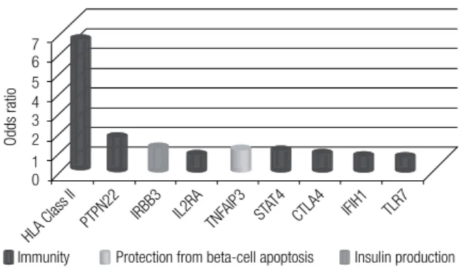
ype 1 diabetes mellitus (T1DM), which accounts for 5%-10% of all cases of diabetes mellitus, is charac terized by severe insulin deiciency secondary to autoimmune destruction of pancreatic beta-cells. Con-sequently, subjects with T1DM are usually dependent on insulin injections for life (1-3). Markers of autoim-mune beta-cell destruction include autoantibodies to insulin, to the islets of Langerhans, to glutamic acid decarbo xylase (GAD65), to beta-cell-speciic zinc transporter ZnT8, and to tyrosine phosphatases IA-2 and IA-2b. One – but usually more – of these anti-bodies are pre sent in 85%-90% of patients when fasting hyperglycemia is initially detected.Inlammation of the islets of Langerhans (insulitis) probably develops within the context of a “dialog” be-tween immune cells and beta-cells. This dialog is me-diated partly by cytokines and chemokines, which are released both by immune cells and by the beta-cells themselves, as well as by other immunogenic signals delivered by dying beta-cells. This may lead to induc-tion and ampliicainduc-tion of the inlammatory process, but in some cases, may lead to the resolution of insulitis, instead (2). The course of beta-cell inlammation and its potential progression to clinical T1DM depends on a complex interaction between a strong genetic com-ponent and a variety of environmental triggers (4,5). Among the various loci associated with T1DM, the hu-man leukocyte antigen(HLA) class II locus on chromo-some 6p21 is, by far, the leading genetic risk factor for T1DM, accounting for 30%-50% of genetic risk for the condition (6). Other genes are associated with relatively minor effects on T1DM risk compared with HLA, such as the insulin gene, the cytotoxic T-lymphocyte asso-ciated protein 4 (CTLA4) gene, the protein tyrosine phosphatase, nonreceptor type 22 (PTPN22) gene, the interleukin 2 receptor – alpha (IL2RA) gene, the interferon induced with helicase C domain 1 (IFIH1) gene, and other genes recently discovered by means of genome-wide association studies (GWAS) (Figure 1) (6,7). Winkler and cols. (8) evaluated how the com-bined allele frequency of 12 T1DM susceptibility genes could stratify T1DM risk in children of parents with T1DM, followed up from birth until the develop-ment of islet autoantibodies and diabetes. The authors showed that non-HLA gene combinations were highly effective in discriminating T1DM, and were most ef-fective in children with a high-risk HLA genotype.
The greatest T1DM discrimination was obtained by the sum of risk alleles of IFIH1, CTLA4, PTPN22, IL-18RAP, SH2B3, KIAA0350, COBL, and ERBB3 genes in HLA-risk children.
The environmental triggers and potentiators of autoimmune beta-cell destruction include viral infec-tions, dietary exposure during childhood (e.g. to cow milk), vaccination, and toxins (9,10). There is sub-stantial evidence that viral pathogens, such as entero-viruses, rubella virus, mumps virus, rotaentero-viruses, par-voviruses, and cytomegalovirus, play a major role in triggering the autoimmune destruction of pancreatic beta-cells (11,12). Among these viral strains, particu-lar attention is given to enteroviruses, which exhibit speciic tropism for the pancreas and have been as-sociated with the development of T1DM in humans (13,14). Epidemiological studies of the seasonality of development of anti-beta-cell antibodies in a group of subjects at increased risk of T1DM showed an in-creased incidence of autoantibodies during winter, which correlated with a period of increased enterovi-ral infection rates (10). Furthermore, coxsackieviruses isolated from patients with T1DM were able to induce diabetes in susceptible mice (15). In a recent study, the Coxsackie B4 virus was identiied in 50% of sam-ples collected from patients with T1DM and was also able to infect human islets in vitro, impairing insulin secretion in response to glucose (16).
Based on the studies presented above, enteroviruses appear to be associated with a fraction of T1DM cas-es. Nevertheless, if enteroviruses play a major role in T1DM pathogenesis, how could we explain the increase in T1DM incidence in countries where exposure to these microorganisms has been dropping, such as
Fin-7 6 5 4 3
Immunity Protection from beta-cell apoptosis Insulin production 2
1 0
Odds ratio
HLA Class II
PTPN22 IL2RA TNF AIP3
CTLA4
STAT4 IFIH1 TLR7 IRBB3
Figure 1. Genes associated with type 1 diabetes mellitus. Odds ratios for
susceptibility alleles of nine genes associated with type 1 diabetes
Cop
yright
© ABE&M t
odos os dir
eit
os r
eser
vados
.
land? Are the data showing that T1DM can be caused by viral infections compatible with the hygiene hypo-thesis (17)? Interestingly, data in NOD mice revealed that coxsackieviruses provoke diabetes only when a preexis ting mass of insulitis has accumulated. When ad-ministered earlier in the life, inoculation is associated with a strong protection against diabetes (18). Taking into account the studies in NOD mice, Coppieters and cols. (18) suggested that the lack of exposure to en-teroviruses in developed countries results in a reduced frequency of subjects with protective immunity caused by early childhood infections. When islet inlammation occurs in these subjects, they would be more suscep-tible to an enteroviral infection that has the potential to initiate autoreactivity and beta-cell damage.
Microbial recognition by the mammalian immune system relies on components of both innate and adap-tive immunity. Innate immunity is the irst line of de-fense against bacteria, fungi, and viruses. Detection of invading microorganisms is carried out by a wide range of cell receptors of the pattern-recognition recep-tors (PRRs) family, which recognize highly conserved pathogen-associated molecular patterns (PAMPs), such as the double-stranded RNA (dsRNA) generated dur-ing viral RNA replication and transcription (19-21). Innate immune system cells, such as macrophages and dendritic cells, kill invading microorganisms by phago-cytosis or induction of cytokine production. Further-more, innate immunity activates the adaptive immune system, consisting of B lymphocytes, which produce speciic antibodies against the invading pathogen, and T lymphocytes, which secrete cytokines that will induce elimination of infected cells by exerting cytotoxic ef-fects or by signaling to B lymphocytes (21).
Some studies have shown that certain PRRs, such as IFIH1, play a role in the development of T1DM in animal models (2,22). The IFIH1 gene, also known as the melanoma differentiation-associated gene-5 (MDA-5), encodes a cytoplasmic receptor that recog-nizes dsRNA and is involved in the innate immune response triggered by viral infection (23). Binding of dsRNA to IFIH1 triggers the release of proinlamma-tory cytokines, particularly interferons (IFNs), by im-mune cells, thus inducing apoptosis of virus-infected cells (19,24). Therefore, IFIH1 is a candidate gene for T1DM susceptibility. Within this context, the objective of the present review was to address the role of IFIH1
in the development of T1DM.
THE ROLE OF PATTERN RECOGNITION RECEPTORS
(PRRS) IN THE RESPONSE TO VIRAL INFECTION
As mentioned above, recognition of pathogens by the innate immune system relies on PRRs, which constitu-te the irst line of defense against microbial infection (25,26). Recent studies have identiied three groups of PRRs: toll-like receptors, retinoic acid-inducible gene I (RIG-I)-like helicases (RLHs), and nucleotide-binding oligomerization domain (NOD)-like receptors (NLRs) (27,28). The RLH class comprises the helicases IFIH1/ MDA-5, retinoic acid-inducible gene 1 (RIG-I), and Laboratory of Genetics and Physiology-2 (LGP2).
During viral infection, dsRNA or single-stranded RNA (ssRNA) are recognized by speciic PRRs pre-sent on infected cells, which undergo conformational changes and activate signaling cascades that ultimately drive the production of several proinlammatory cyto-kines, chemocyto-kines, and type I IFNs (IFN-I) (19,29). IFN-I is a cytokine produced by most cells during viral infection that promotes the expression of several genes involved with antiviral response in target cells, and acts as a modulator of the adaptive immune system by acti-vating dendritic cells, T lymphocytes, and B lympho-cytes (30). This IFN exhibits potent antiviral action, protecting uninfected cells and inducing apoptosis of infected ones, which is partially caused by endoplasmic reticulum stress (27). Interestingly, high levels of IFN-I are found in the pancreas of patients with T1DM, and IFN-a is known to contribute to the development of experimental viral-induced diabetes (2). Furthermore, IFN-I treatment appears to protect against the deve-lopment of T1DM and reduces the incidence of the disease in NOD mice (31).
Cop
yright
© ABE&M t
odos os dir
eit
os r
eser
vados
.
and lymphocytic choriomeningitis virus, were similar in wild-type and TLR3 knockout mice(38). Indeed, more recent studies show that, whereas NF-κB and IRF-3 acti vation by extracellular dsRNA is TLR3-dependent, activation by intracellular dsRNA, a product of viral replication in the cytoplasm, also occurs by means of activation of RIG-1 and IFIH1 (34,35). Activation of NF-κB and IRF-3 triggers production of IFN-a and IFN-b, leading to activation of the Jak/STAT-1 path-way and triggering the expression of MHC class I an-tigens and a variety of chemokines (26,33,35,39). This complex molecular response leads to the attraction of immune cells, which will release more proinlamma-tory cytokines, such as IFN-g, IL-1b, and TNF. Local inlammation and activation of antiviral defenses seek to eradicating infection and triggering apoptosis of in-fected cells. However, in some genetically susceptible individuals, this defense system fails to work properly, inducing excessive, progressive inlammation and pro-longed death of beta-cells, instead, thus predisposing to the development of T1DM (2).
THE INTERFERON INDUCED WITH HELICASE C
DOMAIN 1 (IFIH1) RECEPTOR
The IFIH1 protein belongs to a family of helicases that also comprises two other members: the RIG-I and LGP2 receptors (26). RNA helicases are highly conser-ved enzymes that use energy dericonser-ved from ATP hydroly-sis to bind dsRNA, destabilizing and unwinding it (40). IFIH1 and RIG-I contain two CARD (N-terminal cas-pase activation and recruitment domains) effector do-mains essential to their signaling activity. Furthermore, all three helicases contain a DExD/H-box-type RNA helicase domain, which is also essential to their func-tion. A C-terminal domain was recently identiied as the site of dsRNA binding in all three helicases (26,41,42). The LGP2 helicase does not contain CARD domains and is probably unable to activate downstream signaling pathways. LGP2 also recognizes dsRNA, but appears to act as a negative regulator, interfering with viral RNA recognition by IFIH1 and RIG-I (42). Depending on the type of virus, LGP2 may also act as a positive regu-lator of its other RLH counterparts (21).
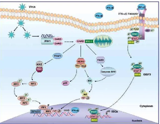
After binding with virus-derived dsRNA, IFIH1 and RIG-I interact, via their CARD domains, with the IPS-1 adaptor molecule (IFN-b promoter stimulator-1, also known as Cardif, MAVS or VISA), which recruits intermediary signaling molecules, such as IKK-a, -b, -e,
and TBK1, ultimately leading to NF-κB and IRF-3 ac-tivation (20,26,41,42). Furthermore, TRAF3, an anti-viral molecule implicated in the production of IFN-I, also plays a role in these intracellular signaling events (37). As described elsewhere, activation of NF-κB and IRF-3 induces expression of IFN-b and several genes regulated by these transcription factors, which will di-rect inlammatory and antiviral responses against infec-tion (Figure 2) (26,42).
IFIH1 and RIG-I share a sequence homology of 25% within the CARD domains and 40% within the helicase domain (41). Experimental data suggest that these two helicases use similar intracellular signaling mechanisms to induce an antiviral response. However, despite their structural similarities, IFIH1 and RIG-I appear to be non-redundant, and are involved in the recognition of different types of dsRNA and ssRNA vi-rus. Whereas RIG-I recognizes the hepatitis C virus, several paramyxoviruses (mumps virus, varicella zoster virus, respiratory syncytial virus, parainluenza virus), vesicular stomatitis virus and inluenza A virus, IFIH1 recognizes Picornaviridae, such as rhinoviruses, echo-viruses, enteroecho-viruses, and encephalomyocarditis virus (23). Furthermore, Iih1 knockout mice (Iih1-/-) fail to produce IFN-a in response to the exposure to synthet-ic dsRNA polyinosinsynthet-ic:polycytidylsynthet-ic acid (PIC), whsynthet-ich demonstrates that IFIH1 is the primary cytoplasmic sensor for long PIC (23). Interestingly, these two heli-cases appear to recognize reoviruses, the West Nile vi-rus, and the dengue virus (26). Experiments involving PIC infection have shown that short dsRNA segments (< 2000 bp) activate RIG-I, whereas long dsRNA segments (> 2000 bp) are best recognized by IFIH1 (43,44). Nevertheless, the mechanisms responsible for distinguishing long dsRNA segments from short ones have yet to be elucidated.
Cop
yright
© ABE&M t
odos os dir
eit
os r
eser
vados
.
Figure 2. Antiviral signaling by IFIH1. Double-stranded RNA (dsRNA) derived from viral replication is detected by the cytoplasmic RNA helicase IFIH1, activating the adaptor protein IPS-1 via CARD domain interactions. IPS-1 then induces intracellular signaling pathways that result in the activation of the
transcriptions factors IRF-3 and NF-κB, leading to the production of IFNa/b by infected cells. IFNa/b is then shown signaling through the IFNa/b receptor
and the Jak-STAT pathway to drive interferon-stimulated genes (ISGs) expression and an innate immune response. See text for further details. Adapted from Wilkins and Gale (26).
5’ 3’
rs1990760 rs3747517
rs35744605
rs35744605
rs35667974
rs35732034 rs13422767
rs2111485
Figure 3. Map of human IFIH1 locus on chromosome 2 (region 2q24.3).
The sixteen exons (boxes) are numbered from left to right according to the transcriptional region. The arrows show the main common polymorphisms
associated with type 1 diabetes mellitus. Figure adapted from Chistiakov
and cols. (42).
Hühn and cols. (47) found that Iih1-/- knockout mice exhibit increased susceptibility to Coxsackie B3 virus infection. Loss of Iih1 enabled faster viral replica-tion, leading to hepatomegaly, pancreatic injury, and high mortality rates in these animals. The authors also found that Iih1 is not required for induction of IFN-I,
Cop
yright
© ABE&M t
odos os dir
eit
os r
eser
vados
.
(CCL2, CCL5 and CXCL10), suggesting that Iih1 is not essential for PIC-induced beta-cell death, but rath-er regulates important inlammatory signaling mecha-nisms in these cells (30).
In mice, increased gene expression of Iih1 leads to a state of chronic IFN-I production, characterized by resistance to lethal viral infections (49). Hultcrantz and cols. (32) showed that, in human pancreatic is-lets, IFN-induced antiviral defenses provide a powerful protective mechanism against replication of coxsacki-eviruses. Treatment with IFN-a is known to increase gene expression of the chemokine CXCL10 in human islets (32). This chemokine, when produced during in-fection, leads to T-cell recruitment to the islet site and appears to play a key role in host defense against islet-tropic viruses in individuals susceptible to T1DM (32).
These studies further strengthen the hypothesis that
IFIH1 plays a key role in the regulation of local islet inlammation during viral infection.
IFIH1 gene polymorphisms and their association with
T1DM
The association between IFIH1 and T1DM was irst re-ported in 2006, by Smyth and cols.(50), who conduc-ted a GWAS in European families affecconduc-ted by T1DM within a large Caucasian population cohort from the United Kingdom, for a total of over 10,000 subjects. Several IFIH1 polymorphisms were associated with T1DM, with the rs1990760 (G/A) polymorphism, which substitutes an alanine to valine in codon 946 of exon 15, being most strongly associated with pro-tection against development of the disease (odds ratio [OR] = 0.86, P = 1.42 x 10⁻¹⁰ for the G allele) (50).
Associations between IFIH1 polymorphisms and T1DM have been replicated in some populations (29,51-55), but not in others (56-58) (Table 1). Jermendy and cols. (52) studied the rs1990760 polymorphism in Hungarians and Finns and conducted a meta-analysis of 5 studies that analyzed this polymorphism in T1DM patients and nondiabetic controls, including their own study. In Hungarians, the A allele of this polymorphism was strongly associated with T1DM (OR = 1.29; P = 0.002). Furthermore, the meta-analysis showed a sig-niicant association between the A allele and risk of de-veloping T1DM (OR = 1.18; P = 5.3 x 10⁻¹⁵).
Qu and cols. (53) assessed three single nucleotide polymorphisms (SNPs) located in the IFIH1 gene or its adjacent intergenic regions (rs1990760, rs3747517,
and rs2111485) in 589 French Canadian nuclear family trios. The rs1990760 and rs3747517 polymorphisms showed a trend toward association with T1DM as re-ported by other studies, but the effect did not reach statistical signiicance, most likely due to weak statistical power (53). Conversely, the A allele of SNP rs2111485 was associated with protection for T1DM (OR = 0.84, P < 0.05). Yang and cols. (55), in a study of Han Chi-nese subjects, also failed to ind an association between the rs1990760 polymorphism and T1DM, but did ind an association between the rs3747517 polymorphism and the condition (P > 0.001).
A GWAS of Caucasian subjects in the United States (Georgia and Denver populations) showed that two SNPs in the coding region of IFIH1 (rs1990760 and rs35744605) and two SNPs in the adjacent 3’ inter-genic region (rs2111485 and rs13422767) were asso-ciated with increased risk of T1DM (OR = 1.7-1.9), but only in the Georgia population, with the lowest P-value obtained for the rs1990760 polymorphism (P = 8 x 10⁻⁸) (51). Interestingly, the G/G genotype of SNP rs1990760 was associated with increased levels of
IFIH1 expression in the peripheral blood mononuclear cells of 374 subjects (187 patients with T1DM and 187 nondiabetic controls) (51). The most common homo-zygous genotypes for the three other polymorphisms of interest were also associated with increased IFIH1
expression, which suggests that increased expression of this gene may be associated with greater susceptibil-ity to T1DM development (51). Nejentsev and cols.
(29) reported that four rare variants of the IFIH1
gene (rs35337543, rs35667974, rs35744605 and rs35732034), as well as the rs1990760 polymorphism, were independently associated with protection against T1DM in a British population (OR = 0.51-0.84; P = 1.3 x 10⁻³ to 2.1 x 10⁻¹⁶).
Cop
yright
© ABE&M t
odos os dir
eit
os r
eser
vados
.
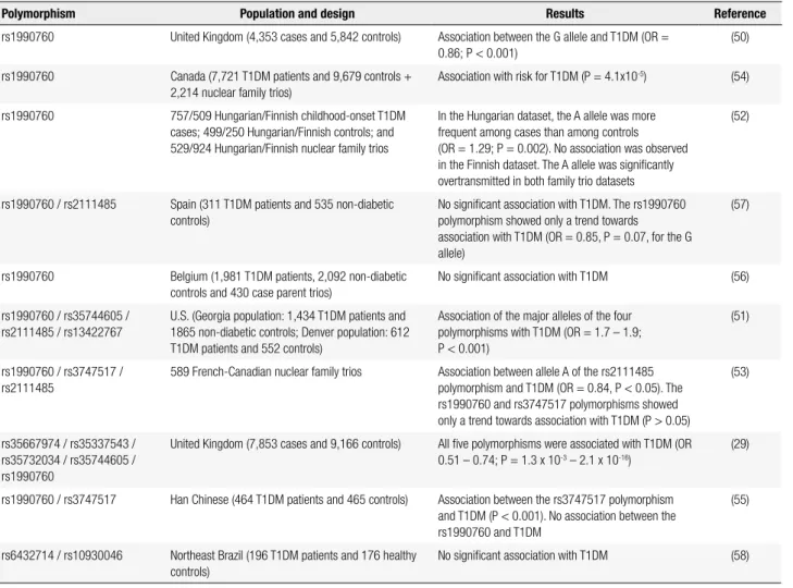
Table 1. Studies of polymorphisms in the IFIH1 gene and type 1 diabetes mellitus (T1DM)
Polymorphism Population and design Results Reference
rs1990760 United Kingdom (4,353 cases and 5,842 controls) Association between the G allele and T1DM (OR = 0.86; P < 0.001)
(50)
rs1990760 Canada (7,721 T1DM patients and 9,679 controls + 2,214 nuclear family trios)
Association with risk for T1DM (P = 4.1x10⁻⁵) (54)
rs1990760 757/509 Hungarian/Finnish childhood-onset T1DM cases; 499/250 Hungarian/Finnish controls; and 529/924 Hungarian/Finnish nuclear family trios
In the Hungarian dataset, the A allele was more frequent among cases than among controls (OR = 1.29; P = 0.002). No association was observed in the Finnish dataset. The A allele was signiicantly overtransmitted in both family trio datasets
(52)
rs1990760 / rs2111485 Spain (311 T1DM patients and 535 non-diabetic controls)
No signiicant association with T1DM. The rs1990760 polymorphism showed only a trend towards association with T1DM (OR = 0.85, P = 0.07, for the G allele)
(57)
rs1990760 Belgium (1,981 T1DM patients, 2,092 non-diabetic controls and 430 case parent trios)
No signiicant association with T1DM (56)
rs1990760 / rs35744605 / rs2111485 / rs13422767
U.S. (Georgia population: 1,434 T1DM patients and 1865 non-diabetic controls; Denver population: 612 T1DM patients and 552 controls)
Association of the major alleles of the four polymorphisms with T1DM (OR = 1.7 – 1.9; P < 0.001)
(51)
rs1990760 / rs3747517 / rs2111485
589 French-Canadian nuclear family trios Association between allele A of the rs2111485 polymorphism and T1DM (OR = 0.84, P < 0.05). The rs1990760 and rs3747517 polymorphisms showed only a trend towards association with T1DM (P > 0.05)
(53)
rs35667974 / rs35337543 / rs35732034 / rs35744605 / rs1990760
United Kingdom (7,853 cases and 9,166 controls) All ive polymorphisms were associated with T1DM (OR 0.51 – 0.74; P = 1.3 x 10⁻³ – 2.1 x 10⁻¹⁶)
(29)
rs1990760 / rs3747517 Han Chinese (464 T1DM patients and 465 controls) Association between the rs3747517 polymorphism and T1DM (P < 0.001). No association between the rs1990760 and T1DM
(55)
rs6432714 / rs10930046 Northeast Brazil (196 T1DM patients and 176 healthy controls)
No signiicant association with T1DM (58)
polymorphism may affect the sequence of the HNF-3b transcription factor binding site in the IFIH1 gene, whereas the rs3747517 polymorphism may alter the binding site for the AP-1 transcription factor. As both polymorphisms are located 45-50kb from the start co-don, it has yet to be determined whether these variants really play a role in the regulation of IFIH1 expression or are merely in linkage disequilibrium with a function-al polymorphism in this region (42,51).
The rs13422767 and rs2111485 polymorphisms are located within the 3’ intergenic region adjacent to the IFIH1 gene, 23kb and 13kb from the end of the 3’-UTR of the gene respectively. These polymorphisms do not change any known transcription factor bind-ing sites, and it is not known whether they contrib-ute to the regulation of IFIH1 gene expression (51). Rare va riants, however, are predicted to have signiicant biological effects on the IFIH1 gene, whether by re-sulting in a truncated protein product by generating a stop codon in exon 10 (rs35744605), affecting splicing
positions (rs35337543 and rs35732034, position +1 of introns 8 and 14 respectively), or changing a highly conserved amino acid (rs35667974 in exon 14) (29).
Cop
yright
© ABE&M t
odos os dir
eit
os r
eser
vados
.
In short, the above-cited studies suggest that more than one IFIH1 polymorphism contributes to T1DM susceptibility in several populations. Additional re-search is required to ascertain the biological effects of these polymorphisms.
Associations between IFIH1 and other autoimmune conditions
Autoimmune diseases are distinct clinical syndromes characterized by changes in normal immune responsi-veness due to loss of tolerance to one or more host constituents (61). Furthermore, it is widely known that genetic factors play a substantial role in the pathoge-nesis of autoimmune conditions. Accordingly, studies of several loci related to the immune system, such as the IFIH1 gene, have been conducted in an attempt to provide better understanding of the pathogenesis of these diseases (62).
Interestingly, some studies have shown strong as-sociations between the rs1990760 polymorphism of the IFIH1 gene and other autoimmune diseases, such as psoriasis (63,64), chronic periodontitis (63), poly-myositis (65), multiple sclerosis (57,66), systemic lupus erythematosus (67), and Graves’ disease (68).
These associations between IFIH1 and autoim-mune conditions unrelated to viral infection, such as Graves’ disease, suggest that IFIH1 may also play an endogenous immunoregulatory role unrelated to its function as a viral receptor (68). Additional studies are needed to identify the role of IFIH1 in the pathogen-esis of these conditions.
CONCLUSIONS
The IFIH1 gene plays a major role in the innate immu-ne response triggered by viral infection. Binding of viral replication-derived dsRNA to IFIH1 triggers the rele-ase of proinlammatory cytokines by immune system cells. This local inlammation and activation of antiviral defenses aims at eradicating infection and trigger apop-tosis of virus-infected cells. However, in some geneti-cally susceptible individuals, this defense system fails to work properly, inducing excessive, progressive inlam-mation and prolonged death of beta-cells instead and, thus, predisposing them to the development of T1DM. Hence, IFIH1 is a good candidate gene for T1DM. Indeed, several studies conducted in different popula-tions suggest that more than one IFIH1 polymorphism
is associated with T1DM. The rs1990760 polymor-phism has also been associated with other autoimmune conditions, such as Graves’ disease and systemic lupus erythematosus. Additional studies are required to elu-cidate the molecular mechanisms underlying the asso-ciation between these polymorphisms and T1DM and other autoimmune diseases. Knowledge of the factors associated with T1DM development will enable a kee-ner understanding of its pathogenesis and may provide more effective approaches for the treatment and pre-vention of T1DM.
Acknowledgements: this study was supported by grants from the Conselho Nacional de Desenvolvimento Cientíico e Tecnológi-co (CNPq), Fundação de Amparo à Pesquisa do Estado do Rio Grande do Sul (Fapergs), Coordenação de Aperfeiçoamento de Pessoal de Nível Superior (Capes) and Fundo de Incentivo à Pes-quisa e Eventos (Fipe) at Hospital de Clínicas de Porto Alegre. The authors would like to thank Dr. Máikel L. Colli, Dr. Cristia-ne B. Leitão and Dr. Ticiana C. Rodrigues for the suggestions made to improve the quality of this manuscript.
Disclosure: no potential conlict of interest relevant to this article was reported.
REFERENCES
1. American Diabetes Association. Diagnosis and classiication of diabetes mellitus. Diabetes Care. 2013;36(Suppl 1):S67-74. 2. Eizirik DL, Colli ML, Ortis F. The role of inlammation in insulitis
and beta-cell loss in type 1 diabetes. Nat Rev Endocrinol. 2009;5(4):219-26.
3. Pirot P, Cardozo AK, Eizirik DL. Mediators and mechanisms of pancreatic beta-cell death in type 1 diabetes. Arq Bras Endocrinol Metabol. 2008;52:156-65.
4. Jahromi MM, Eisenbarth GS. Cellular and molecular pathogenesis of type 1A diabetes. Cell Mol Life Sci. 2007;64(7-8):865-72. 5. Kim MS, Polychronakos C. Immunogenetics of type 1 diabetes.
Horm Res. 2005;64(4):180-8.
6. Steck AK, Rewers MJ. Genetics of type 1 diabetes. Clin Chem. 2011;57(2):176-85.
7. Todd JA, Walker NM, Cooper JD, Smyth DJ, Downes K, Plagnol V, et al. Robust associations of four new chromosome regions from genome-wide analyses of type 1 diabetes. Nat Genet. 2007;39(7):857-64.
8. Winkler C, Krumsiek J, Lempainen J, Achenbach P, Grallert H, Giannopoulou E, et al. A strategy for combining minor genetic susceptibility genes to improve prediction of disease in type 1 diabetes. Genes Immun. 2012;13(7):549-55.
9. Pearl-Yafe M, Kaminitz A, Yolcu ES, Yaniv I, Stein J, Askenasy N. Pancreatic islets under attack: cellular and molecular effectors. Curr Pharm Des. 2007;13(7):749-60.
10. Knip M, Veijola R, Virtanen SM, Hyöty H, Vaarala O, Akerblom HK. Environmental triggers and determinants of type 1 diabetes. Diabetes. 2005;54(Suppl 2):S125-36.
Cop
yright
© ABE&M t
odos os dir
eit
os r
eser
vados
.
12. Hober D, Sauter P. Pathogenesis of type 1 diabetes mellitus: interplay between enterovirus and host. Nat Rev Endocrinol. 2010;6(5):279-89.
13. Jaidane H, Hober D. Role of coxsackievirus B4 in the pathogenesis of type 1 diabetes. Diabetes Metab. 2008;34(6 Pt 1):537-48. 14. Tauriainen S, Oikarinen S, Oikarinen M, Hyoty H. Enteroviruses
in the pathogenesis of type 1 diabetes. Semin Immunopathol. 2011;33(1):45-55.
15. van der Werf N, Kroese FG, Rozing J, Hillebrands JL. Viral infections as potential triggers of type 1 diabetes. Diabetes Metab Res Rev. 2007;23(3):169-83.
16. Dotta F, Censini S, van Halteren AG, Marselli L, Masini M, Dionisi S, et al. Coxsackie B4 virus infection of beta cells and natural killer cell insulitis in recent-onset type 1 diabetic patients. Proc Natl Acad Sci U S A. 2007;104(12):5115-20.
17. Bach JF, Chatenoud L. The hygiene hypothesis: an explanation for the increased frequency of insulin-dependent diabetes. Cold Spring Harb Perspect Med. 2012;2(2):a007799.
18. Coppieters KT, Boettler T, von Herrath M. Virus infections in type 1 diabetes. Cold Spring Harb Perspect Med. 2012;2(1):a007682. 19. Randall RE, Goodbourn S. Interferons and viruses: an interplay
between induction, signalling, antiviral responses and virus countermeasures. J Gen Virol. 2008;89(Pt 1):1-47.
20. Meylan E, Tschopp J, Karin M. Intracellular pattern recognition receptors in the host response. Nature. 2006;442(7098):39-44. 21. Kumar H, Kawai T, Akira S. Toll-like receptors and innate immunity.
Biochem Biophys Res Commun. 2009;388(4):621-5.
22. Zipris D. Innate immunity in type 1 diabetes. Diabetes Metab Res Rev. 2011;27(8):824-9.
23. Kato H, Takeuchi O, Sato S, Yoneyama M, Yamamoto M, Matsui K, et al. Differential roles of MDA5 and RIG-I helicases in the recognition of RNA viruses. Nature. 2006;441(7089):101-5. 24. Rotondi M, Chiovato L, Romagnani S, Serio M, Romagnani P. Role
of chemokines in endocrine autoimmune diseases. Endocr Rev. 2007;28(5):492-520.
25. Pichlmair A, Reis e Sousa C. Innate recognition of viruses. Immunity. 2007;27(3):370-83.
26. Wilkins C, Gale M Jr. Recognition of viruses by cytoplasmic sensors. Curr Opin Immunol. 2010;22(1):41-7.
27. Marinou I, Montgomery DS, Dickson MC, Binks MH, Moore DJ, Bax DE, et al. The interferon induced with helicase domain 1 A946T polymorphism is not associated with rheumatoid arthritis. Arthritis Res Ther. 2007;9(2):R40.
28. Wang Q, Miller DJ, Bowman ER, Nagarkar DR, Schneider D, Zhao Y, et al. MDA5 and TLR3 initiate pro-inlammatory signaling pathways leading to rhinovirus-induced airways inlammation and hyperresponsiveness. PLoS Pathog. 2011;7(5):e1002070. 29. Nejentsev S, Walker N, Riches D, Egholm M, Todd JA. Rare
variants of IFIH1, a gene implicated in antiviral responses, protect against type 1 diabetes. Science. 2009;324(5925):387-9.
30. Colli ML, Moore F, Gurzov EN, Ortis F, Eizirik DL. MDA5 and PTPN2, two candidate genes for type 1 diabetes, modify pancreatic beta-cell responses to the viral by-product double-stranded RNA. Hum Mol Genet. 2010;19(1):135-46.
31. Swiecki M, McCartney SA, Wang Y, Colonna M. TLR7/9 versus TLR3/MDA5 signaling during virus infections and diabetes. J Leukoc Biol. 2011;90(4):691-701.
32. Hultcrantz M, Huhn MH, Wolf M, Olsson A, Jacobson S, Williams BR, et al. Interferons induce an antiviral state in human pancreatic islet cells. Virology. 2007;367(1):92-101.
33. Ylipaasto P, Kutlu B, Rasilainen S, Rasschaert J, Salmela K, Teerijoki H, et al. Global proiling of coxsackievirus- and cytokine-induced gene expression in human pancreatic islets. Diabetologia. 2005;48(8):1510-22.
34. Dogusan Z, Garcia M, Flamez D, Alexopoulou L, Goldman M, Gy-semans C, et al. Double-stranded RNA induces pancreatic beta--cell apoptosis by activation of the toll-like receptor 3 and inter-feron regulatory factor 3 pathways. Diabetes. 2008;57(5):1236-45. 35. Rasschaert J, Ladriere L, Urbain M, Dogusan Z, Katabua B, Sato
S, et al. Toll-like receptor 3 and STAT-1 contribute to double-stranded RNA+ interferon-gamma-induced apoptosis in primary pancreatic beta-cells. J Biol Chem. 2005;280(40):33984-91. 36. Liu D, Cardozo AK, Darville MI, Eizirik DL. Double-stranded RNA
cooperates with interferon-gamma and IL-1 beta to induce both chemokine expression and nuclear factor-kappa B-dependent apoptosis in pancreatic beta-cells: potential mechanisms for viral-induced insulitis and beta-cell death in type 1 diabetes mellitus. Endocrinology. 2002;143(4):1225-34.
37. Meylan E, Tschopp J. Toll-like receptors and RNA helicases: two parallel ways to trigger antiviral responses. Mol Cell. 2006;22(5):561-9.
38. Edelmann KH, Richardson-Burns S, Alexopoulou L, Tyler KL, Flavell RA, Oldstone MB. Does Toll-like receptor 3 play a biological role in virus infections? Virology. 2004;322(2):231-8.
39. Rasschaert J, Liu D, Kutlu B, Cardozo AK, Kruhøffer M, ØRntoft TF, et al. Global proiling of double stranded RNA- and IFN-gamma-induced genes in rat pancreatic beta cells. Diabetologia. 2003;46(12):1641-57.
40. Luking A, Stahl U, Schmidt U. The protein family of RNA helicases. Crit Rev Biochem Mol Biol. 1998;33(4):259-96.
41. Barral PM, Sarkar D, Su ZZ, Barber GN, DeSalle R, Racaniello VR, et al. Functions of the cytoplasmic RNA sensors RIG-I and MDA-5: key regulators of innate immunity. Pharmacol Ther. 2009;124(2):219-34.
42. Chistiakov DA. Interferon induced with helicase C domain 1 (IFIH1) and virus-induced autoimmunity: a review. Viral Immunol. 2010;23(1):3-15.
43. Takahasi K, Yoneyama M, Nishihori T, Hirai R, Kumeta H, Narita R, et al. Nonself RNA-sensing mechanism of RIG-I helicase and acti-vation of antiviral immune responses. Mol Cell. 2008;29(4):428-40. 44. Kato H, Takeuchi O, Mikamo-Satoh E, Hirai R, Kawai T, Matsushi-ta K, et al. Length-dependent recognition of double-stranded ri-bonucleic acids by retinoic acid-inducible gene-I and melanoma differentiation-associated gene 5. J Exp Med. 2008;205(7):1601-10. 45. Huang F, Adelman J, Jiang H, Goldstein NI, Fisher PB.
Differentiation induction subtraction hybridization (DISH): a strategy for cloning genes displaying differential expression during growth arrest and terminal differentiation. Gene. 1999;236(1):125-31.
46. Kang DC, Gopalkrishnan RV, Lin L, Randolph A, Valerie K, Pestka S, et al. Expression analysis and genomic characterization of human melanoma differentiation associated gene-5, MDA-5: a novel type I interferon-responsive apoptosis-inducing gene. Oncogene. 2004;23(9):1789-800.
47. Hühn MH, McCartney SA, Lind K, Svedin E, Colonna M, Flodström-Tullberg M. Melanoma differentiation-associated protein-5 (MDA-5) limits early viral replication but is not essential for the induction of type 1 interferons after Coxsackievirus infection. Virology. 2010;401:42-8.
48. McCartney SA, Vermi W, Lonardi S, Rossini C, Otero K, Calderon B, et al. RNA sensor-induced type I IFN prevents diabetes caused by a beta cell-tropic virus in mice. J Clin Invest. 2011;121(4):1497-507. 49. Crampton SP, Deane JA, Feigenbaum L, Bolland S. Iih1 gene
dose effect reveals MDA5-mediated chronic type I IFN gene signature, viral resistance, and accelerated autoimmunity. J Immunol. 2012;188(3):1451-9.
Cop
yright
© ABE&M t
odos os dir
eit
os r
eser
vados
.
identiies a type 1 diabetes locus in the interferon-induced helicase (IFIH1) region. Nat Genet. 2006;38(6):617-9.
51. Liu S, Wang H, Jin Y, Podolsky R, Reddy MV, Pedersen J, et al. IFIH1 polymorphisms are signiicantly associated with type 1 diabetes and IFIH1 gene expression in peripheral blood mononuclear cells. Hum Mol Genet. 2009;18(2):358-65.
52. Jermendy A, Szatmari I, Laine AP, Lukacs K, Horvath KH, Korner A, et al. The interferon-induced helicase IFIH1 Ala946Thr polymorphism is associated with type 1 diabetes in both the high-incidence Finnish and the medium-incidence Hungarian populations. Diabetologia. 2010;53(1):98-102.
53. Qu HQ, Marchand L, Grabs R, Polychronakos C. The association between the IFIH1 locus and type 1 diabetes. Diabetologia. 2008;51(3):473-5.
54. Concannon P, Onengut-Gumuscu S, Todd JA, Smyth DJ, Pociot F, Bergholdt R, et al. A human type 1 diabetes susceptibility locus maps to chromosome 21q22.3. Diabetes. 2008;57(10):2858-61. 55. Yang H, Wang Z, Xu K, Gu R, Chen H, Yu D, et al. IFIH1 gene
polymorphisms in type 1 diabetes: genetic association analysis and genotype-phenotype correlation in Chinese Han population. Autoimmunity. 2012;45(3):226-32.
56. Aminkeng F, Van Autreve JE, Weets I, Quartier E, Van Schravendijk C, Gorus FK, et al. IFIH1 gene polymorphisms in type 1 diabetes: genetic association analysis and genotype-phenotype correlation in the Belgian population. Hum Immunol. 2009;70(9):706-10. 57. Martinez A, Santiago JL, Cenit MC, de Las Heras V, de la Calle H,
Fernandez-Arquero M, et al. IFIH1-GCA-KCNH7 locus: inluence on multiple sclerosis risk. Eur J Hum Genet. 2008;16(7):861-4. 58. Moura R, Araujo J, Guimarães R, Crovella S, Brandão L. Interferon
induced with helicase C domain 1 (IFIH1): trends on helicase domain and type 1 diabetes onset. Genes Immun. 2013;516(1):66-8. 59. Shigemoto T, Kageyama M, Hirai R, Zheng J, Yoneyama M, Fujita
T. Identiication of loss of function mutations in human genes
encoding RIG-I and MDA5: implications for resistance to type I diabetes. J Biol Chem. 2009;284(20):13348-54.
60. Bonifacio E, Warncke K, Winkler C, Wallner M, Ziegler AG. Cesa-rean section and interferon-induced helicase gene polymorphis-ms combine to increase childhood type 1 diabetes risk. Diabetes. 2011;60(12):3300-6.
61. Lee SJ, Kavanaugh A. 4. Autoimmunity, vasculitis, and autoanti-bodies. J Allergy Clin Immunol. 2006;117(2 Suppl):S445-50. 62. Pearce SH, Merriman TR. Genetic progress towards the molecular
basis of autoimmunity. Trends Mol Med. 2006;12(2):90-8. 63. Chen G, Zhou D, Zhang Z, Kan M, Zhang D, Hu X, et al. Genetic
variants in IFIH1 play opposite roles in the pathogenesis of psoriasis and chronic periodontitis. Int J Immunogenet. 2012;39(2):137-43.
64. Li Y, Liao W, Cargill M, Chang M, Matsunami N, Feng BJ, et al. Carriers of rare missense variants in IFIH1 are protected from psoriasis. J Invest Dermatol. 2010;130(12):2768-72.
65. Gono T, Kawaguchi Y, Sugiura T, Furuya T, Kawamoto M, Hanaoka M, et al. Interferon-induced helicase (IFIH1) polymorphism with systemic lupus erythematosus and dermatomyositis/ polymyositis. Mod Rheumatol. 2010;20(5):466-70.
66. Enevold C, Oturai AB, Sorensen PS, Ryder LP, Koch-Henriksen N, Bendtzen K. Multiple sclerosis and polymorphisms of innate pattern recognition receptors TLR1-10, NOD1-2, DDX58, and IFIH1. J Neuroimmunol. 2009;212(1-2):125-31.
67. Gateva V, Sandling JK, Hom G, Taylor KE, Chung SA, Sun X, et al. A large-scale replication study identiies TNIP1, PRDM1, JAZF1, UHRF1BP1 and IL10 as risk loci for systemic lupus erythematosus. Nat Genet. 2009;41(11):1228-33.