Cop
yright
© ABE&M t
odos os dir
eit
os r
eser
vados
.
The unusual association of Graves’
disease, chronic spontaneous
urticaria, and premature ovarian
failure: report of a case and HLA
haplotype characterization
A associação incomum entre doença de Graves, urticária crônica idiopática e insuiciência ovariana prematura: relato de caso clínico e tipagem HLA
Rosaria Maddalena Ruggeri1, Giuseppe Vita2, Anna Grazia D’Angelo3, Paolina Quattrocchi3, Rosaria Certo1, Salvatore Benvenga1,4,
Salvatore Cannavò1, Sebastiano Gangemi3,5
SUMMARY
Chronic spontaneous urticaria (CSU), deined as the occurrence of spontaneous wheals for more than six weeks, has been associated with autoimmune diseases. Herein, we report the unusual as-sociation of CSU, Graves’ disease, and premature ovarian failure. Human leukocyte antigen (HLA) studies were performed. A 36-year-old woman presented symptoms and signs of hyperthyroidism for three months. In the same period, the patient complained of widespread urticarial wheals, intensely itchy, and poorly responsive to therapy with antihistaminic agents. Hyperthyroidism was conirmed biochemically, and treatment with methimazole was started. As hyperthyroidism improved, a marked improvement in her urticaria was also observed. However, the patient continued to complain of ame-norrhea. Endocrine evaluation, at the age 38, was consistent with premature ovarian failure. This is the irst report of coexistence of GD, CSU, and POF. The genetic background of such unusual associa-tion is a speciic combinaassocia-tion of HLA. Arq Bras Endocrinol Metab. 2013;57(9):748-52
SUMÁRIO
A urticária crônica idiopática, caracterizada pelo aparecimento de pápulas espontâneas e persisten-tes por pelo menos seis semanas, tem sido associada a doenças autoimunes. Apresentamos aqui o caso da associação incomum entre urticária crônica idiopática, doença de Graves e falência ovariana prematura. Foram conduzidos estudos de tipagem HLA. Uma mulher de 36 anos apresentou sinais e sintomas de hipertireoidismo por três meses. No mesmo período, a paciente queixou-se do apareci-mento de pápulas urticariformes generalizadas que coçavam intensamente e não eram responsivas ao tratamento com anti-histamínicos. O hipertireoidismo foi conirmado bioquimicamente, e o trata-mento com metimazol foi iniciado. Assim que os valores hormonais se normalizaram, observou-se uma melhoria signiicativa do quadro de urticária. No entanto, a paciente continuou a apresentar amenorreia. A avaliação endocrinológica, com a idade de 38 anos, mostrou falência ovariana pre-matura. Este é o primeiro caso de associação entre doença de Graves, urticária idiopática crônica e falência ovariana prematura. A base genética dessa associação incomum é representada por combi-nações especíicas de haplótipos HLA. Arq Bras Endocrinol Metab. 2013;57(9):748-52
1 Unit of Endocrinology,
Department of Clinical and Experimental Medicine, University of Messina, Italy
2 Unit of Tissue Typing,
Department of Pathology and Experimental Microbiology, University of Messina, Italy
3 School and Unit of Allergy and
Clinical Immunology, Department of Clinical and Experimental Medicine, University of Messina, Italy
4 Interdepartmental Program
of Molecular and Clinical Endocrinology & Women’s Health; A.O.U. Policlinico “G. Martino”, Messina Italy
5 Institute of Biomedicine and
Molecular Immunology-National Research Council, Palermo, Italy
Correspondence to: Rosaria Maddalena Ruggeri Dipartimento di Medicina Clinica e Sperimentale, Unità di Endocrinologia
Padiglione H, 4 piano – AOU Policlinico “G. Martino” via Consolare Valeria, 1 98125 – Messina, Italy [email protected]
Received on July/11/2013 Accepted on Aug/7/2013
INTRODUCTION
C
hronic urticaria is a common condition character-ized by itchy wheals and lare-type skin reactions (hives) lasting more than six weeks (1). The term idio-pathic is used when the causes remain unknown, whichCop
yright
© ABE&M t
odos os dir
eit
os r
eser
vados
.
At any given time, 0.5%-1.0% of the population is suf-fering from CSU (1). Nevertheless, its etiopathogenesis is not well understood yet (1,2). At least in a subset of patients, CSU has an autoimmune basis, as demonstrat-ed by the presence of antibodies against the α-subunit of the high-afinity IgE receptor (FcεRIα), or against IgE itself in the serum of 30%-40% of such patients (1-3). The association of CSU with thyroid autoimmu-nity further supports its autoimmune origin (4). CSU patients frequently suffer from Hashimoto’s thyroiditis and increased serum levels of anti-thyroid antibod-ies were reported in 10% to 33% CSU patients (4,5). Coexisting Graves’ disease (GD) has been reported less frequently than HT, with an estimated preva lence ranging from 1% to 4% (6). Moreover, CSU may also be associated with autoimmune conditions other than thyroid diseases (7). Herein, we report the unusual as-sociation of GD, CSU, and premature ovarian failure (POF) of autoimmune origin. Human leukocyte anti-gen (HLA) studies were performed.
PATIENT
A 36-year-old woman referred to our outpatient clinic because of fatigue, palpitations, tremors, nervousness and irritability, insomnia, oligo-amenorrhea, sweating, and weight loss for three months. In the same period, she complained of the repeated occurrence of short-lived cutaneous wheals accompanied by redness and itching. Individual wheals lasted less than 24 hours and were poorly responsive to regular antihistamine ther-apy. GD was diagnosed based on clinical symptoms/ signs, TSH level of < 0.001 mIU/L (normal values, 0.27-4.2) with elevated free triiodothyronine (FT3, 17.39 pg/mL, normal values, 2-4.4) and free thyrox-ine (FT4, 38.3 pmol/L, normal values, 12-22), and positivity of anti-TSH-receptor antibodies (TRAb, 19 IU/L, normal values, < 1.5), as well as of anti-thyroid peroxidase antibodies (TPOAb, 178 U/L; normal values, < 35). Ultrasound (US) examination showed a diffuse enlargement of the thyroid, associated with hypoechogenicity and increased vascularity. The 131I
thyroid scan revealed an enlarged gland with diffuse increased uptake of radio-iodine at 6 and 24 hours. Therapy with methimazole (MMI, 30 mg/day) was started, with progressive attenuation of symptoms and signs of thyrotoxicosis and normalization of FT3 and FT4 values. As thyroid function improved, a marked
Cop
yright
© ABE&M t
odos os dir
eit
os r
eser
vados
.
Of note, HLA-B44 and HLA DR3-DQ2-DQ7 confer high predisposing risk of CSU (9) and GD (10-12), re-spectively, while the HLA B8/DR3(DR52) and HLA DRB1*11/DQB1*03 haplotype is reported as predis-posing for autoimmune POF (13,14).
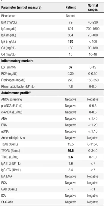
Table 1. Serum biochemical data of our patient at the time of the second
episode of urticaria*
Parameter (unit of measure) Patient Normal ranges
Blood count Normal
IgM (mg/dL) 79 40-230
IgG (mg/dL) 804 700-1600
IgA (mg/dL) 364 70-400
IgE (mg/dL) 170 < 100
C3 (mg/dL) 130 90-180
C4 (mg/dL) 15 10-40
Inlammatory markers
ESR (mm/h) 37 0-15
RCP (mg/dL) 0.30 0-0.50
Fibrinogen (mg/dL) 270 150-350
Rheumatoid factor (IU/mL) 7.8 0-8.0
Autoimmune proile§
ANCA screening Negative Negative
p-ANCA (EU/mL) Negative 0-0.5
c-ANCA (EU/mL) Negative 0-0.5
ANA Negative < 1:40
ENA Negative < 1.20
nDNA Negative < 1:10
Anticardiolipin Abs Negative Negative
TgAb (IU/mL) 15.5 0-115.0
TPOAb (IU/mL) 39.5 0-34.0
TRAB (IU/mL) 2.6 0-1.0
IgA tTG (IU/mL) 1.6 < 7
IgG tTG (IU/mL) 3.4 < 7
IgA EMA Negative Negative
PCA Negative Negative
GAD (IU/mL) < 1 < 1
ICA Negative Negative
St-C-Abs Negative Negative
* Boldface values indicate abnormality. § p- and c-ANCA: perinuclear and classic anti-neutrophil cytoplasmic antibodies. ANA: anti-nuclear antibodies; ENA: anti-extractable nuclear antigens antibodies; n-DNA: anti-native DNA. TgAb: thyroglobulin antibodies. TPOAb: thyroid peroxidase antibodies. TRAb: TSH receptor antibodies tTG: anti-tissue transglutaminase antibodies. EMA: anti-endomysial antibodies. PCA: anti- gastric parietal cell antibodies. GAD: anti-glutamic acid decarboxylase antibodies. ICA: anti-islet cell antibodies. St-C-Abs: anti-steroid-cell antibodies. Anticardiolipin Abs, ANA, ENA, n-DNA, tTG, PCA, St-C-Abs were assayed by indirect immunoluorescence (IIF). p- and c-ANCA were assayed by both IIF and ELISA techniques. TgAb, TPOAb, TRAb and GAD were measured by immunoradiometric assay. EMA were assayed by ELISA.
Table 2. Thyroid function tests and thyroid autoantibodies of the patient at the time of the second episode of urticaria, and six months after that*
Parameter# (unit
of measure) At admittance 6 months later Normal ranges
TSH (mU/L) 0.35 < 0.005 0.27-4.2
FT3 (pg/mL) 3.66 32.52 2.0-4.4
FT4 (pmol/L) 16.96 7.21 12-22
TgAb (IU/mL) 15.5 145.5 0-115.0
TPOAb (IU/mL) 39.5 235.5 0-34.0
TRAB (IU/L) 2.6 10.10 0-1.0
* Boldface values indicate abnormality.
# FT4: free thyroxine; FT3: free triiodothyronine; TSH: thyroid stimulating hormone. Serum FT4,
FT3 and TSH concentrations were measured by immunoenzymatic methods (commercial kits by Medical Systems [Genoa, Italy]). Thyroglobulin antibodies (TgAb) and thyroid peroxidase antibodies (TPOAb) were measured by the corresponding immunoradiometric assay kit by DiaSorin (Saluggia, Italy). TSH receptor antibodies (TRAb) were measured by using a second-generation radioreceptor assay (Dynotest TRAK Human; B.R.A.M.S., Henningsdorf, Germany).
DISCUSSION
dis-Cop
yright
© ABE&M t
odos os dir
eit
os r
eser
vados
.
Table 3. Genomic and serological human leukocyte antigens (HLA) haplotype of our patient§
HLA loci
HLA-A HLA-B HLA-Cw HLA-DRB HLA-DQ
Genomic *24:02/*26:01 *08:01/*44:02 *07:01/*12:03 B1*03:01/*11:01 B3*03:06/ 03:06
A1*05:01/*05:01 B1*02:01/*03:01
Serological 24(9)/26(10) 8/44(12) 7/- a 17(3)/11(5)
52
2/7(3)
§ We studied HLA expression on lymphocytes of our patient by both a serological and a molecular approach, employing the micro-lymphocytotoxicity test (Biotest AG, Dreieich, Germany) and the
PCR–SSOreverse assay (HLA Kits by Innogenetics N.V., Gent, Belgium), respectively, according to the manufacturer’s instructions. The nomenclature of HLA alleles was based on Marsh SG, Albert ED, Bodmer WF, et al. Nomenclature for factors of the HLA system, 2010. Tissue Antigens 2010;75:291-455 (www.ebi.ac.uk/imgt/hla/).
a The minus sign indicates “not expressed”.
equilibrium with DQB1*0201 and DQA1*0501, both of which are strongly associated with Graves’ disease (10-12). Finally, the DRB1*11/ DQB1*0301 geno-type is associated with aPOF (13,14). Thus, the HLA haplotype of our patient (see Table 3) is consistent with those frequently found in these autoimmune disorders and confers genetic susceptibility to all three diseases.
Besides the genetic background, we believe that there is a pathogenic link between the three diseases. From the lesson learned with other autoimmune disea-ses, a reaction against a common antigen shared by the involved tissue, autoantibodies targeting similar epitopes or a cross-reaction between autoantibodies may be hypothesized. For instance, Altrichter and cols. have recently demonstrated that a sizeable subgroup of CSU patients exhibits IgE antibodies against thyroid peroxidase. These IgE-anti-TPO autoantibodies, when bound to the IgE receptor on the surface of mast cells, could cause mast cell activation and degranulation, thus playing an active role in the pathogenesis of CSU (17). Moreover, increased serum levels of IgE have been re-ported in one third of GD patients, and serum levels of TRAb are signiicantly related with total IgE, sug-gesting that a close cross-talk between the allergic and immune reactions exist in GD (18,19).
Another important inding of our report is the clinical evidence of improvement of CSU when thyroid function was restored. In our case, the irst manifesta-tions of CSU appeared while the patient was hyperthy-roid, before starting MMI, and were poorly responsive to antihistamine therapy alone. During MMI therapy, CSU quickly improved as thyroid function normal-ized, and become responsive to standard antihistamine treatment. Few years later, during a period of increased stress, the patients experienced again urticarial symp-toms, and within six months of the onset of urticaria, hyperthyroidism recurred. Once again, urticaria
im-proved signiicantly as thyroid function recovered with methimazole. A similar improvement in skin manifesta-tions of CSU after therapeutic correction of hyperthy-roidism has been reported by other authors (20-22). Unfortunately, these are anecdotal reports regarding only one or a few number of patients, so they do not enable us to establish the real role of anti-thyroid drugs on the course of urticaria. Obviously, the ameliora-tion of urticarial symptoms observed may be a result of the reduction in sweating, itching, and heat intoler-ance experienced by the patient when hyperthyroidism was corrected. However, anti-thyroid drugs not only decrease the synthesis (and serum levels) of thyroid hormones, but they may also exert immunosuppressive effects, that contribute to GD remission (23). It is also conceivable that they may inluence the course of CSU of autoimmune origin. In our patient, improvement of urticaria was associated with a decrease of serum levels of TPO-Ab and TRAb that persisted negative during remission. When CSU recurred, a rise in TPOAb and TRAb titers was noted.
In conclusion, the main indings of this report are the coexistence of GD, CSU, and POF, which have not been previously reported; the detection of a haplotype associated with susceptibility to all three diseases; and the clinical evidence of improvement of CSU when thy-roid function was restored.
Disclosure: no potential conlict of interest relevant to this article was reported.
REFERENCES
1. Zuberbier T, Asero R, Bindslev-Jensen C, Church Mk, Giménez-Arnau A, Grattan CE, et al. EAACI/GA(2)LEN/EDF/WAO guideline: deinition, classiication and diagnosis of urticaria. Allergy. 2009;64:1417-26.
Cop
yright
© ABE&M t
odos os dir
eit
os r
eser
vados
.
spontaneous urticaria. A GA²LEN task force report. Allergy. 2011;66:317-30.
3. Sabroe R, Poon E, Orchard G, Lane D, Francis DM, Barr RM, et al. Cutaneous inlammatory cell iniltrate in chronic idiopathic urticaria: comparison of patients with and without anti-FcεRI or anti-IgE antibodies. J Allergy Clin Immunol. 1999;103:484-93. 4. Bagnasco M, Minciullo PL, Saraceno GS, Gangemi S, Benvenga S.
Urticaria and thyroid autoimmunity. Thyroid. 2011;21(4):401-10. 5. Gangemi S, Saitta S, Lombardo G, Patai M, Benvenga S. Serum
thyroid autoantibodies in patients with idiopathic either acute or chronic urticaria. J Endocrinol Inves. 2009;32(2):107-10.
6. Ruggeri RM, Imbesi S, Saitta S, Campennì A, Cannavò S, Trimarchi F, et al. Chronic idiopathic urticaria and Graves’ disease. J Endorinol Invest. 2013;36(7):531-6.
7. Conino-Cohen R, Chodick G, Shalev V, Leshno M, Kimhi O, Goldberg A. Chronic urticaria and autoimmunity: associations found in a large population study. J Allergy Clin Immunol. 2012;129 (5):1307-13.
8. Sabroe RA, Grattan CE, Francis DM, Barr RM, Kobza-Black A, Greaves MW. The autologous serum skin test: a screening test for autoantibodies in chronic idiopathic urticaria. Br J Dermatol. 1999;140 (3):446-52.
9. Bozek A, Krajewska J, Filipowska B, Polanska J, Rachowska R, Granzka A, et al. HLA status in patients with chronic spontaneous urticaria. Int Arch Allergy Immunol. 2010;153(4):419-23.
10. Barlow ABT, Wheatcroft N, Watson P, Weetman AP. Association of HLA-DQA*0501 with Graves’ disease in English Caucasian men and women. Clin Endocrinol. 1996;44 (1):73-7.
11. Zamani M, Spaepen M, Bex M, Bouillon R, Cassiman JJ. Primary role of the HLA class II DRB1*0301 allele in Graves’ disease. Am J Med Genet. 2000;95(5):432-7.
12. Ramos Lopez E, Fernandez-Balsells M, Kahles H, Seidl C, Ferrer J, Badenhoop K. HLA-DQ haplotypes in Spanish and German families with Graves’ disease: contribution to DQA1*0501-DQB1*0301 mediated genetic susceptibility from fathers. Thyroid. 2007;17(11):1131-5.
13. Arif S, Underhill JA, Donaldson P, Conway GS, Peakman M. Hu-man leukocyte antigen-DQB1* genotypes encoding aspartate at
position 57 are associated with 3b-hydroxysteroid dehydrogena-se autoimmunity in premature ovarian failure. J Clin Endocrinol Metab. 1999;84(3):1056-60.
14. Ferraù F, Gangemi S, Vita G, Trimarchi F, Cannavò S. Pregnancy after azathioprine therapy for ulcerative colitis in a woman with autoimmune premature ovarian failure and Addison’s disease: HLA haplotype characterization. Fertil Steril. 2011;95(7):2430.e15-7. 15. Betterle C, Presotto F. Autoimmune polyendocrine syndromes (APS) or multiple autoimmune syndrome (MAS). In: Walker S, Jara LJ, editors. Handbook of systemic autoimmune diseases. Endocrine manifestations of systemic autoimmune diseases. Amsterdam: Elsevier; 2008, p. 135-48.
16. Thorsby E, Lie BA. HLA associated genetic predisposition to autoimmune diseases: genes involved and possible mechanisms. Transpl Immunol. 2005;14(3-4):175-82.
17. Altrichter S, Peter HJ, Pisarevskaja D, Metz M, Martus P, Maurer M. IgE mediated autoallergy against thyroid peroxidase--a novel pathomechanism of chronic spontaneous urticaria? PLoS One. 2011;6(4):e14794.
18. Komiya I, Yamada T, Sato A, Kouki T, Nishimori T, Takasu N. Remission and recurrence of hyperthyroid Graves’ disease during and after thiamazole treatment when assessed by IgE and interleukin 13. J Clin Endocrinol Metab. 2001;86(8):3540-4. 19. Yamada T, Komiya I, Miyahara Y, Komatsu M, Shima I, Inazawa T,
et al. Effect of methimazole treatment for 2 years on circulating IL-4, IgE, TBII, and TSAb in patients with hyperthyroid Graves’ disease. Endocr J. 2006;53(6):783-8.
20. Henderson CA, Highet AS. Urticaria associated with thyrotoxico-sis. Clin Exp Dermatol. 1995;20(2):173-4.
21. Gaig P, García-Ortega P, Enrique E, Richart C. Successful treatment of chronic idiopathic urticaria associated with thyroid autoimmunity. J Investig Allergol Clin Immunol. 2000;10(6):342-5. 22. Bansal AS, Hayman GR. Graves’ disease associated with chronic
idiopathic urticaria: 2 case reports. J Investig Allergol Clin Immunol. 2009;19(1):54-6.