Cop
yright
© ABE&M t
odos os dir
eit
os r
eser
vados
.
A rare case of Cushing syndrome
by cyclic ectopic-ACTH
Um caso raro de síndrome de Cushing associada a ACTH-ectópico cíclico
Mariana Farage1, Mario Alberto da Dantas Loures Costa2, Amélio Fernando Godoy-Matos1
SUMMARY
ACTH-dependent Cushing syndrome (CS) due to ectopic ACTH production is most times di-ficult to manage. The identiication of the source of ACTH may take many years. Surgery or chemotherapy for the primary tumor is not always possible. Control of Cushing symptoms is many times achieved using medication, or bilateral adrenalectomy in refractory cases. This case presents a Brazilian male who showed severe hypertension, mood changes, muscle weak-ness, darkening of skin, and increased abdominal fat. An investigation for Cushing syndrome was carried out and, after a four-year follow-up, a carotid glomus tumor (chemodectoma) was conirmed, a rare ectopic ACTH-producing tumor. Besides, the patient presented cyclic Cushing syndrome that was exacerbated by diverticulitis episodes. This case presents interesting pitfalls on diagnosis and management of ACTH-dependent CS. This is the only report of a chemodecto-ma that produced ACTH in the literature. Arq Bras Endocrinol Metab. 2012;56(5):324-30
SUMÁRIO
A síndrome de Cushing ACTH-dependente causada por produção ectópica de ACTH é, muitas vezes, difícil de diagnosticar e conduzir. A identiicação da fonte produtora de ACTH pode demo-rar muitos anos. A cirurgia ou quimioterapia para o tumor primário nem sempre é possível, sen-do o controle sen-do hipercortisolismo alcançasen-do com uso de fármacos ou adrenalectomia bilateral, nos casos refratários. Este caso apresenta um homem com hipertensão grave, mudança de humor, fraqueza proximal, escurecimento da pele e aumento de gordura abdominal. A investi-gação para síndrome de Cushing foi feita e, após quatro anos de acompanhamento, conirmou--se um tumor de glomus carotídeo (quemodectoma), causa rara de tumor secretor de ACTH. Nesse período, o paciente apresentou síndrome de Cushing cíclica, exacerbada por crises de diverticulite. O caso ilustra pontos importantes no diagnóstico, no acompanhamento e na con-dução da síndrome de Cushing ACTH-dependente, sendo este o único caso de tumor de glomus de carótida produzindo ACTH descrito na literatura médica. Arq Bras Endocrinol Metab. 2012;56(5):324-30 1 Serviço de Metabologia,
Instituto Estadual de Diabetes e Endocrinologia (IEDE/RJ), Rio de Janeiro, RJ, Brazil 2 Oncoclínica, Rio de Janeiro, RJ, Brazil
Correspondence to:
Amélio Fernando Godoy-Matos Rua Moncorvo Filho, 90
20211-340 – Rio de Janeiro, RJ, Brazil [email protected]
Received on Aug/5/2011 Accepted on Feb/15/2012
INTRODUCTION
N
umerous types of endocrine and non-endocrine tumors acquire the ability to secrete substances that are not usually produced by the normal tissue from which the tumor is originated (1). Various solid tumors, mainly of neuroendocrine origin, are as well recognized as ACTH-secreting ones, causing ectopic-ACTH secre-tion (EAS) (2). Either benign or malignant tumors may be the cause of EAS. Malignant tumors, nevertheless, have been associated with extremely high circulating ACTH and cortisol levels, and short duration ofsymp-toms of Cushing syndrome (CS) besides atypical clinical phenotype, when compared with pituitary-dependent Cushing (1). Identiication of the source of ACTH can be challenging, as sometimes the primary lesion is not identiied even after prolonged and repeated follow-up (3). When the patient has CS characteristics, with tests indicating an ectopic source and the primary tumors are not identiied, it is called occult EAS (1).
Cop
yright
© ABE&M t
odos os dir
eit
os r
eser
vados
.
medullary thyroid carcinomas. Other miscellaneous tu-mors associated with EAS are paragangliomas, neuro-blastomas, as well as prostate, breast, kidney, stomach, ovary, colon, anorectal, and other cancers (1).
Diagnosis of ACTH ectopic secretion involves two basic steps that cannot be omitted to prevent misdiagno-sis: conirmation of hypercortisolism and determination of its etiology (differential diagnosis) (4). After conir-mation of hypercortisolism in ACTH-dependent matter, bilateral sinus sampling (BIPSS) is the best single test to differentiate between a central and a peripheral source of ACTH (2,5). Once EAS is conirmed, identiication of the site of the primary tumor may begin with chest high-resolution computerized tomography (CT) scan. If results are normal, extensive abdominal CT should be carried out (4). In the case of negative scans, somatosta-tin receptor scintigraphy (SRS) or luorine-18-luorode-oxyglucose positron emission tomography scan ([18-F]-FDG-PET) may be used. The rationale for SRS is the presence of large number of high-afinity somatostatin receptors in many neuroendocrine tumors. The poten-tial advantage in relation to conventional radiology is that it gives information about the whole body, enabling
primary and metastatic lesions to be visualized (4). Fluo-rine-18-luorodeoxyglucose PET is widely used to iden-tify malignant (high metabolism) tumors. In small series, [18-F]-FDG-PET failed to identify tumors that were occult on CT or magnetic resonance imaging. It seems to confer no beneit in the detection of ectopic ACTH-secreting tumors beyond conventional imaging (6).
CASE REPORT
A 49-year-old Brazilian male presented with severe hy-pertension, mood changes mainly characterized by insta-bility, alternating between aggressiveness and depression. Progressive muscle weakness, darkening of skin, and in-creased abdominal fat were observed. His symptoms and laboratory tests conirmed an ACTH-dependent Cush-ing syndrome (CS) (Table 1, Figure 1). Chest X-ray and computed tomography scan (CT), as well as pituitary CT and magnetic resonance imaging (MRI) were normal. Abdominal CT revealed bilateral adrenal enlargement. Bilateral simultaneous inferior petrosal sinus sampling af-ter desmopressin (DDAVP) stimulus (BIPSS) suggested ectopic ACTH production (Table 2).
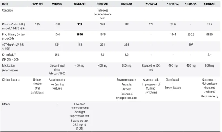
Table 1. Laboratory results and corresponding clinical manifestations during the irst four years of disease
Date 06/11/91 2/10/92 01/04/93 03/05/93 28/02/94 25/04/94 10/12/94 18/01/95 18/04/95
Condition High-dose
dexamethasone test Plasma Cortisol (8h)
mcg/dL* (NR 5 -25)
125 13.8 303 370 184 177 23.9 - 41.7
Free Urinary Cortisol (mcg) 24h
10.4 1540 1546 - - 1444 230.6 9860
ACTH (pg/mL)# (NR
< 160)
124 113 238 238 - - 397
K+ mEq/L##
(NR 3,5 – 5,3)
5.0 3.5 3.5 - - - 2.4
Medication (ketoconazole)
Discontinued since February/1992
400 mg 400 mg 600 mg Reduced to 200 mg
400 mg 400 mg 800 mg
Clinical features Urinary infection Oral candidiasis
Assyntomaptic No Cushing
features
Severe myopathy Anorexia
Anxiety Cutaneous hyperpigmentation
Asymptomatic Improvement of
Cushing’ symptoms
Ciproloxacin + Metronidazole
Garamicyn + Metronidazole (inpatient treatment) Hemicolectomy
Others - Low dose dexamethasone
overnight suppression test
Plasma cortisol 28,5 ng/mL
(5-25)
Cop
yright
© ABE&M t
odos os dir
eit
os r
eser
vados
.
Table 2. Patient’s bilateral simultaneous inferior petrosal sinus sampling (BIPSS) after DDAVP stimulation
ACTH measurement* after DDAVP stimulus during bilateral simultaneous inferior petrosal sinus sampling (BIPSS)
Right Left
Central 675 pg/mL 665 pg/mL
Peripheral Jugular Vein
870 pg/mL 880 pg/mL
Subclavic vein 985 pg/mL 795 pg/mL
* Immunoradiometric (IRMA) assay.
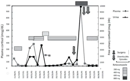
Figure 1. Plasma and free-urinary cortisol variations in the irst three years of CS onset (before identiication of primary tumor site identiication).
Interestingly, this patient presented a quite variable level of serum cortisol and free urinary cortisol that were followed by different symptoms of hypercortisolism. Several times, episodes of diarrhea, fever and abdomi-nal pain preceded worsening of hypercortisolism symp-toms. During these episodes, an astonis hing increase in serum and urinary cortisol were noticed (such as 9,850 mcg free urinary cortisol/24h, as shown in Table 1). In contrast, antibiotics plus ketoconazole therapy was al-ways accompanied by a sharp decrease in cortisol levels, amelioration of Cushing stigmata, even with disappear-ance of hyperpigmentation. Diverticulitis was suspect-ed, and later on, diagnosed. In general, ketoconazole in a daily doses between 200 and 600 mg controlled hypercortisolemia until normalization. Ketoconazole had to be discontinued many times due to symptoms of adrenal insuficiency.
During four years, this patient was followed up and kept showing periodic hypercortisolism. Then
keto-conazole was reintroduced and periods of quite normal cortisol status followed. MRI or CT scans were per-formed every 6 months and did not succeed in locating the source of ACTH. Colonoscopies were done twice. Few intestinal polyps were found and removed. No ma-lignancy was demonstrated, and immunohistochemis-try showed no evidence of ACTH production. Finally, hemicolectomy for diverticular disease was performed, and no tumor was found.
Cop
yright
© ABE&M t
odos os dir
eit
os r
eser
vados
.
after the initial diagnosis) the therapy protocol barely helped to control symptoms. Bilateral adrenalectomy was programmed.
In the end, patient developed bilateral pneumonia complicated by sepsis. In this setting, extremely high cortisol levels resulted in severe hypertension, hypoka-lemia, hypomagnesemia, hypocalcemia, and hypergly-cemia. Ketoconazole therapy was attempted to control CS symptoms but, at this time, it resulted in severe liver toxicity (ALS and ALT increased 100 times above normal range). After improvement of the septic state, videoendoscopic bilateral adrenalectomy was success-fully carried out. During anesthesia procedures tracheal invasion by the tumor was documented (Figure 3).
The patient died from a massive hemorrhagic episo-de due to carotid artery istulae to the trachea.
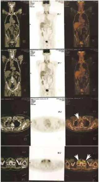
Figure 2. Fluorodeoxyglucose positron emission tomography scan (FDG-PET CT) demonstrating tumor recurrence (18).
Figure 3. Bronchoscope image showing invasion of the posterior wall of the trachea by the tumor (arrow; kindly provided by Dr. Fernando Pacheco).
DISCUSSION
This case presents an ACTH-dependent CS with a cyclic pattern, most of the time related with intestinal infection episodes. This pattern kept the medical team’s attention on the intestinal tract, which emphasizes the dificulties that may be associated with the investigation and management of ectopic ACTH-producing tumors. Besides, as far as we are concerned, a chemodectoma producing ACTH in a cyclic fashion has never been described before in the literature.
Cyclic CS is a rare disorder, characterized by repe-ated episodes of cortisol excess interspersed by periods of normal cortisol secretion (7). The so-called cycles of hypercortisolism can occur regular or irregularly with intercyclic phases ranging from days to years. To for-mally diagnose cyclic CS, three peaks and two troughs of cortisol production should be demonstrated (7). In a review of cyclic CS cases in the literature published in English from 1960 to 2007, 65 cases were found (7). Cushing’s disease (CD) was the underlying cause in 54%, ectopic secretion of ACTH in 26%, and prima-ry adrenal CS in 11% of these cases of cyclic Cushing. Considering CS in general, the corresponding preva-lence, reported in the literature is 68%, 12%, and 20% respectively (8). Thus, the occurrence of ectopic ACTH syndrome seems to be more frequent in patients with cyclic CS (7).
Cop
yright
© ABE&M t
odos os dir
eit
os r
eser
vados
.
carotid body tumors are rare. They may be single, bila-teral or multicentric. In a review of 88 familial and 835 non-familial cases, bilateral disease was more frequent in the familial (31.8%) than in the non-familial (4.4%) cases (9).
Chemodectomas are usually benign and slow-gro-wing tumors of the parasympathetic ganglia with an incidence of roughly 1:30,000 – 1:100,000 cases in the overall population. Risk factors for HNPGLs include conditions associated with chronic hypoxia, such as li-ving at a high altitudes, respiratory or heart diseases with chronic arterial hypoxemia, which were not the case with this patient. However, in 7%-10% to 50% of the cases, genetic predisposition has been suspected ba-sed on positive family history and/or development of bilateral or multiple primary tumors (10-12); more re-cently, the proportion of tumors caused by an inherited predisposition has been identiied as close to 35% (13). Hereditary susceptibility to HNPGLs was recogni-zed at least two decades ago. It enabled the identii-cation, using linkage analysis, of three loci on chro-mosome 11 and 1, named PGL1 on 11q23, PGL2 on 11q11.3, and PGL3 on 1q21-23 (14). We could not attribute a hereditary background to this patient, but a mutation at the SDH loci is possible, considering the malignant characteristic and bilateralism. SDH is an enzyme complex composed by four subunits en-coded by four nuclear genes (SDHA, SDHB, SDHC and SDHD), located on chromosomes 1, 5 and 11. Heterozygous mutations of the last three subunits have been associated with a genetic predisposition to HNPGLs and adrenal/extra-adrenal pheochro-mocytomas (PHEOs) (15,16). Therefore, an autos-somic dominant mutation on succinyl dehydrogenase gene (SDH) was suspected to be responsible for both the bilateral and malignant characteristic of our patient tumor. However, genetic testing for this mutation was not available at that time.
The association between chemodectoma and phe-ochromocytoma is well recognized (17). In fact it is recommended that all patients with chemodectomas should be screened for PHEOs. Urinary catecholamine in the present case was always negative, as well as serum cromogranin A.
Ectopic production of ACTH by chemodectomas is a rare event. As far as we know, there is only one case reported in literature associating a neck paraganglioma with CS (18). Another patient with CS has been repor-ted, in whom resection of a chemodectoma following
bilateral adrenalectomy resulted in decreased ACTH levels. Nonetheless, there was no evidence of cyclic ACTH production (19).
Identiication of the primary tumor responsible for the ectopic ACTH production may be troublesome. CT, PET-FDG, and octreoscan have equal, but low sensitivities for this purpose. They must complement each other in dificult cases. A variety of factors may inluence the ability of FDG-PET to locate a tumor. Increased metabolic rate and glucose transport through tumor cell membranes are necessary for increased up-take of FDG (20). Classically, tumors from the carotid body are PGLs, predominantly with chief cells delimi-ted by trabeculae, and present low mitotic index. Theo-retically, PET-FDG is not a good method for the scree-ning and follow-up of this kind of tumor. Our patient, on the other hand, had a very aggressive and persistent tumor that, despite long term evolution, was invasive and very avid for FDG.
TREATMENT AND MANAGEMENT
The choice of treatment for ectopic ACTH syndrome depends on tumor identiication, location, and classii-cation. The most effective treatment option is surgical resection and cure, although this is not always possi-ble (e.g. in metastatic disease or in the case of occult tumors). Tumor-directed therapy involves an indivi-dualized approach, and can include somatostatin ana-logues, systemic chemotherapy, interferon-α, chemo-embolization, radiofrequency ablation, and radiation therapy (21-24). In the present case, surgery for tumor resection was attempted three times, but the tumor was locally invasive since diagnosis. Radiotherapy was also attempted, with poor response and hypoparathyroi-dism as a consequence. Chemotherapy with both anti--neoplastic drugs and, later on, somatostatin analogues (ostreotide-LAR) was carried out during follow-up.
Cop
yright
© ABE&M t
odos os dir
eit
os r
eser
vados
.
Most studies with steroidogenesis inhibitors have been carried out with metyrapone and ketoconazole (27-31). Metyrapone treatment leads to marked inhi-bition of aldosterone biosynthesis, and accumulation of aldosterone precursors with weak mineralocorticoid ac-tivity. Blood pressure levels and electrolyte balance vary individually with the degree of aldosterone inhibition and 11-deoxycorticosterone (DOC) stimulation. Ad-verse effects due to increased DOC levels (hypokalemia, edema, hypertension) are infrequent (31). Metyrapone is not commercially available in Brazil. Ketoconazole is usually the irst choice. Widely available, and generally well tolerated, it was our choice to control Cushing symptoms since the beginning of the case. Mild eleva-tion in liver enzymes (up to three-fold normal levels), which are transient, is not a contraindication to medical therapy with ketoconazole. Liver function should be carefully monitored because of the rare complication of liver failure (32). In our case, for several years, this drug was well tolerated and actually controlled hypercorti-solemia. However, on the very last days, liver enzymes rose 100 times above the normal range. It is possible that drug interaction contributed to this outcome.
Mitotane (o,p’-DDD) may prove to be highly effec-tive in the long-term suppression of hypercortisolism in the majority of patients with ACTH-dependent CS, because of its speciic adrenolytic action. However, the onset of its action is slow (weeks or months), and the adverse effects associated with mitotane therapy (main-ly digestive and neurological) require careful monito-ring of drug levels. This kind of therapy is routinely used in only a few centers (33), and is more useful for adrenocarcinoma treatment.
Intravenous etomidate therapy may be considered in situations where rapid control of cortisol levels is re-quired and oral therapy is not advisable (34). It can be a bridge to deinitive treatment, such as surgery for primary tumor, bilateral adrenalectomy, or until the ac-tion of a slow-onset adrenolytic agent or steroidogene-sis inhibitor starts.
Neuroendocrine tumors that cause EAS often ex-press functional somatostatin (SS) receptors (35). Smaller studies and case reports have been published on the use of octreotide in patients with EAS. Interestin-gly, octreotide was eficacious in lowering cortisol levels in a signiicant number of these patients, as opposed to studies performed in patients with CS (36,37). This in-ding can be justiied by the fact that many patients with EAS have positive lesions on 111In-pentereotide scan
(Octreoscan), whereas most patients with CS do not (38). The observation that many of the EAS-producing neuroendocrine tumors have functional sst2 receptors, despite chronic hypercortisolism, could be explained by aberrant glucocorticoid receptor signaling in these tu-mor cells. One of the main concerns with the use of SS analogs in EAS, however, appears to be the long-term control of hypercortisolism. Although initial responses to octreotide are frequent, these are not always sustai-ned, and unresponsiveness to treatment is common, due to a number of possible mechanisms of tachyphyla-xis (39). A trial with octreotide-LAR was also done in the presented case; nonetheless, Cushing symp-toms were not properly controlled.
Our patient stayed under ketoconazole treatment for many years, with good control of Cushing symp-toms. The only reasonable option, considering tumor treatment failure and late ketoconazole adverse effects, was bilateral adrenalectomy.
CONCLUSION
ACTH-dependent Cushing syndrome due to ectopic ACTH production most of times is dificult to mana-ge. The identiication of the source of ACTH may take many years until inal diagnosis.
We report a case of a cyclic ACTH-dependent Cushing syndrome due to a chemodectoma located in the carotid glomus. A chemodectoma producing ACTH, as far as we know, has never been described before in the literature. Experience acquired with this patient suggests that a practitioner facing an ACTH--dependent Cushing syndrome must invest all availa-ble resources in shortening ACTH production site lo-cation. Several distinct locations, and association with others neuroendocrine tumors warrants such a stre-nuous effort. Moreover, therapy directed to the ACTH source seems obvious, but not always possible or cura-tive. Meanwhile, ketoconazole to control hypercortiso-lism demonstrated to be eficacious and safe for several years, underscoring its appropriateness. Determination of the genetic proile looking for SDH mutations is a new tool, which might be considered for prognostic stratiication.
Acknowledgements: we would like to thank Dr. Fernando Pache-co, who kindly offered the bronchoscope image.
Cop
yright
© ABE&M t
odos os dir
eit
os r
eser
vados
.
REFERENCES
1. Isidori AM, Lenzi A. Ectopic ACTH syndrome. Arq Bras Endocrinol Metabol. 2007;51(8):1217-25.
2. Newell-Price J, Trainer P, Besser M, Grossman A. The diagnosis and differential diagnosis of Cushing’s syndrome and pseudo--Cushing’s states. Endocr Rev. 1998;19(5):647-72.
3. Isidori AM, Kaltsas GA, Grossman AB. Ectopic ACTH syndrome. Front Horm Res. 2006;35:143-56.
4. Vilar L, Freitas Mda C, Faria M, Montenegro R, Casulari LA, Naves L, et al. Pitfalls in the diagnosis of Cushing’s syndrome. Arq Bras Endocrinol Metabol. 2007;51(8):1207-16.
5. Newell-Price J, Bertagna X, Grossman AB, Nieman LK. Cushing’s syndrome. Lancet. 2006;367(9522):1605-17.
6. Pacak K, Ilias I, Chen CC, Carrasquillo JA, Whatley M, Nieman LK. The role of [(18)F]luorodeoxyglucose positron emission tomo-graphy and [(111)In]-diethylenetriaminepentaacetate-D-Phe-pen-tetreotide scintigraphy in the localization of ectopic adrenocor-ticotropin-secreting tumors causing Cushing’s syndrome. J Clin Endocrinol Metab. 2004;89(5):2214-21.
7. Meinardi JR, Wolffenbuttel BH, Dullaart RP. Cyclic Cushing’s syn-drome: a clinical challenge. Eur J Endocrinol. 2007;157;245-54. 8. Orth DN. Cushing’s syndrome. N Engl J Med. 1995;332:791-803. 9. Grufferman S, Gillman MW, Pasternak LR, Peterson CL, Young Jr
WG. Familial carotid body tumors: case report and epidemiologic review. Cancer. 1980;46:2116-22.
10. Sobol SM, Dailey JC. Familial multiple cervical paragangliomas: report of a kindred and review of the literature. Otolaryngol Head Neck Surg. 1990;102:382-90.
11. Drovdlic CM, Myers EN, Peters JA, Baysal BE, Brackmann DE, Slattery WH 3rd, et al. Proportion of heritable paraganglioma cases and associated clinical characteristics. Laryngoscope. 2001;111:1822-7.
12. Amar L, Servais A, Gimenez-Roqueplo AP, Zinzindohoue F, Cha-tellier G, Plouin PF. Year of diagnosis, features at presentation, and risk of recurrence in patients with pheochromocytoma or se-creting paraganglioma. J Clin Endocrinol Metab. 2005;90: 2110-6. 13. Goldstein RE, O’Neill JA Jr, Holcomb GW 3rd, et al. Clinical ex-perience over 48 years with pheochromocytoma. Ann Surg. 1999;229:755-64; discussion 764-756 (1999).
14. Maher ER, Eng C. The pressure rises: update on the genetics of phaeochromocytoma. Hum Mol Genet. 2002;11:2347-54. 15. Niemann S, Muller U. Mutations in SDHC cause autosomal
domi-nant paraganglioma, type 3. Nat Genet. 2000;26:268-70. 16. Astuti D, Latif F, Dallol A, Dahia PL, Douglas F, George E, et al.
Gene mutations in the succinate dehydrogenase subunit SDHB cause susceptibility to familial pheochromocytoma and to fami-lial paraganglioma. AM J Hum Genet. 2001;69:49-54.
17. Becker M, Aron DC. Ectopic ACTH syndrome and CRH-media-ted Cushing’s syndrome. Endocrinol Metab Clin North Am. 1994;23:585-606.
18. Martin E, Rutishauser E, Bach J. [Cushing syndrome in a patient having had a tumor of the carotid paraganglion.]. Ann Endocrinol (Paris). 1952;13:938-42.
19. Boscaro M, Merola G, Sonino N, Menegus AM, Sartori F, Mantero F. Evidence for ectopic ACTH production years after bilateral adre-nalectomy for Cushing’s syndrome: in vivo and in vitro studies. J Endocrinol Invest. 1985;8:417-21.
20. Seregni E, Chiti A, Bombardieri E. Radionuclide imaging of neu-roendocrine tumours: biological basis and diagnostic results. Eur J Nucl Med. 1998;25:639-58.
21. Aniszewski JP, Young WF Jr, Thompson GB, Grant CS, van Heer-den JA. Cushing syndrome due to ectopic adrenocorticotropic hormone secretion. World J Surg. 2001;25:934-40.
22. Isidori AM, Kaltsas GA, Pozza C, Frajese V, Newell-Price J, Reznek RH, et al. The ectopic adrenocorticotropin syndrome: clinical fea-tures, diagnosis, management, and long-term follow-up. J Clin Endocrinol Metab. 2006;91:371-7.
23. Mansi L, Rambaldi PF, Panza N, Esposito D, Esposito V, Pastore V. Diagnosis and radioguided surgery with 111In-pentetreotide in a patient with paraneoplastic Cushing’s syndrome due to a bron-chial carcinoid. Eur J Endocrinol. 1997;137:688-90.
24. von Werder K, Muller OA, Stalla GK. Somatostatin analogs in ec-topic corticotropin production. Metabolism. 1996;45:129-31. 25. Pivonello R, Ferone D, Lamberts SW, Colao A. Cabergoline
plus lanreotide for ectopic Cushing’s syndrome. N Engl J Med. 2005;352;2457-8.
26. Winquist EW, Laskey J, Crump M, Khamsi F, Shepherd FA. Keto-conazole in the management of paraneoplastic Cushing’s syndro-me secondary to ectopic adrenocorticotropin production. J Clin Oncol. 1995;13:157-64.
27. Beardwell CG, Adamson AR, Shalet SM. Prolonged remission in lorid Cushing’s syndrome following metyrapone treatment. Clin Endocrinol (Oxf). 1981;14:485-92.
28. Boscaro M, Sonino N, Rampazzo A, Mantero F. Response of pitui-tary-adrenal axis to corticotrophin releasing hormone in patients with Cushing’s disease before and after ketoconazole treatment. Clin Endocrinol (Oxf). 1987;27:461-7.
29. Burrin JM, Yeo TH, Ashby MJ, Bloom SR. Effect of ketoconazole on adrenocorticotrophic hormone secretion in vitro and in vivo. J Endocrinol. 1986;108:37-41.
30. Sonino N. The use of ketoconazole as an inhibitor of steroid pro-duction. N Engl J Med. 1987;317:812-8.
31. Sonino N, Boscaro M. Medical therapy for Cushing’s disease. En-docrinol Metab Clin North Am. 1999;28:211-22.
32. Biller BMK, Grossman AB, Stewart PM, Melmed S, Bertagna X, Bertherat J, et al. Treatment of adrenocorticotropin-dependent Cushing’s syndrome: a consensus statement. J Clin Endocrinol Metab. 2008;93:2454-62.
33. Luton JP, Mahoudeau JA, Bouchard P, Thieblot P, Hautecouverture M, Simon D, et al. Treatment of Cushing’s disease by O,p’DDD. Survey of 62 cases. N Engl J Med. 1979;300:459-64.
34. Johnson TN, Canada TW. Etomidate use for Cushing’s syndrome caused by an ectopic adrenocorticotropic hormone-producing tu-mor. Ann Pharmacother. 2007;41:350-3.
35. de Bruin C, Feelders RA, Lamberts SW, Holand LJ. Somatosta-tin and dopamine receptors as targets for medical treatment of Cushing’s Syndrome. Rev Endocr Metab Disord. 2009;10:91-102. 36. Bertagna X, Favrod-Coune C, Escourolle H, Beuzeboc P,
Christofo-rov B, Girard F, et al. Suppression of ectopic adrenocorticotropin secretion by the long-acting somatostatin analog octreotide. J Clin Endocrinol Metab. 1989;68:988-91.
37. Vignati F, Loli P. Additive effect of ketoconazole and octreotide in the treatment of severe adrenocorticotropin-dependent hyper-cortisolism. J Clin Endocrinol Metab. 1996;81:2885-90.
38. de Herder WW, Krenning EP, Malchoff CD, Holand LJ, Reubi JC, Kwekkeboom DJ, et al. Somatostatin receptor scintigraphy: its value in tumor localization in patients with Cushing’s syndrome caused by ectopic corticotropin or corticotropin-releasing hormo-ne secretion. Am J Med. 1994;96:305-12.