119
Revista da Sociedade Brasileira de Medicina Tropical 44(1):119-121, jan-fev, 2011
Case Report/Relato de Caso
1. Internal Medicine Residency, Federal University of Minas Gerais, Belo Horizonte, MG. 2. Department of Pathology, Faculty of Medicine, Federal University of Minas Gerais, Belo Horizonte, MG. 3. Department of Internal Medicine, Faculty of Medicine, Federal University of Minas Gerais, Belo Horizonte, MG. 4. Graduate Program in Health Sciences, Department of Internal Medicine: Infectology and Tropical Medicine, Faculty of Medicine, Federal University of Minas Gerais, Belo Horizonte, MG.
Address to: Dra. Luciana Cristina dos Santos Silva. Depto Clínica Médica/
FM/UFMG. Av. Prof. Alfredo Balena 190, sala 246, Santa Eigênia, 30130-100 Belo Horizonte, MG.
Phone: 55 31 3409-9746
e-mail: [email protected] Received in 05/08/2010 Accepted in 26/10/2010
INTRODUCTION
Progressive multifocal leukoencephalopathy as an AIDS-deining
condition in a patient with high CD4
+T-lymphocyte count
Leucoencefalopatia multifocal progressiva como condição deinidora de AIDS em paciente
com contagem alta de linfócitos T CD4
+Roberta Oliveira de Paula e Silva
1, Rafaela Cabral Gonçalves Fabiano
1, Moisés Salgado Pedrosa
2,
José Roberto Lambertucci
3,4and Luciana Cristina dos Santos Silva
3,4ABSTACT
We present the case of a 31-year-old man with acute manifestation of progressive multifocal leukoencephalopathy(PML) as an AIDS-deining disease. he patient presented with a three-day history of neurological disease, brain lesions without mass efect or contrast uptake and a slightly increased protein concentration in cerebrospinal luid. A serological test for HIV was positive and the CD4+ T-cell count was 427/mm3. Histological
examination of the brain tissue revealed abnormalities compatible with PML. he disease progressed despite antiretroviral therapy, and the patient died three months later. PML remains an important cause of morbidity and mortality among HIV-infected patients.
Keywords: Progressive multifocal leukoencephalopathy, AIDS, HIV, CD4+ T-lymphocytes, JC virus.
RESUMO
Apresentamos o caso de um homem de 31 anos com leucoencefalopatia multifocal progressiva (LMP) de manifestação aguda como doença deinidora de AIDS. O paciente apresentou-se com doença neurológica com três dias de evolução, lesões encefálicas sem efeito de massa ou captação de contraste e leve aumento de proteínas no líquor. Sorologia para o HIV foi positiva e a contagem de linfócitos T CD4+ era de 427/mm3. O exame
histológico de tecido cerebral revelou alterações compatíveis com LMP. A doença progrediu a despeito da terapia antirretroviral, e o paciente morreu após três meses. LMP permanece como causa relevante de mortalidade e morbidade em pacientes infectados pelo HIV.
Palavras-chaves: Leucoencefalopatia multifocal progressiva, AIDS, HIV, Linfócitos T CD4+, Vírus JC.
Progressive multifocal leukoencephalopathy (PML) is a demyelinating disease of immunocompromised patients caused by John Cunningham virus ( JCV), which is a human polyomavirus. It has become more frequent due mainly to AIDS1,2. he prevalence
of PML in HIV-infected patients ranges from 3 to 5% and it is an AIDS-deining illness in as many as 57% of patients presenting with PML1,3.
John Cunningham virus predominantly infects oligodendrocytes and astrocytes, resulting in multifocal cell lysis and demyelination, which can develop in all central nervous system regions, but especially in cerebral white mater1,4. he classic presentation begins with focal deicits that vary depending on the location of the lesion, and these worsen with time, relecting the spread of individual lesions. Mono or hemiparesis are the most common symptoms (52%), followed by cognitive (45%) and speech deicits (31%)2. Seizures are less common, and headache and fever are usually absent.
Diagnosis is based on clinical suspicion, image identiication and etiological conirmation by means of cerebrospinal luid (CSF) or brain tissue analysis. PML should be considered as a possible diagnosis in any HIV-infected patient with focal neurological signs4,5. Imaging techniques on the brain provide additional and more speciic diagnostic information. Magnetic resonanceimaging (MRI) has been found to be more sensitive than computed tomography (CT) for detecting the extent of the disease. CT and MRI on the brain usually show multiple foci in subcortical white-matter regions, without mass efect or contrast enhancement. Lesions show low attenuation on CT and are hyperintense on T2-weighted and hypointense on T1-weighted MRI sequences, thus indicating white mater destruction4.
Routine analysis on CSF is usually unhelpful for diagnosing PML. Evaluation of CSF for the presence of JCV by means of the polymerase chain reaction (PCR) has been the least invasive procedure for etiological identiication, and this is the method of choice for PML diagnosis6. When no JCV DNA identiication from CFS is available, or the analysis is inconclusive and clinical suspicion remains, brain biopsy is desirable4. PML is identified from the characteristic tissue histopathology and virological identiication.
The differential diagnosis with PML includes diseases that can cause neurological deicits in HIV-infected patients, such as infectious conditions (toxoplasmic encephalitis, cryptococcal meningoencephalitis, cytomegalovirus infection or brain abscess), neoplasms (primary or metastatic) and vascular brain complications (ischemic events or vascular malformations)5,7.
120
Paula e Silva RO et al - PML as an AIDS-deining condition with high CD4+ T-lymphocyte count
CASE REPORT
he aim of this article is to present the case of a man with a CD4+ T-lymphocyte count of 427/mm3, who presented acute illness due to severe PML as an AIDS-deining condition.
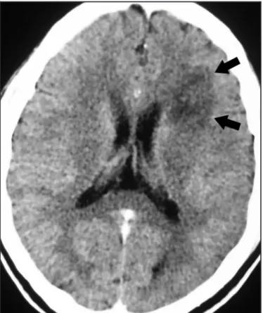
A previously healthy 31-year-old man was admited to the Risoleta Tolentino Neves Hospital in Belo Horizonte, State of Minas Gerais, Brazil, presenting with a three-day history of confusion and alteration of motricity and coordination. Neurological examination showed right-sided hemiparesis and aphasia. Computed tomography showed irregular hypodense lesions without mass efect or contrast enhancement throughout the subcortical white mater of the let frontal lobe and basal ganglia (Figure 1). Initial blood cell counts reveled 4,600 leukocytes/ mm3 (neutrophils 55%, lymphocytes 25%, eosinophils 9% and monocytes 10%). Cerebrospinal luid analysis retrieved ive cells with 64% lymphomonocytes; protein levels of 81mg/dl and glucose assay of 70mg/dl. Cerebrospinal luid was negative for bacterial, fungal, and parasitic microorganisms. A positive test for HIV was obtained, and the CD4+ T-cell count was 427/mm3. Serological tests for cytomegalovirus and human T cell lymphotropic virus (HTLV I-II) were negative, while the patient was negative for IgM and positive for IgG for toxoplasmosis.
Even though the CD4+ T-cell count was higher than 200/mm3, empirical treatment for toxoplasmic encephalitis was started, using sulfadiazine, pyrimethamine and folinic acid. The neurological symptoms progressed, culminating in coma, and empirical treatments for bacterial abscess, herpes, tuberculosis and cryptococcosis were ofered. A new CT showed expansion of the previous lesions, without mass efect or contrast enhancement. he lack of clinical response to the empirical treatment instituted, associated with compatible CT images, suggested PML. MRI was performed in order to beter characterize the lesions. MRI showed difuse irregular hypointense lesions without mass efect or contrast enhancement, throughout the sub-cortical white mater on T1-weighted images (Figure 2). he lesions were hyperintense in T2-weighted sequences. A cerebellar lesion was also noted. Since JCV DNA identification was not
FiGURE 1 - Axial computed tomography of the brain ater contrast administration. An irregular hypodense lesion without mass efect or contrast enhancement in the sub-cortical white mater of let frontal lobe (arrows) is observed.
available, the deinitive diagnosis of PML was achieved consequent to pathological examination of brain tissue that was obtained through biopsy. Histological examination revealed white mater with loss of myelin, vacuolization, presence of foamy macrophages and reactive astrocytes (Figure 3).
Combined antiretroviral therapy with zidovudine, lamivudine and efavirenz was instituted, but no improvement was observed. he patient did not recover from the coma; developed respiratory infections associated with hospital microorganisms; and died three months ater hospitalization.
121
Rev Soc Bras Med Trop 44(1):119-121, jan-fev, 2011
DISCUSSION
FiGURE 3 - Histological examination of brain specimen obtained by stereotatic biopsy of the frontal lobe, stained with hematoxilin and eosin, magniication 400x. White mater shows loss of myelin, vacuolization, presence of foamy macrophages (M) and reactive astrocytes (A), compatible with leukoencephalopathy.
he case reported here described a man who was acutely ill with severe PML, as an AIDS-deining condition, and with a CD4+ T-lymphocyte count higher than 200/mm3. he CD4+ T-lymphocyte count in HIV-infected patients with PML is typically < 100/mm3, and is inversely correlated with the likelihood of development of this opportunistic disease2. he patient described in this case developed PML with 437 CD4+ T-cells/mm3, which favored the possibility of other diagnoses such as an ischemic event, brain abscess or cerebral tuberculosis5,7. However, PML has been described in patients with CD4+ T-cell counts greater than 200/ mm3, especially in those who are starting combined antiretroviral therapy, and, more rarely, in patients with full viral suppression resulting from long-term treatment4. he development of PML in patients with a high CD4+ T-lymphocyte count is partly explained by the diferences between TH1 and TH2 responses in HIV-infected patients. In PML, JCV is associated with reduction of lymphocyte proliferation and reduced production of the TH1 cytokine interferon-γ. Production of TH2 cytokine IL-10 is usually elevated in PML. hese indings point towards suppression of the T-helper function of the TH1 type2,8. Patients who demonstrate major histocompatibility complex class I speciic cytotoxic CD8+ T cell immune response, called JCV-speciic cytotoxic T lymphocytes, show a prolonged survival time. In contrast, in the case described here, the patient presented only three months of survival.
he diagnosis of severe PML is based on clinical features and imaging signs followed by cerebrospinal luid or brain tissue analysis. he patient described in this case presented with clinical and imaging signs compatible with the disease. he diagnosis was conirmed by means of brain histology, since the less invasive alternative procedure of PCR for JCV in CSF was not available. Among HIV-positive patients who were not being treated with highly active antiretroviral therapy (HAART) and who presented neurological diseases, the diagnostic sensitivity of PCR for JCV in CSF has been found to be 72-92% and the speciicity, 92-100%9. herefore, a positive result is seen as diagnostic in the appropriate clinical context. Because the ability to detect JCV DNA increases with progression of the disease, lumbar puncture is usually repeated if the irst PCR analysis is negative but suspicion remains high4. However, recent evidence suggests that the sensitivity in patients undergoing HAART is lower, ranging from 58% to 81%10. he brain histological analysis in the present case was diagnostic. It revealed loss of myelin in the white mater, vacuolization and presence of reactive
astrocytes, which was compatible with leukoencephalopathy. Foamy macrophages that formed in response to myelin breakdown were also seen, although these are not speciic to the disease1,4.
he main approach to treatment of HIV-related PML involves combined antiretroviral therapy with the objective of reversing the immunological defect that interferes with the normal host response to JCV4. he currently available data suggest that prolonged survival is associated with immunological parameters (CD4+ T-cell count and JCV-speciic cytotoxic T lymphocytes) and virological parameters (HIV-RNA plasma level and JCV load), rather than with treatment approaches that have the intention of being directly toxic to JCV2. HAART is the only treatment that has proven efective in reducing mortality4. he survival of patients with PML has increased substantially over the past ten years, from 0-30% one year ater diagnosis in the period before HAART to 38-62% since its introduction. However, the understanding of the factors associated with survival in PML cases remains incomplete. Patients with PML who harbor in their blood JCV-speciic CD8+ cytotoxic T lymphocytes have a beter clinical outcome.
In summary, PML results from brain infection with the JC virus and is usually rapidly fatal in cases of advanced HIV infection. his disease usually occurs in individuals with low CD4+ T-lymphocyte counts, but as in the case described here, it has been seen in HIV-positive individuals with counts of as high as 500 CD4+ T-cells. JCV-speciic T cell response seems to be a critical determinant in PML cases, irrespective of the CD4+ T-lymphocyte levels. here is no speciic therapy for PML, and HAART remains the only option. However, a signiicant number of cases appear unresponsive to antiretroviral therapy. hus, PML remains an important cause of morbidity and mortality in HIV-infected patients.
REFERENCES
1. Mamidi A, DeSimone JA, Pomerantz RJ. Central nervous system infections in individuals with HIV-1 infection. J Neurovirol 2002; 8:158-167.
2. Weber T. Progressive multifocal leukoencephalopathy. Neurol Clin 2008; 26:833-854.
3. San-Andres FJ, Rubio R, Castilla J, Pulido F, Palao G, de Pedro I, et al. Incidence of acquired immunodeiciency syndrome-associated opportunistic diseases and the efect of treatment on a cohort of 1115 patients infected with human immunodeiciency virus, 1989-1997. Clin Infect Dis 2003; 36:1177-1185. 4. Cinque P, Koralnik IJ, Gerevini S, Miro JM, Price RW. Progressive multifocal
leukoencephalopathy in HIV-1 infection. Lancet 2009; 9:625-636.
5. Kaplan JE, Benson C, Holmes KH, Brooks JT, Pau A, Masur H. Guidelines for prevention and treatment of opportunistic infections in HIV-infected adults and adolescents: recommendations from CDC, the National Institutes of Health, and the HIV Medicine Association of the Infectious Diseases Society of America. MMWR Recomm Rep 2009; 58:1-207.
6. Weber T, Major EO. Progressive multifocal leukoencephalopathy: molecular biology, pathogenesis and clinical impact. Intervirol 1997; 40:98-111. 7. Lambertucci JR, Rayes AA, Nunes F, Landazuri-Palacios JE, Nobre V. Fever of
undetermined origin in patients with the acquired immunodeiciency syndrome in Brazil: report on 55 cases. Rev Inst Med Trop Sao Paulo 1999; 41:27-32. 8. Koralnik I.J. Overview of the cellular immunity against JC virus in progressive
multifocal leukoencephalopathy. J Neurovirol 2002; 8:59-65.
9. Cinque P, Scarpellini P, Vago L, Linde A, Lazzarin A. Diagnosis of central nervous system complications in HIV-infected patients: cerebrospinal luid analysis by the polymerase chain reaction. AIDS 1997; 11:1-17.