22
Revista da Sociedade Brasileira de Medicina Tropical 44(1):22-25, jan-fev, 2011
Article/Artigo
INTRODUCTION
1. Dermatology Department, Manaus Oncology Control Foundation Center, Manaus, AM, Brazil. 2. Evandro Chagas Clinical and Research Institute, Oswaldo Cruz Foudation, Rio de Janeiro, RJ, Brazil. 3. Dermatology Department, Amazonas Tropical Medicine Foundation, Manaus, AM, Brazil.
Address to: Dr. Fabio Francesconi. Depto Dermatologia/FCECOM. Rua Francisco Orellana 215, Planalto,
69040-010 Manaus, AM, Brazil. Phone: 55 92 3655-4600
e-mail: [email protected]
Received in 11/04/2010
Accepted in 06/10/2010
Long-term outcome of neuroparacoccidioidomycosis treatment
Resultado de longo prazo no tratamento da neuroparacoccidioidomicose
Fabio Francesconi
1, Marcus Tulius Teixeira da Silva
2, Regina Lana Braga Costa
2, Valeska Albuquerque Francesconi
1,
Eleonora Carregal
2, Sinésio Talhari
3and Antonio Carlos Francesconi do Valle
2ABSTACT
Introduction: Neuroparacoccidioidomycosis (NPCM) is a term used to describe the invasion of the central nervous system by the pathogenic fungus Paracoccidioides brasiliensis. NPCM has been described sporadically in some case reports and small case series, with litle or no focus on treatment outcome and long-term follow-up. Methods: All patients with NPCM from January 1991 to December 2006 were analyzed and were followed until December 2009. Results: Fourteen (3.8%) cases of NPCM were identiied out of 367 patients with paracoccidioidomycosis (PCM). A combination of oral luconazole and sulfamethoxazole/ trimethoprim (SMZ/TMP) was the regimen of choice, with no documented death due to
Paracoccidioides brasiliensis infection. Residual neurological deicits were observed in 8 patients. Residual calciication was a common inding in neuroimaging follow-up. Conclusions: All the patients in this study responded positively to the association of oral luconazole and sulfamethoxazole/trimethoprim, a regimen that should be considered a treatment option in cases of NPCM. Neurological sequela was a relatively common finding. For proper management of these patients, anticonvulsant treatment and physical therapy support were also needed.
Keywords: Paracoccidioides brasiliensis. Neuroparacoccidioidomycosis.Treatment. Central nervous system infection.
RESUMO
Introdução: Neuroparacoccidioidomicose (NPCM) é um termo utilizado para descrever a invasão do sistema nervoso central pelo fungo patogênico Paracoccidioides brasiliensis. NPCM é descrita, esporadicamente, em relatos de casos ou pequenas séries de casos com pouco ou nenhum enfoque no tratamento ou acompanhamento de longo prazo. Métodos: Todos os pacientes com diagnóstico de NPCM entre janeiro de 1991 a dezembro de 2006 foram acompanhados até dezembro de 2009. Resultados: Foram identiicados 14 (3,8%) casos de NPCM de 367 pacientes com paracoccidioidomicose (PCM). Regime combinando luconazol oral e sulfamethoxazol/trimetoprim (SMZ/TMP) foi o tratamento de escolha. Não houve nenhum caso de óbito causado pelo fungo Paracoccidioides brasiliensis.Sequela neurológica foi identiicada em 8 pacientes. Durante o seguimento, calciicação residual foi um achado comum de neuroimagem. Conclusões: Todos os pacientes deste estudo responderam de forma favorável a associação do luconazol com o sulfamethoxazol/trimetoprim, um esquema terapêutico que deve ser considerado nos casos de NPCM. Sequela neurológica foi um achado relativamente comum, desta forma, a utilização de anticonvulsivantes, assim como foi necessário suporte isioterápico para um manejo adequado destes pacientes.
Palavras-chaves: Paracoccidioides brasiliensis. Neuroparacoccidioidomicose. Tratamento. Infecção do sistema nervoso central.
Paracoccidioidomycosis (PCM) is a systemic fungal disease caused by the dimorphic fungus
Paracoccidioides brasiliensis, with an annual incidence rate of 1 to 3 cases per 100.000 inhabitants in endemic areas1. It is endemic in several Latin
American countries, especially in Brazil, which is responsible for at least 80% of all reported cases2.
Non-autochthonous cases of PCM are considered to be imported disease from endemic countries.
Primary infection caused by P. brasiliensis is usually asymptomatic and normally occurs in the irst two decades of life ater inhaling the fungus conidia from the soil3. Some of these individuals develop PCM
disease, but illness commonly occurs months or years ater primary infection, the so-called chronic or adult-type PCM4. In 10% of cases or less, the disease
develops acutely or sub-acutely soon ater primary infection, namely acute or juvenile-type PCM5.
Following primary infection, the lungs are compromised in at least 90% of all chronic cases. Ater hematogenous and or lymphatic dissemination, any other organ or system may be affected, principally the mucous membranes of the aerodigestive system, lymph nodes, skin and adrenal glands. he juvenile-type PCM usually manifests with extra-pulmonary disease afecting the mononuclear phagocytic system5.
Central nervous system (CNS) disease caused by
P. brasiliensis is mainly described as a manifestation of the chronic PCM. Its real prevalence is unknown, though case series report neurological disease in 4.1% to 13.9% of PCM patients6-9. In this study, all
23
Francesconi F et al - Neuroparacoccidioidomycosis treatment outcome
RESULTS METHODS
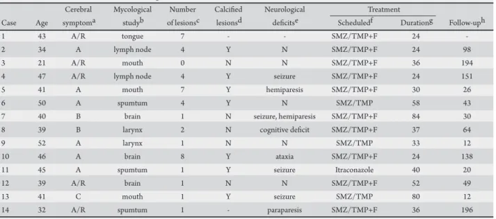
TABLE 1 - Clinical and laboratorial data of the patients.
Cerebral Mycological Number Calciied Neurological Treatment
Case Age symptoma studyb of lesionsc lesionsd deicitse Scheduledf Durationg Follow-uph
1 43 A/R tongue 7 - - SMZ/TMP+F 24
-2 34 A lymph node 4 Y N SMZ/TMP+F 24 98
3 21 A/R mouth 0 N N SMZ/TMP+F 36 194
4 47 A/R lymph node 4 Y seizure SMZ/TMP+F 24 151
5 41 A mouth 7 Y hemiparesis SMZ/TMP+F 30 26
6 50 A spumtum 4 Y N SMZ/TMP 58 43
7 40 B brain 1 N seizure, hemiparesis SMZ/TMP+F 84 30
8 39 B larynx 2 N cognitive deicit SMZ/TMP+F 37 64
9 52 A larynx 1 N N SMZ/TMP 33 12
10 46 A brain 8 Y ataxia SMZ/TMP+F 24 138
11 45 A spumtum 1 Y seizure Itraconazole 40 20
12 39 A/R brain 1 N N SMZ/TMP+F 52 49
13 41 C mouth 1 Y seizure SMZ/TMP 80 12
14 32 A/R spumtum 1 - paraparesis SMZ/TMP+F 36 196
a: onset of neurological disease in relation with systemic PCM, b: place where samples were taken for PCM diagnosis, c: number of lesions observed at irst brain computed tomography scan, d: calciied lesions present in computed tomography scan performed ater treatment, e: Residual deicit at the last visit, f: treatment used, g: length of treatment in months, h: follow-up ater withdrawal of treatment (in months). A: ater, B: before, C: concomitantly, R: cerebral PCM during relapse, Y: yes, N: no, SMZ/TMP: sulfamethoxazole/trimethoprim, F: luconazole.
All patients diagnosed with NPCM at FIOCRUZ, from January 1991 to December 2006, were included in this study and followed until December 2009. As a routine, all PCM patients are submited to clinical, dermatological, neurological and otolaryngological examination. he routine laboratorial tests include HIV serology, ACTH stimulation test, sputum examination for mycological and bacterial analysis, stool examination and serum double-immunodifusion serology for P. brasiliensis. Further investigation with neuroimaging was performed in all cases with neurological symptoms or complaints. Cerebrospinal luid (CSF) analysis was performed in selected cases.
he criteria used to deine NPCM were evidence of P. brasiliensis
infection in the CNS (brain biopsy or positive double-immunodifusion test in CSF) or unequivocal evidence of P. brasiliensis infection in other organs associated with one or more CNS lesions in neuroimaging and posterior involution with speciic treatment.
he treatment protocol used was sulfamethoxazole (2.400mg) plus trimethoprim (480mg) for 2 months followed by sulfamethoxazole (1.600mg) plus trimethoprim (320mg) until the end of the treatment. he dose of luconazole was 200mg a day from beginning to end of treatment.
Between January 1991 and December 2006, 367 consecutive PCM patients were admited to the FIOCRUZ. Fourteen patients (3.8%) (Table 1) were diagnosed with NPCM. One out of 14 was classiied as juvenile-type PCM (patient 3)10; the remaining 13 were
classiied as adult-type PCM.
All patients came from endemic areas and reported a prior history of soil related labor. he median age at diagnosis was 41.78 years-old, ranging from 21 to 57 years of age. hirteen patients were addicted to tobacco cigars.
All patients presented clinical evidence of P. brasiliensis, with positive mycological culture in at least one other organ or tissue (multifocal disease) (Table 1). Evidence of pulmonary disease was detected in 13 (93%) patients. Other organs/systems afected were mucous membranes of the aerodigestive system (11 patients), adrenal glands (7 patients), lymph nodes (5 patients), skin (3 patients) and bone, tongue and liver (1 patient each). Systemic symptoms (fever, weight loss, and anorexia) were present in 12 patients.
Considering symptom chronology, 11 patients presented systemic symptoms before neurological complaints, with a mean period of 24 months (7 to 43). In 5 of these cases, CNS disease occurred ater relapse of the PCM during irregular treatment. CNS disease presented concomitantly with symptoms in other organ in one patient and was the index manifestation in two (Table 1).
The most common neurological manifestation was seizure (57%) followed by hemiparesis (29%), headache (21%) and ataxia (21%). One patient presented spinal cord compression due to an intramedullary, nodular-enhanced lesion at D12 (patient 14)11.
Another patient presented a subacute meningoencephalitis with difuse cortical contrast enhancement without focal brain lesions in brain computed tomography (CT) scan (patient 3)10. In two patients, the
neurological disease initially manifested with psychiatric symptoms. CSF analysis was performed on 5 patients. It was only useful in case 3 (patient with subacute meningoencephalitis), where the liquor double-immunodifusion test presented positive titers of 1:6410.
Computed tomography scan diagnosed a total of 39 brain lesions in 12 patients. Granulomatous, pseudotumoral form was observed in 11 patients; in 3 meningeal enhancement was also observed. Brain lesions varied from 8.5 to 35mm in size (median of 18.8mm) and 95% of these exhibited contrast enhancement (ring-enhancement in 32 and nodular enhancement in 7 lesions). Supratentorial lesions were seen in 64% of patients and in 21% of cases the lesions were located both supra and infratentorially.
24
Rev Soc Bras Med Trop 44(1):22-25, jan-fev, 2011
DISCUSSION
was maintained for a mean period of 41.57 months (24 to 84 months). he criteria for ceasing treatment were absence of disease activity based on systematic clinical examination of the patient, a reduction in serology titers and improvement in radiological studies. In addition to clinical treatment, surgery was indicated for the patient with spinal cord lesion.
One patient presented severe allergy to ketoconazole, amphotericin B, luconazole and sulfamethoxazole/trimethoprim. He was treated with itraconazole, based on a publication writen by Villa et al12.
Posttreatment follow-up was maintained for a mean period of 73.78 months (12 to 196 months). One patient died of a
non-PCM related cause during follow-up (patient 1; cardiac shock). Residual neurological deicits were observed in eight patients, with seizure and motor deicit the most common symptoms reported (Table 1). he use of anticonvulsants was necessary in six patients and this was maintained in four.
A control CT scan was performed in nine patients during the irst year of follow-up (27 brain lesions) and in seven patients during the second year (22 brain lesions). he size and the intensity of edema were the irst signs to show alteration under the treatment, followed by diminished lesions. Approximately one third of lesions calciied ater 23 months. In seven patients, at least one normal brain CT scan was performed before the appearance of a calciied lesion (Figure 1).
FIGURE 1 - Brain computed tomography scan of a patient with cerebral paracoccidioidomycosis showing a hypodense, ring-enhanced lesion in the let thalamus surrounded by mild edema (1A - arrow). A dense calciication was observed several months ater treatment (1B - thin arrow).
he real prevalence of cerebral PCM is unknown. In the present cohort, cerebral PCM was diagnosed in 14 out of 367 PCM patients, showing a prevalence of 3.8%; in other clinical series the prevalence ranged from 4.1% to 13.9%. On the other hand, imaging studies with computed tomography (CT) showed a prevalence of NPCM in 12.5% of cases (5 out of 40 asymptomatic patients) and magnetic resonance (MR) in 40% of cases (10 out of 25 patients, 8 of which were asymptomatic). Although these radiological studies involved few patients, they suggest that neurological disease may be more common than routinely thought6-9,13-15.
he cases in this work all presented multifocal disease and in all cases, it was possible to isolate P. brasiliensis from the afected organs (Table 1). he notion of multifocal disease in PCM and the rarity of unifocal neurological disease should prevent unnecessary brain biopsy in patients with suspicion of NPCM.
It is still unclear what the treatment of choice for NPCM should be. A systematic review of all published cases of NPCM up to 2007
showed that sulfonamide-based treatment, amphotericin B and surgery were the most common treatment approaches used, with an overall mortality rate of 44.1%16. Although amphotericin B is the
drug of choice for severe cases, it is well known that this drug does not have good penetration into the blood-brain barrier and should ideally be used intrathecally for fungal infections of the CNS. Voriconazole is a good therapeutic option for NPCM, due to its action against P. brasiliensis and its good penetration into the CNS. Costs limit their routine use. Itraconazole also does not have adequate penetration into the CNS5.
25
FINANCIAL SUPPORT ACKNOWLEDGMENTS
REFERENCES
he authors declare that there are no conlicts of interest.
CONFLICT OF INTEREST
In ive patients, the neurological disease appeared ater a period of irregular treatment schedule. his fact suggests that the CNS may represent a sanctuary to the fungus and contribute to disease relapse in nonadherent patients.
his study is the irst to highlight the importance of neurological sequelae in the management of NPCM, emphasizing the need to control seizures and physiotherapy support. Ater completing the speciic antifungal therapy, anticonvulsive treatment was maintained in four patients. hree patients were unable to regain muscle strength, despite physiotherapy.
he evolution of neurological imaging is also noteworthy. his paper shows that calciication can appear ater a period of apparently normal CT scans and NPCM should be considered in the diferential diagnosis of cerebral calciication of unknown cause in endemic areas of PCM.
his study has certain weaknesses. First of all, the treatment schedule is based on irst-hand experience of the professionals at our service and should be tested in a multicentric, prospective, double-blind study. he association of luconazole and SMZ/TMP should also be tested in well planned pharmacological studies. On the other hand, NPCM occurs in many poor areas with no access to certain drugs or neuroimaging exams, making the ideal study diicult to conduct. he period of treatment should be programmed individually, based on clinical, radiological, serological and laboratorial data. Neurological sequelae are also an important morbidity that should be considered by all professionals that deal with PCM.
Considering patients who live in rural endemic regions for PCM, NPCM should be included in the diferential diagnosis regarding any neurological clinical picture. he association of luconazole with SMZ/TMP appears to be a viable option in the treatment of patients with NPCM and should at least be considered in cases where other treatment schedules are not working as expected. Anticonvulsive therapy and physiotherapy are necessary to correctly manage the patients alicted by neurological symptoms, at any stage of the disease, including active disease, symptomatic central nervous system calciication or neurological sequelae.
he authors are grateful to Professor Joseph Zunt for kindly reviewing the manuscript.
his study was partially supported by the Ministério da Saúde and Fundação Oswaldo Cruz.
1. Wanke B, Londero A. Epidemiology of paracoccidioidomycosis infection.
In: Franco M, Lacaz C, Restrepo M, editors. Paracoccidioidomycosis. Boca Raton: CRC Press; 1994.p.109-117.
2. Restrepo A. he ecology of Paracoccidioides brasiliensis: a puzzle still unsolved. Sabouraudia1985;23:323-334.
3. Albornoz MB, Albornoz R. Isolation of Paracoccidioides brasiliensis from rural soil in Venezuela. Sabouraudia 1971;9:248-253.
4. Montenegro MR, Franco M. Pathology. In: Franco M, Lacaz C, Restrepo-Moreno A, Del Negro G, editors. Paracoccidioidomycosis London: CRC Press; 1994; p.131-150.
5. Shikanai-Yasuda MA, Telles Filho F de Q, Mendes RP, Colombo AL, Moreti ML. Guidelines in paracoccidioidomycosis. Rev Soc Bras Med Trop2006; 39:297-310.
6. De Almeida SM, Queiroz-Telles F, Teive HA, Ribeiro CE, Werneck LC. Central nervous system paracoccidioidomycosis: clinical features and laboratorial indings. J Infect 2004;48:193-198.
7. Paniago AMM, Oliveira PA, Aguiar ESA, Aguiar JIA, Cunha RV, Leme LM, et al. Neuroparacoccidioidomycosis: analysis of 13 cases observed in an endemic area in Brazil. Trans R Soc Trop Med Hyg 2007;101:414-420.
8. P l a M P, Ha r t u ng C, Me n d oz a P, St u k a n o f f A , Mo re n o M J. Neuroparacoccidioidomycosis: case reports and review. Mycopathologia 1994;127:139-144.
9. Elias Jr J, dos Santos AC, Carloti Jr CG, Colli BO, Canheu A, Matias C, et al. Central nervous system paracoccidioidomycosis: diagnosis and treatment. Surg Neurol2005;63(suppl 1):S13-21.
10. Francesconi F, do Valle ACF, Silva MTT, Costa RLB, Carregal E, Talhari S. Meningoencephalitis due to Paracoccidioides brasiliensis. Neurology 2008;71:e65-e67.
11. do Valle AC, Skacel M, Costa RL, Ribeiro CT, Montagna NA, da Cruz LC. A case report of intraspinal paracoccidioidomycosis. Rev Inst Med Trop Sao Paulo 1998;40:203-207.
12. Villa LA, Tobón A, Restrepo A, Calle D, Rosero DS, Gómez BL. Central nervous system paracoccidioidomycosis. Report of a case successfully treated with itraconazol. Rev. Inst. Med. Trop. Sao Paulo 2000;42:231-234.
13. Hutzler RU, Brussi ML, Capitani Cde M, Lima SS. Neurological involvement of paracoccidioidomycosis, evaluated by computerized skull tomography. Rev Paul Med 1985;103:243-244.
14. Fagundes-Pereyra WJ, Carvalho GT, Miranda Goes A, Chagas Lima e Silva F, Sousa AA. Central nervous system paracoccidioidomycosis: analysis of 13 cases. Arq Neuropsiquiatr 2006; 64:269-276.
15. de Castro CC, Benard G, Ygaki Y, Shikanai-Yasuda MA. MRI of head and neck paracoccidioidomycosis. Br J Radiol 1999; 72:717-722
16. Pedroso VS, Vilela Mde C, Pedroso ER, Teixeira AL. Paracoccidioidomycosis compromising the central nervous system: a systematic review of the literature. Rev Soc Bras ed Trop 2009; 42:691-697.