26
Revista da Sociedade Brasileira de Medicina Tropical 44(1):26-29, jan-fev, 2011
Article/Artigo
INTRODUCTION
Oropharyngeal histoplasmosis: report of eleven cases and review of
the literature
Histoplasmose orofaríngea: relato de onze casos e revisão da literatura
Vicente Sperb Antonello
1, Vanice Ferrazza Zaltron
2, Marcela Vial
3, Flávio Matos de Oliveira
4and Luiz Carlos Severo
4,51. Department of Infectious Diseases, Hospital Nossa Senhora da Conceição, Porto Alegre, RS, Brazil. 2. Department of Internal Medicine, Complexo Hospitalar Santa Casa de Porto Alegre, Porto Alegre, RS, Brazil. 3. Faculty of Medicine, University of Passo Fundo, Passo Fundo, RS, Brazil. 4. Department of Mycology, Complexo Hospitalar Santa Casa de Porto Alegre, Porto Alegre, RS, Brazil. 5. Department of Internal Medicine, Federal University of Rio Grande do Sul, Porto Alegre, RS, Brazil.
Address to: Dr. Vicente Sperb Antonello. Deptº. SCIH/HF. Rua Mostardeiro 17, Bairro Independência, 91430-001 Porto Alegre, RS.
Tel: 55 51 3314-5239
e-mail: [email protected]
Received in 29/04/2010
Accepted in 26/10/2010
ABSTACT
Introduction: Histoplasmosis is a systemic mycosis endemic in Brazil, especially in the State of Rio Grande do Sul, where Histoplasma capsulatum was isolated from the soil. H. capsulatum
may compromise unusual areas, including the oropharynx, particularly in patients presenting disseminated histoplasmosis; which is associated with a state of immunosuppression, such as AIDS. Methods: During database analysis of a total of 265 cases of histoplasmosis, the medical records of 11 patients with histological or microbiological diagnoses of oral histoplasmosis (OH) between 1987 and 2008 were retrospectively reviewed. Results: his work reports 11 cases of OH, the majority presenting histopathological or microbiological evidence of disseminated histoplasmosis (DH). In the patients with DH, OH was the irst manifestation of histoplasmosis. Five of the 11 patients discussed were HIV-seropositive with clinical and laboratory indings of AIDS. Four patients presented active pulmonary tuberculosis concomitant with histoplasmosis. Treatment was based on the use of itraconazole and amphotericin B deoxycholate. Eight patients responded successfully to therapy ater one year, two did not come back for reevaluation and one died despite adequate therapy.
Conclusions: Oral histoplasmosis is closely associated with immunosuppression status, especially in patients presenting AIDS; moreover, in many cases, OH is the irst sign of disseminated histoplasmosis.
Keywords: Histoplasmosis. Histoplasma capsulatum. HIV infection. AIDS.
RESUMO
Introdução: Histoplasmose é uma micose sistêmica, endêmica no Brasil, especialmente no Estado do Rio Grande do Sul, onde Histoplasma capsulatum foi isolado do solo. H. capsulatum
pode acometer áreas não-usuais, como cavidade orofaríngea, particularmente em pacientes com histoplasmose disseminada, por sua vez, associada com estado de imunossupressão, como na AIDS. Métodos: A partir de 265 casos de histoplasmose em um banco de dados de um laboratório de micologia, foram analisados retrospectivamente 11 prontuários de pacientes com diagnóstico histológico ou microbiológico de histoplasmose oral (HO) entre 1987 e 2008.
Resultados: Reportamos neste trabalho onze casos de HO, a grande maioria com evidências histopatológicas e microbiológicas de histoplasmose disseminada (HD). Nos pacientes com HD, HO foi a primeira manifestação de histoplasmose. Cinco dos onze casos relatados eram portadores do vírus do HIV, todos com diagnóstico clínico e laboratorial de AIDS. Quatro pacientes do total tinham concomitantemente tuberculose pulmonar e histoplasmose. Tratamento foi baseado no uso de itraconazol e anfotericina B principalmente. Oito pacientes tiveram sucesso terapêutico após um ano, dois não retornaram para reavaliação e um faleceu apesar da adequada terapia antifúngica. Conclusões: Histoplasmose oral está associada muitas vezes com estado de imunossupressão, especialmente em pacientes com AIDS. Em muitos casos pode representar o primeiro sinal indicativo de histoplasmose disseminada.
Palavras-chaves: Histoplasmose. Histoplasma capsulatum. Infecção pelo HIV. AIDS.
Histoplasmosis is a worldwide systemic mycosis caused by Histoplasma capsulatum, a dimorphic fungus that has been isolated from soil contaminated with bird or bat droppings in endemic areas, as reported in Brazil and central United States1-4.
Infection occurs almost exclusively by inhalation of airborne conidia or mycelial fragments1-4.
he course of histoplasmosis can be inluenced by the immune status of the host and by the quantity of infective propagules the host is exposed to1,5,6.
Disease manifestations range from asymptomatic infection in the normal host with low-inoculum exposure, to rapidly fatal, disseminated infection in the severely immunocompromised host, emphasizing the importance of cellular immunity in defense against H. capsulatum1.
Disseminated histoplasmosis (DH) refers to the relentless growth of the fungus in multiple organ systems. It can develop by re-exposure to a large inoculum of the fungus or by reactivation of dormant endogenous foci1,6. he most important risk factors
are immunosuppression, including transplantation, chronic renal disease, prolonged use of corticosteroids, acquired immune deficiency syndrome (AIDS) and age over 54 years-old1,7. In the last few years,
AIDS has contributed to the increased incidence of DH, described in some series as up to 25%1,6,8.
Occasionally, as a consequence of DH, though less prevalent as a unique manifestation,
H. capsulatum may involve unusual areas, such as the oropharynx. he most common sites of oral cavity afected are the tongue, palate and buccal mucosa8. he clinical signiicance of oropharyngeal
lesions is primarily diagnostic. Goodwin et al showed that up to two-thirds of chronic patients with DH presented oropharyngeal involvement, which was almost invariably part of the clinical status leading to a diagnosis6. Oropharyngeal involvement is
more frequently observed in immunocompromised patients, who oten develop DH1,5.
27
Antonello VS et al - Oropharyngeal Histoplasmosis
RESULTS METHODS
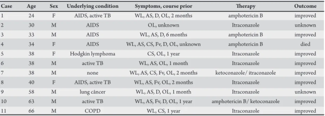
TABLE 1 - Clinical features of 11 patients with oropharyngeal histoplasmosis.
Case Age Sex Underlying condition Symptoms, course prior herapy Outcome
1 24 F AIDS, active TB WL, AS, D, OL, 2 months amphotericin B improved
2 30 M AIDS OL, unknown Itraconazole unknown
3 33 M AIDS WL, AS, D, 6 months amphotericin B improved
4 34 F AIDS WL, AS, CS, Fv, D, OL, unknown amphotericin B died 5 38 F Hodgkin lymphoma CS, OL, 1 year Itraconazole improved 6 38 M active TB WL, AS, OL, 1 month Itraconazole improved 7 38 M none WL, AS, CS, Fv, OL, 2 months ketoconazole/ itraconazole improved 8 40 F AIDS, active TB WL, AS, Fv, OL, 2 months Itraconazole improved 9 58 M lung câncer WL, AS, D, OL, 1 month Itraconazole unknown 10 63 M active TB WL, AS, Fv, D, OL, 1 year amphotericin B/ ketoconazole improved
11 66 M COPD WL, CS, 1 year Itraconazole improved
M: male, F: female, TB: tuberculosis, AIDS: acquired immunodeficiency syndrome, COPD: chronic obstructive pulmonary disease, WL: weight loss, AS: asthenia, Fv: fever, CS: cough with sputum, D: dysphagia, OL: oral lesion.
over from 1987 to 2008 selected from the medical records of the Laboratory of Mycology, Santa Casa Hospital Complex (Complexo Hospitalar Santa Casa), Porto Alegre, Brazil.
During database analysis of a total of 265 cases of histoplasmosis, the medical records of 11 patients with histological or microbiological diagnoses of OH between 1987 and 2008 were retrospectively reviewed. The patients were referred from five public hospitals in Porto Alegre, Brazil, for deinitive diagnosis. Diagnoses were determined in all cases by a combination of the following criteria: direct visualization of oropharyngeal lesion; biopsy of the lesion; and presence of characteristic yeast structures of H. capsulatum. Serology for H. capsulatum was performed on almost all patients. Patients with DH were deined as those with evidence of any other focus of infection other than the oropharynx, as analyzed by clinical examination, laboratory results and radiological data present in their records.
Baseline characteristics
Between March 1987 and October 2008, 11 patients (seven men and four women; age range, 24-66 years-old; mean 42 years-old) with
H. capsulatum oral lesions positive by histological or microbiological methods, were identiied in the iles of the laboratory of mycology of our institution. he baseline characteristics of these patients are shown in Table 1. Five (45.5%) patients had clinical and laboratory diagnosis of AIDS before conirmation of DH and none of them were taking highly active antiretroviral therapy (HAART) regularly. CD4 counts were not available in all patients presenting with AIDS. In three cases, patients presented CD4 < 100/μL. Of the 11 patients, only two presented no evidence other than histoplasmosis in the oropharynx. In seven cases, clinical or microbiological pulmonary involvement was observed concomitant with oral histoplasmosis. Regarding patients with AIDS, four of the ive described presented microbiological or histopathological evidence of DH, proving the strong association between DH and immunocompromised patients, as reported in literature1,3,7. Four (36.4%) patients had a history of
active pulmonary tuberculosis when the diagnosis of DH was made.
Signs and symptoms
The most common presenting symptoms were weight loss (72.7%), asthenia (63.6%), dysphagia (45.5%), chronic cough (36.4%) and fever (36.4%). All patients presented oral lesions. he most commonly involved sites of oropharyngeal histoplasmosis were buccal mucosa (54.5%), tongue (45.5%) and palate (18.2%). One case presented with gingival histoplasmosis, two cases presented concomitant tongue and buccal mucosa involvement and one case with gingival and buccal mucosa involvement by histoplasmosis.
Microscopy, culture, histopathology and immunodifusion for Histoplasma capsulatum
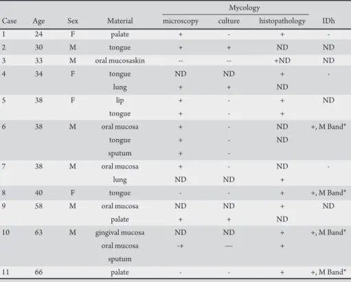
Regarding microbiology and pathology, the organisms were morphologically consistent with the diagnosis of H. capsulatum in the biopsies performed from all cases reported, as shown in Table 2.
Treatment
Patients were treated with amphotericin B deoxycholate, itraconazole or ketoconazole. All patients were administered therapy for at least 6 months despite diferent treatment, except for two cases, treated exclusively with amphotericin B deoxycholate for 3 weeks. Four patients used amphotericin B deoxycholate for induction, with duration ranging from 2 to 4 weeks. Of these patients, one received ketoconazole for a further 6 months, one continued with amphotericin B deoxycholate twice weekly for a further 6 months and one patient stopped therapy ater 3 weeks of initial induction. hese three cases responded well to treatment. One patient, presenting with AIDS, died from systemic sepsis during therapy, despite initial induction with amphotericin B deoxycholate for 3 weeks. Seven patients received itraconazole during treatment. Duration ranged from 6 to 12 months. Five of them responded to therapy. Two patients did not return for reevaluation. In only one case, initial induction was performed with ketoconazole for two weeks, with posterior introduction of itraconazole.
Outcome
28
Rev Soc Bras Med Trop 44(1):26-29, jan-fev, 2011
DISCUSSION
TABLE 2 - Sites where Histoplasma capsulatum was isolated.
Mycology
Case Age Sex Material microscopy culture histopathology IDh
1 24 F palate + - +
-2 30 M tongue + + ND ND
3 33 M oral mucosaskin -- -- +ND ND
4 34 F tongue ND ND +
-lung + + ND
5 38 F lip + - + ND
tongue + - +
6 38 M oral mucosa + - ND +, M Band*
tongue + - ND
sputum + -
7 38 M oral mucosa + - ND
-lung ND ND +
8 40 F tongue - - + +, M Band*
9 58 M oral mucosa ND ND + ND
palate + + ND
10 63 M gingival mucosa ND ND + +, M Band*
oral mucosa -+ — +
sputum
11 66 palate - - + +, M Band*
M: male, F: female, IDh: immunodifusion for histoplasmosis, ND: not done, +: positive, -: negative. *M Band indicates recent contact with the fungus.
Histoplasmosis is a systemic mycosis endemic in Brazil, particularly in the State of Rio Grande do Sul, where H. capsulatum
was isolated from the soil8,9. While most infections are asymptomatic
or self-limiting, some individuals develop acute pulmonary infections or severe and progressive disseminated infection. Although hematogenous dissemination probably occurs in most patients during acute infection, progressive illness is unusual, except in immunocompromised hosts and those at the extremes of age1.
he clinical manifestations of DH and the timing of presentation difer based on host immunodeiciency and degree of exposure to the fungus5. hus, diagnosis is made based on a combination of clinical
indings, serology, light microscopy and microbiological culture of the organism10.
Up to 12% of DH involves the gastrointestinal tract. Lesions more oten involve the colon and ileum, though can occur from the anus to the mouth7. Oral compromise is fairly unusual, with most of
cases presented in patients with DH, as a consequence of the spread of respiratory mycosis acquired through aspiration of airborne spores found in the excreta of birds10-11.
Clinical presentation in the oropharyngeal area includes ulceration, nodular-ulcerative lesions, granulomas, verrucous and plaque-like lesions. Even following the outbreak of AIDS, reports of oral involvement by histoplasmosis have been rare in America and Europe2,10-12.
his study reports 11 cases of oropharyngeal histoplasmosis, mostly with histopathological or microbiological evidence of DH. In nine cases, oral histoplasmosis was the irst manifestation of DH. Five of the 11 patients presented were HIV-seropositive with clinical and laboratory indings of AIDS. Oral lesions of histoplasmosis were
also the irst manifestations of DH in four of the ive cases presenting AIDS.
Six patients of a total of 11 were HIV-seronegative. Four patients presented active pulmonary tuberculosis concomitant with histoplasmosis (two were HIV-seropositive), one presented with lung cancer, one with Hodgkin lymphoma and one patient with chronic obstructive pulmonary disease. Only one patient had no history of concomitant illness at the time of diagnosis of histoplasmosis.
Diagnosis of histoplasmosis was achieved in all cases by fungal identiication from the oral lesion. In seven cases, cultures were performed, with positive results in three cases. Immunodifusion for Histoplasma capsulatum was performed in 10 cases, four of which were positive.
he most common symptoms presented in the cases were weight loss, asthenia, dysphagia, chronic cough and fever. he sites involved in order of frequency of oral histoplasmosis were buccal mucosa, tongue and palate.
Treatment was based on the use of itraconazole (seven patients) and amphotericin B deoxycholate (four patients). Eight patients responded well to therapy ater one year, two did not return for reevaluation and one died despite adequate therapy.
In conclusion, oral histoplasmosis is closely associated with immunosuppression status, especially in patients presenting AIDS1,2,6,10-13. In some cases, it is the initial indication of AIDS, as
reported in literature11,12. Moreover, in the majority of cases, oral
histoplasmosis is the irst sign of DH, as observed in nine of the 11 cases presented in this article.
29
REFERENCEShe authors declare that there are no conlicts of interest. CONFLICT OF INTEREST
1. Deepe Jr GS. Histoplasma capsulatum. In: Mandell GL, Bennet JE, Dolin R, editors. Principles and Practice of Infectious Diseases. 7th ed. Philadelphia: Churchill Livingstone; 2009. p.3305-3318.
2. Severo LC, Petrillo VF, Camargo JJ, Geyer GR, Porto NS. Acute Pulmonary Histoplasmosis and First Isolation of Histoplasma Capsulatum from soil of Rio Grande do Sul, Brazil. Rev Inst Med Trop Sao Paulo1986; 28:51-55. 3. Valle ACF, Moreira LC, Almeida-Paes R, Moreira JS, Pizzini CV, Muniz MM,
et al. Chronic disseminated histoplasmosis with lesions restricted to the mouth: case report. Rev Inst Med Trop Sao Paulo2006; 48:113-116.
4. Lamps LW, Molina CP, West AB, Haggitt RC, Scott MA. The pathologic spectrum of gastrointestinal and hepatic histoplasmosis. Am J Clin Pathol 2000; 113:64-72.
5. Wheat LJ. Diagnosis and management of histoplasmosis. Eur J Clin Microbiol Infect Dis 1989; 8:480-490.
6. Goodwin Jr RA, Shapiro JL, Thurman GH, Thurman SS, Des Prez RM. Disseminated Histoplasmosis: Clinical and Pathologic Correlations. Medicine (Baltimore) 1980; 59:1-33.
7. d´Ávila SCGP. Histopathology and Immunocytochemistry of cutaneous and mucous disseminated histoplasmosis associated with AIDS. Rev Soc Bras Med Trop 1997; 30:429-430.
8. Assi M, McKinsey DS, Driks MR, O’Connor MC, Bonacini M, Graham B, et al. Gastrointestinal histoplasmosis in the acquired immunodeiciency syndrome: report of 18 cases and literature review. Diagn Microbiol Infect Dis 2006; 55:195-201.
9. Severo LC, Oliveira FM, Irion K, Porto NS, Londero AT. Histoplasmosis in Rio Grande do Sul, Brazil: a 21-year experience. Rev Inst Med Trop Sao Paulo 2001; 43:183-187.
10. Rocha MM, Severo LC. Disseminated histoplasmosis in patients with acquired immunodeiciency syndrome (AIDS). Study of 25 cases. Rev Inst Med Trop Sao Paulo1994; 36:167-170.
11. Warnakulasuriya A, Harrison JD, Johnson NW, Edwards S, Taylor C, Pozniak AL. Localised oral histoplasmosis lesions associated with HIV infection. J Oral Pathol Med 1997; 26:294-296.
12. Solari R, Corti M, Cangelosi D, Escudero M, Negroni R, Saccheri C, et al. Disseminated histoplasmosis with lesions restricted to the larynx in a patient with AIDS. Report of a case and review of the literature. Rev Iberoam Micol 2007; 24:164-166.
13. Ferreira OG, Cardoso SV, Borges AS, Ferreira MS, Loyola AM. Oral histoplasmosis in Brazil. Oral Surg Oral Med Oral Pathol Oral Radiol Endod 2002; 93:654-659.